
 Worksheets
Histology - Spinee
Total questions: 49
Worksheet time: 49mins
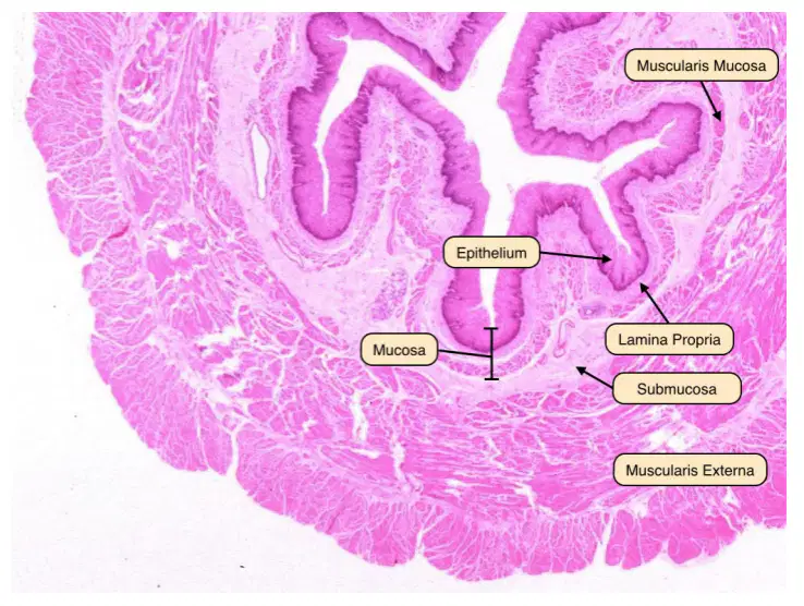
ID the organ
(a)
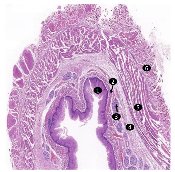
a. ID the organ
(a)
ID the labeled layers/ tissues
1.
(a)
ID the labeled layers/ tissues
2.
(a)
ID the labeled layers/ tissues
3.
(a)
ID the labeled layers/ tissues
4.
(a)
ID the labeled layers/ tissues
5.
(a)
ID the labeled layers/ tissues
6.
(a)
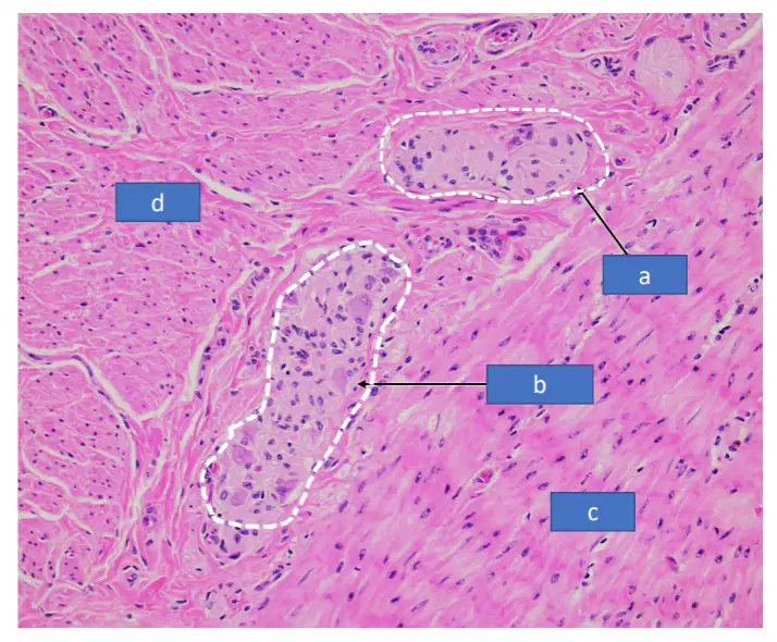
ID the labeled structures
a.
(a)
ID the labeled structures
b.
(a)
ID the labeled structures
c.
(a)
ID the labeled structures
d.
(a)
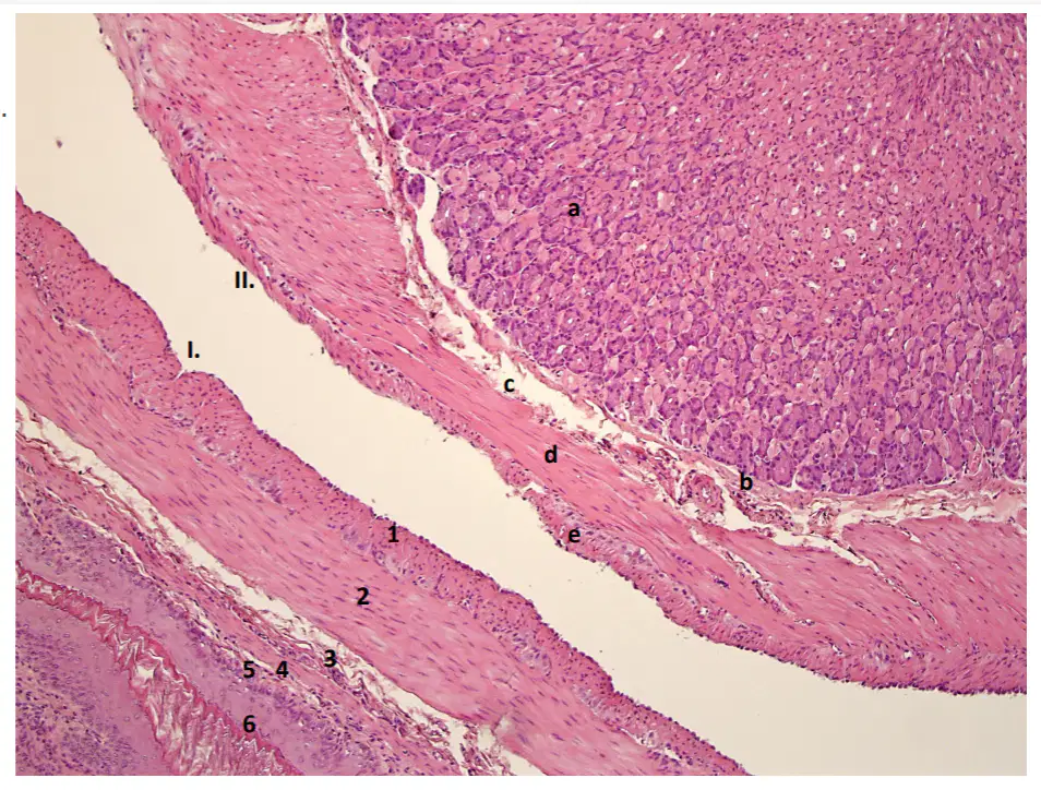
ID the organ
I.
(a)
ID the organ
II.
(a)
Label the image
1.
(a)
Label the image
2.
(a)
Label the image
3.
(a)
Label the image
4.
(a)
Label the image
5.
(a)
Label the image
6.
(a)
Label the image
a.
(a)
Label the image
b.
(a)
Label the image
c.
(a)
Label the image
d.
(a)
Label the image
e.
(a)
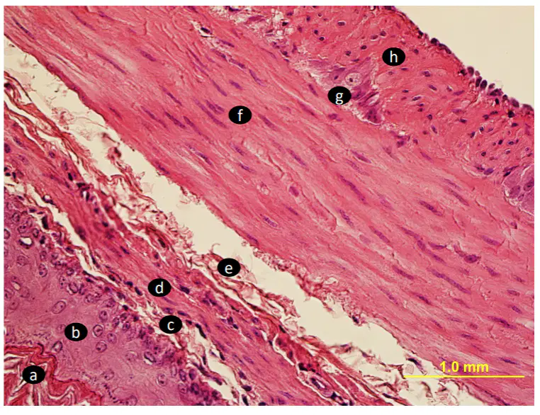
This section is taken at one side of a typical tubular organ, ID labeled regions, cell layer
a.
(a)
This section is taken at one side of a typical tubular organ, ID labeled regions, cell layer
b.
(a)
This section is taken at one side of a typical tubular organ, ID labeled regions, cell layer
c.
(a)
This section is taken at one side of a typical tubular organ, ID labeled regions, cell layer
d.
(a)
This section is taken at one side of a typical tubular organ, ID labeled regions, cell layer
e.
(a)
This section is taken at one side of a typical tubular organ, ID labeled regions, cell layer
f.
(a)
This section is taken at one side of a typical tubular organ, ID labeled regions, cell layer
g.
(a)
This section is taken at one side of a typical tubular organ, ID labeled regions, cell layer
h.
(a)
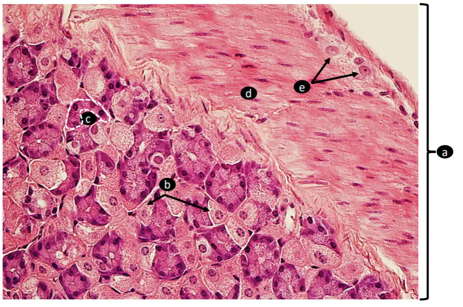
a. ID the segment of the stomach the section is taken from.
(a)
b. ID the specific labeled cells
(a)
c. ID the specific labeled cells
(a)
d. ID the layer
(a)
e. ID the specific cell type
(a)
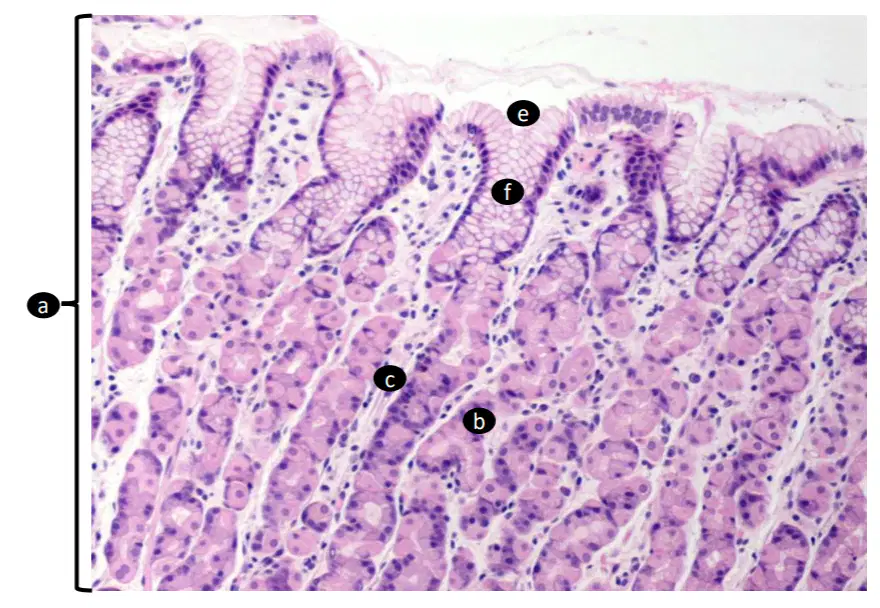
a. ID the specific part of the stomach.
(a)
b. Classify the gland
(a)
c. ID the specific region
(a)
d. ID type of tissue of (c.)
(a)
e. ID the specific region of the gland
(a)
f. ID the specific region of the gland
(a)
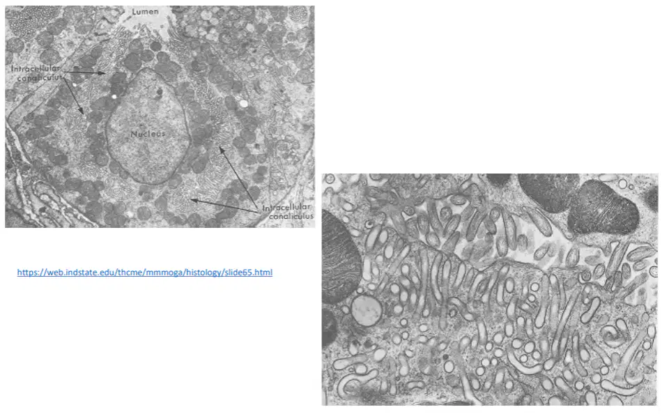
TEM taken within the fundic stomach showing parietal cells. ID as many structures as possible.
(a)
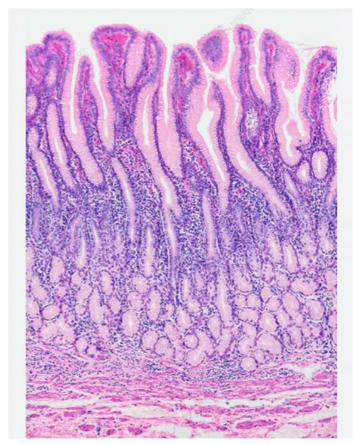
a. ID the specific site this section is taken from.
(a)
b. ID type of epithelium, seen in this entire section
(a)
c. ID type of glands, seen in this entire section
(a)
d. ID the specific layer, seen in this entire section
(a)

