
 Worksheets
Cardiac and Blood Vessel Anatomy and Physiology
Total questions: 137
Worksheet time: 2hrs 28mins
Veins get smaller when they get closer to the heart.
True
False
Capillaries are large blood vessels.
True
False
What is the function of capillaries?
Transports deoxygenated blood
Transport oxygenated blood
Site for exchange of materials
What are present in veins but absent in arteries?
smooth elastic tissues
muscular tissues
valves
How are capillaries adapted for their function?
Have partially permeable walls
One-cell thick endothelium
Presence of valves
Large lumen
Which of the following is NOT a type of blood vessel?
Artery
Capillary
Vein
Ventricle
Which of the following is NOT a major branch of the aorta?
brachiocephalic
right subclavian
left common carotid
left subclavian
What helps blood flow back to the heart? (check all that apply)
Contraction of the diaphragm.
Pumping action of the skeletal muscles.
Valves in the veins.
The narrowing of blood vessels is?
vasoconstriction
vasodilation
The expanding of blood vessels is?
vasoconstriction
vasodilation
Which carries blood away from the heart and is under high pressure?
Artery
Vein
Capillary
Which allows exchange of substances between blood and body tissues?
Artery
Vein
Capillary
Which is under low pressure and contains valves?
Artery
Vein
Capillary
Which is the only vein to carry oxygenated blood?
Aorta
Pulmonary Artery
Pulmonary Vein
Superior and Inferior Vena Cava

Smooth muscles in arteries help
to push blood along through peristalsis.
to prevent the blood vessel from bursting.
to maintain blood pressure.
the blood vessel in elastic recoil in order to push the blood along during diastole.
The highly branched nature of capillaries results in
the blood vessel being very delicate.
a high surface area for material exchange.
a slow blood flow to allow more time for material exchange.
all of the above.

Large amounts of elastic fibers help arteries to
not burst under high pressure.
stretch and recoil to help push blood along it.
help maintain blood pressure.
carry out peristalsis.

Skeletal muscles help blood to move towards the heart when it
contract.
relax.
Low blood pressure in veins is due to
their large lumen relative to the diameter.
lower blood volume.
a lot of pressure had been lost at the capillary bed.
All of the above.

A difference between veins and arteries is that veins have narrow lumens and arteries have wide lumens
True
False

Left ventricle
1
2
3
4

Aorta
1
2
4
5
The function of valves in the human circulatory system is to
stimulate the heart beat.
accelerate the flow of blood.
prevent the backward flow of blood.
serve as a cushion to prevent friction
Which is the correct flow of blood?
R. atrium --> R. Ventricle --> pulmonary artery
R. atrium --> L. atrium --> aorta
L. ventricle --> pulmonary artery --> aorta
L. ventricle --> L. atrium --> aorta
In the walls of the heart, there are two thin layers that form a sandwich around a thick layer of muscle called the
epithelial tissue.
pericardium.
connective tissue.
myocardium.

Pulmonary Veins
A
C
F
I
Blood vessels in the body include arteries, veins, and capillaries.
true
false
The upper chambers of the heart are ventricles.
true
false
The blood vessel that has the largest diameter is
vein
artery
capillary
What type of blood do the arteries usually carry?
Oxygenated Blood
Deoxygenated Blood
Which blood vessels take blood AWAY from the heart?
Veins
Arteries
Capillaries
Which blood vessels take blood TO the heart?
Veins
Arteries
Capillaries
Which of the following is NOT a type of blood vessel?
Artery
Capillary
Vein
Ventricle

In a diagram of a heart and/or blood vessels, red represents...
Deoxygenated blood
(blood without oxygen)
Oxygenated blood
(blood with oxygen)
Mixed oxygenated and deoxygenated blood
None of these
In a diagram of a heart and/or blood vessels, blue represents...
Deoxygenated blood
(blood without oxygen)
Oxygenated blood
(blood with oxygen)
Mixed oxygenated and deoxygenated blood
None of these
Capillaries __________ veins and arteries
seperate
connect

Name this structure:
Vena Cava
Pulmonary Artery
Pulmonary Vein
Aorta
If the P wave on an EKG looks abnormal, which structures in the heart are likely damaged?
SA/AV node
Aortic valve
Bundle of His
Purkinje fibers

The p-wave correlates with which event?
atrial depolarization
ventricle depolarization
ventricle repolarization
atrial repolarization
What does depolarization result in?
relaxation
contraction
repulsion
attraction
What does repolarization result in?
relaxation
contraction
attraction
repulsion

Which of the following describes the network that distributes electrical impulses throughout the heart?
cardiac conduction system
electrocardiogram
HECO
action potential network
Intrinsic electrical impulses start at which of the following locations?
AV Node
Bundle of His
Purkinje fibers
SA Node
Which of the following is most likely to occur if there is a blockage to the AV bundle nerve impulse?
the AV Node will not produce electrical impulses
the SA Node will not produce electrical impulses
no effect
the Purkinje fibers will not produce electrical impulses

The function of the atrium
To collect blood
To pump blood to the lungs
To pump blood into systemic circulation
To pump blood to the heart muscle
The Vena Cava delivers blood to the heart into the.....
left atrium
right atrium
left ventricle
right ventricle
Found in the interventricular septum
AV bundle
AV node
SA node
Purkinje fibers
Network found in the ventricular myocardium
Purkinje fibers
AV node
AV bundle
SA node
The P wave of a normal electrocardiogram indicates:
Atrial depolarization
Atrial repolarization
Ventricular depolarization
Ventricular repolarization
The pacemaker cells of the heart are located in the:
SA node
Purkinje fibers
Wall of the left ventricle
AV node
Both the left and right ventricles
The tricuspid valve is closed:
When the ventricle is in systole
While the ventricle is diastole
While the atrium is contracting
By the movement of blood from atrium to ventricle
The deflection waves in an EKG include:
The T wave, which indicates ventricular repolarization.
The P wave, which is present only in patients who have had a heart attack
The Q-T interval, which indicates the time of atrial contraction
The PQRS complex, which follows ventricular contraction
Blood enters which of these vessels during ventricular systole?
Aorta &Pulmonary arteries
Aorta
Pulmonary vein
Superior vena cava
Pulmonary Artery & Inferior vena cava
___ is to slow heart rate as ___ is to fast heart rate
bradycardia; tachycardia
tachycardia; bradycardia
bradycardia; cardiomyopathy
cardiac tamponade; bradycardia
none of the above
In an EKG, the electrical activity associated with atrial repolarization (relaxation) is usually obscured by the QRS complex
True
False
After the AV node is depolarized and the impulse spreads through the atria, there is a slight delay before the impulse spreads to the ventricles. The reason for this delay is to allow:
The atria to finish contracting
The ventricles to repolarize
A greater venous return
Nothing; there is no reason for the delay
An irregular heart rhythm is known as
Arrhythmia
Bradycardia
Tachycardia
Fibrillation
The correct sequential path of a normal action potential in the heart is:
SA node, AV node, bundle of His, bundle branches, Purkinje fibers
SA node, AV bundle, AV node, Purkinje fibers
AV node, SA node, AV bundle, bundle of His
SA node, AV node, bundle branches, AV bundle, Purkinje fibers
Blood pressure in the large systemic arteries is greatest during:
Ventricular systole
Ventricular diastole
Atrial systole
Isovolumetric diastole
1.atria contract
2. AV node
3. Bundle of His
4.Purkinje fibers
5. SA node
6. ventricles contract

The Purkinje fibers are indicated by ________.
A
B
C
D
E
The atrioventricular bundle (bundle of His) is represented by ________.
A
B
C
D
E
The bundle branches are indicated by ________.
A
B
C
D
E


The SA node fires at a rate of
40-60 bpm
80-90 bpm
60-80 bpm
60-100 bpm
The heart layer that keeps blood from sticking to the inside of the heart and forming potentially deadly blood clots is called
endocardium
pericardium
myocardium
epicardium
A heart rate 100 bpm or above is called _______.
bradycardia
tachycardia
arrhythmia
fibrillation
A cardiac cycle is:
ventricular relaxation
ventricular contraction
equivalent to 1 complete heart beat
a heart attack
The pacemaker of the heart
AV node
SA node
Bundle of His
Bundle branches

Which of the following is true about the semi-lunar valves in the heart?
They are between atria and ventricles
They close when pressure in the arteries is higher than pressure in the atria
They prevent backflow of blood into the atria
They open when pressure in the ventricles is higher than pressure in the arteries
During ventricular systole, which of the following occurs?
Atrioventricular valves close and semilunar valves close
Atrioventricular valves close and semilunar valves open
Atrioventricular valves open and semilunar valves are open
Atrioventricular valves open and semilunar valves close
Define diastole
when the heart is relaxing
when the heart is contracting
duration of the cardiac cycle
frequency of the cardiac cycle
Define systole
when part of the heart is contracting
when part of the heart is relaxing
duration of the cardiac cycle
frequency of the cardiac cycle
The cardiac cycle does not require nerve stimulation to contract like other muscles
True
False


Which artery is located in the leg and supplies blood to the lover limbs and feet? (Highlighted in green).
Radial Artery
Renal Artery
Pulmonary Artery
Femoral Artery

Which artery travels up into the head from the aorta? (highlighted in green)
Brachial
Pulmonary
Carotid
Iliac

Which artery is connected directly to the kidneys? (Highlighted in green).
Renal Artery
Femoral Artery
Anterior Tibial Artery
Aorta

Which vein takes blood away from the kidney?
Portal Vein
Renal Vein
Femoral Vein
Antebrachial Vein

Which vein returns blood from the head back to the heart? (Highlighted in green).
Superior Vena Cava
Renal Vein
Jugular Vein
Hepatic Vein

Which vein takes blood from the liver and delivers it to the heart via the inferior vena cava? (Highlighted in green)
Inferior Vena Cava
Hepatic Vein
Portal Vein
Femoral Vein

Which vein takes blood from the intestines and delivers it to the liver to filter toxins? (Highlighted in green)
Hepatic Vein
Mesenteric Vein
Hepatic Portal Vein
Iliac Vein
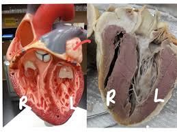
Which side of the heart handles oxygenated blood?
Left side only
Right side only
Both right and left
Neither side
Which specimen did we use in place of studying a human heart?
Swine
Sheep
Bovine
Feline
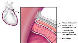
Double layered closed sac that surrounds the heart & anchors it is called:
Pericardium
Pericardial cavity
Myocardium
Papillary muscles

The muscle of the heart is called:
Pericardium
Cardicardium
Myocardium
Endocardium

The front of the hear is recognized by a groove that extends from the right side of broad end of the heart diagonally to a point above, left of the apex. Name this feature.
Interatrial groove
Interventricular gyri
Interventricular sulcus
Interventricular fissure

The atria are the _____________ chambers.
Receiving
Pumping
Discharge
Oxygenated

The ventricles are the _____________ chambers.
Receiving
Pumping/Discharge
Deoxygenated
Thinner

This major vessel is a large artery that supplies blood to your upper & lower body.
Pulmonary
Coronary
Aorta
Interior & superior vena cava

These vessels return oxygenated blood from right and left lungs to the left atrium.
Pulmonary artery
Pulmonary veins
Aorta
Interior/superior vena cava

The long fibers of connective tissue that attach to papillary muscles of the heart that enable the valves to properly open and close are called:
Chordae tendinae
Cardiac tendons
Cardiac ligaments
Myocardial tendinae

This valve allows blood flow from the right atrium into the right ventricle during ______________ (heart is relaxed)
diastole
systole

Within the right ventricle the one way valve that controls blood flow away from the right ventricle is called:
Pulmonary valve
Tricupsid valve
Bicupsid valve
Aortic valve

Identify # 12?
Bicupsid valve
Tricupsid valve
Seminlunar valve
Aortic valve

The valve labeled #7 is
bicuspid
tricuspid
aortic semilunar
pulmonary semilunar

Identify the chamber labeled #1
right atrium
left atrium
left ventricle
right ventricle

Identify the structure labeled #5
interventricular sulcus
apex
left ventricle
septum

The structure labeled #6 is
left auricle
right auricle
left ventricle
adipose tissue
If you were to insert your finger into the superior vena cava, you would be feeling inside which chamber?
right atrium
left atrium
left ventricle
right ventricle
If you inserted a probe into the aorta, which chamber of the heart would it lead to?
left ventricle
right ventricle
left atrium
right atrium
If you insert your finger into the pulmonary vein, you will be feeling inside which chamber?
right atrium
left atrium
left ventricle
right ventricle
The pulmonary artery connects to which heart chamber?
right ventricle
left ventricle
right atrium
left atrium
The first branch of the aorta is the
brachiocephalic artery
left common carotid
left subclavian
pulmonary vein
If you are looking at the dorsal side of the heart you are looking at the (a) side of the heart.

Identify the Left Atrium.
C
D
E
F
Identify the Right Ventricle.
C
D
E
F

Which valve is indicated by the red rectangle?
Pulmonary valve
Aortic valve
Mitral (bicuspid) valve
Tricuspid valve

Which structure is indicated by the red rectangle? Hint - this structure holds the bicuspid valve in place.
Trabeculae carneae
Papillary muscles
Chordae tendineae
Pulmonary valve

The structure indicated by the red dot is known as the __________.
Right ventricle
Right Atrium
Interventricular septum
Left Ventricle
What is the inside lining of the heart called?
pericardium
endercardium
extracardium
endocardium
What is the name of the sac that surrounds the heart?
endocardium
pericardium
intercardium
extracardium
What type of tissue comprises the bulk of the myocardium
skeletal muscle
cardiac muscle
smooth muscle
What layer of an artery consist mostly of smooth muscle?
tunica smoothica
tunica intima
tunica externa
Tunica media

