
NEW
Font size
 Worksheets
EKG for SO
Total questions: 20
Worksheet time: 12mins

Label # 5 in the image
PR interval
QRS complex
T wave
P wave
On an ECG, atrial depolarization is indicated by the
P wave
T wave
QRS complex
U wave
On an ECG, ventricular depolarization is indicated by the
QRS
T wave
P wave
PR interval
Ventricular repolarization is represented on the ECG by the
T wave
QRS
P wave
S wave
What is the bpm range for normal sinus rhythm?
60-100
60-80
80-100
100-120
What is the term for rapid heartbeat?
Angina Pectoris
Fibrillations
Tachycardia
Ischemia
What wave on a rhythm strip indicates the repolarization of the ventricles?
P
Q
R
S
T
What does the T wave represent
The contraction of the ventricles
The contraction of the atria
if the rhythm is normal
The relaxation of the heart.

Which of the following best describes this strip?
Tachycardia
Normal Sinus Rhythm
Bradycardia
Asystole

Which of the following best describes this strip?
Tachycardia
Normal Sinus Rhythm
Bradycardia
Asystole

Which of the following best describes this strip?
Tachycardia
Normal Sinus Rhythm
Bradycardia
Asystole

How many cardiac cycle shown in the diagram
2
4
5
6
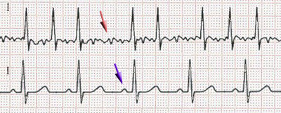
What is the ECG showing (Red Arrow)
Atrial Fibrillation
Ventricular Fibrillation
Asystole
Premature Ventricular Contraction
Heart Block

What is the ECG showing
Atrial Fibrillation
Ventricular Fibrillation
normal
Premature Ventricular Contraction
Heart Block
Common noninvasive diagnostic test which records the electrical activity of the heart
Echocardiogram
Electrocardiogram
Electrophysiology
PR Interval:
represents the time from the beginning of atrial depolarization to the beginning of ventricular depolarization
represents atrial depolarization
represents ventricular depolarization
Located in the proximal intraventricular septum. It merges from the AV node to begin the conduction of the impulse from the AV node to the ventricles.
Bundle of His
ST segment
T wave
Electrode
A small rounded wave on an EKG that indicates atrial contraction.
T wave
AV node
P wave
SA node
A cluster of special muscle fibers located in the right atrial wall of the heart considered to be the primary pacemaker of the heart.
Av node
SA node
SV node
AV node
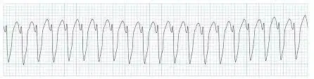
What disorder is indicated on this ECG strip?
Atrial Fibrillation
Ventricular Tachycardia
Pulseless Electrical Activity (PEA)
Normal Sinus Rhythm

