
 Worksheets
NIU Anatomy Review
Total questions: 203
Worksheet time: 2hrs 13mins
The upper chambers of the heart are called (a) .
Which ventricle has the thickest walls?
Left Atrium
Right Atrium
Left Ventricle
Right Ventricle

Letter D is pointing to
Aorta
Right Pulmonary Artery
Superior Vena Cava
Pulmonary Trunk

Letter G is pointing to
Right atrium
Left Atrium
Right ventricle
Left ventricle

Of the four chambers, which one carries deoxygenated blood?
F and J
F and A
A and G
J and G

Which letter is pointing to the tricuspid valve?
M
N
L
O
The mitral valve is another name for the
Aortic valve
Pulmonary valve
Tricuspid valve
Bicuspid valve
What large vessel takes blood to the bottom half of the body?
descending aorta
pulmonary arteries
ascending aorta
both vena cava

The aortic semilunar valve is letter
O
L
M
N
Prevention of backflow is the function of the
pulmonary arteries
valves
septums
vena cava

What is the arrow pointing to?
pulmonary artery
myocardium
pulmonary vein
aorta

What is the arrow pointing to?
pulmonary artery
myocardium
pulmonary vein
aorta


#15
superior vena cava
inferior vena cava
aorta
septum


Pulmonary Semilunar Valve


Chordae Tendineae


Name the fibrous cords that aid in opening/closing the AV valves
Papillary muscles
Capillary muscles
Chordae tendineae
Aortic strings

The long fibers of connective tissue that attach to papillary muscles of the heart that enable the valves to properly open and close are called:
Chordae tendinae
Cardiac tendons
Cardiac ligaments
Myocardial tendinae

Identify the chamber labeled #1
right atrium
left atrium
left ventricle
right ventricle

Identify #9 (a)
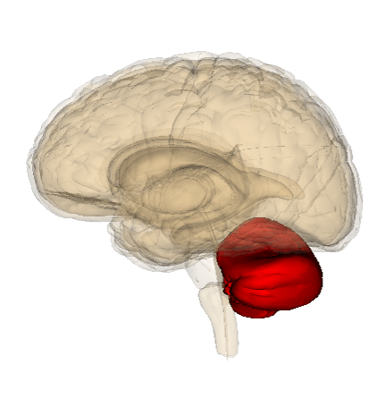


What structure is GREY in this picture?
Cerebrum
Cerebellum
Brainstem
Pons

The cerebellum controls ____________
senses
movement, posture, balance and coordination
What part of the brain is respobsible for visual perception
Occiptial lobe
Frontal lobe
Cerebelum
Thalamus
Damage to the medulla oblongata would most likely result in
A person has a stroke in her occipital lobe. What might happen to her?
thirst and hunger
cerebellum
brain stem
hypothalamus
hippocampus
Which brain structure filters and relays sensory info to the appropriate areas of the cerebral cortex?
thalamus
hypothalamus
hippocampus
cerebellum
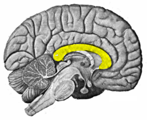

Which part of the brain stem contains visual and auditory reflex centers?
Midbrain
Pons
Medulla oblongata
Thalamus
Which part of the brain stem relays information from the cerebrum to the cerebellum and is also called the bridge?
Midbrain
Pons
Medulla oblongata
Thalamus
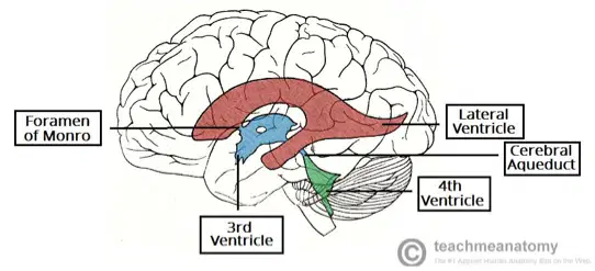
Reorder the following for the Flow of CSF through the ventricles. CSF starts in the Choroid Plexus......
Lateral Ventricles
Foramen of Monro
3rd Ventricle
Cerebral Aquaduct
4th Ventricle
Match the following (hindbrain)
Cerebellum
Motor activity, balance, musical rhythm
Pons
Relay to the cerebellum on opposite side
Medulla oblongata
homeostasis, breathing, heart rate, BP
Label 1-6
Frontal Lobe
Temporal Lobe
Parietal Lobe
Occipital Lobe
Cerebellum
Brain stem
Label A,B,C & D
Hypothalamus
Corpus Callosum
Thalamus
Medulla
Which brain structure is located in the hindbrain?
Hpothalamus
Cerebral cortex
Hippocampus
Substantia Nigra
Pons

Identify #1
Midbrain
Medulla
Cerebellum
Pons
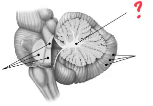
Cerebellum
Vermis
Folia
Arbor vitae
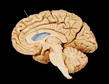
Lateral ventricles
Third ventricle
Fourth ventricle
Choroid plexus
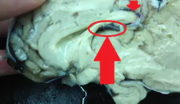
Lateral ventricles
Third ventricle
Fourth ventricle
Choroid plexus
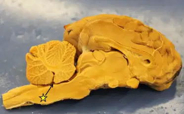
Lateral ventricles
Third ventricle
Fourth ventricle
Choroid plexus
The spleen's surface most hugs which of the following organs?
Stomach
Large Intestine
Kidney
Pancreas
Small Intestine
The spleen is located adjacent to ribs...
11 - 12
9 - 11
8 - 12
10 - 12
8 - 10
The spleen is a __ organ
Red
Deep Red
Purple
Deep Purple
Pink
What is the largest organ of the lymphatic system?
Appendix
Spleen
Thymus
Soup
Which of the following regions of the abdomen is the spleen located in?
Right hypochondrial region
Left hypochondrial region
Right iliac region
Left iliac region
The spleen is often referred to as the "graveyard of RBCs" because:
It is the primary site of RBC production in the body
It stores a large number of RBCs for future use.
It filters the blood and removes microorganisms, including RBCs
It destroys old and fragile red blood cells as they pass through its thin capillaries.
A combining form for urine is:
ur/o
cyst/o
void/o
A combining form meaning kidney is:
nephr/o
ur/o
cyst/o
How many urethras does a person typically have?
one
two
three
How many ureters does a person typically have?
one
two
three
four
Filtering blood to remove excess water and waste products is the job of the:
kidneys
ureters
urinary bladder
Transporting urine from the kidneys to the bladder is the job of the:
kidneys
ureters
urethra
The functional unit of the kidney is the:
Urethra
Ureter
Nephron
Hilus
Which of the following structures carries blood that contains waste?
Renal vein
Renal artery
Loop of Henle
Collecting duct
The functional unit of the kidney is called __________.
a glomerulus
a nephron
a renal corpuscle
a renal capsule

Identify the structure labelled "A" in the diagram.
Kidney
Ureter
Bladder
Urethra
Identify the structure labelled "B" in the diagram.
Kidney
Ureter
Bladder
Urethra
Identify the structure labelled "C" in the diagram.
Kidney
Ureter
Bladder
Urethra
Identify the structure labelled "D" in the diagram.
Kidney
Ureter
Bladder
Urethra
Which of the following is NOT a function of the kidneys?
Regulation of pH
Regulation of body fluid volume
Removal of waste products from the body
Formation of plasma proteins
The outermost region of the kidney is called the:
(a)
Dehydration causes your urine to become _________.
lighter
darker
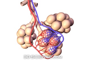
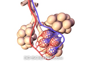
The "windpipe" is also called the...
trachea
bronchi
bronchioles
alveoli
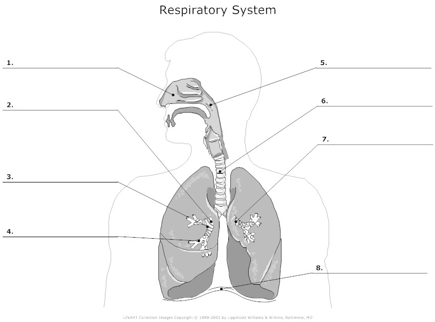
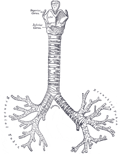

What is #8 pointing to?
nasal passageway
nose
right lung
left lung

What is #1 pointing to?
larynx
pharynx
alveoli
bronchi

Which number is pointing to the diaphragm?
8
7
6
5

Which numbers point to the right and left lungs?
5 and 10
10 and 11
5 and 15
15 and 16
Which two muscles contract and create more space in the chest cavity?
diaphragm and intercostals
rib muscles and esophagus muscles
peristalsis and rib muscles
stomach and rib muscles
Why does the pleural cavity have fluid?
This fluid provides oxygen for the lungs.
This fluid keeps the lungs from getting thirsty.
This fluid provides lubrication for the lungs to expand and contract.
This fluid keeps the lungs clean.
Gaseous exchange happens because of...
Osmosis
Respiration
Diffusion
Semi-permeable membranes
The voicebox is also known as the
(a)


Which parts of the diagram is the small and large intestine?
A~ small intestine
E~ large intestine
D~ small intestine
E~ large intestine
C~ large intestine
B~ small intestine
A~ large intestine
C~ small intestine
E~ small intestine
D~ large intestine

produces enzymes for the small intestine that help break down starches, proteins, and fats
pancreas
large intestine
liver

absorbs water into bloodstream
gall bladder
large intestine
liver

organ that stores bile
gall bladder
large intestine
liver

produces bile to break up fat particles
pancreas
large intestine
liver

Digestive organ where most chemical digestion and absorption of food takes place
pancreas
small intestine
liver

large muscular sac that continues the mechanical and chemical digestion of food
stomach
small intestine
liver

tube connecting the mouth to the stomach
stomach
esophagus
peristalsis

where digestion begins
chemical digestion
mouth
mechanical digestion

body system that breaks down food and absorbs nutrients
digestive system
mouth
mechanical digestion
Breaking food down by way of saliva, stomach acid, and absorbing nutrients is called (a) .
Breaking food down into small pieces by way of the teeth and twisting of the esophagus is called (a) .

Where is the duodenum ?
A
B
C
E
F
Where is the gallbladder ?
A
J
K
I
H

Where is the pancreas located in the body?
a. Above the heart.
b. Underneath the liver and near the stomach.
c. In the chest.
d. Near the kidneys.

The chewing of your food is called what?
Eating
Mastication
Swallowing
Enzymes

Where does MOST of the digestion process occur?
Small intestine
Mouth
Stomach
Large Intestine
Colon

Pick the correct order of digestion
Mouth,stomach,esophagus,large intestine,small intestine
Mouth,esophagus,stomach,small intestine, large intestine
Mouth, esophagus, stomach, large intestine, small intestine, colon
Mouth, stomach, esophagus, small intestine, large intestine

What organ produces Insulin?
Liver
Stomach
Appendix
Pancreas

Another name for the Large Intestine?
Really big intestine
Colon
Pancreas
Massive

What is this body part?
Small Intestine
Large Intestine
Stomach
Brain

What is this body part?
Liver
Heart
Intestine
Pancreas

Which body part is shown here?
Brain
Kidneys
Bladder
Heart
Which part of the small intestine mixes chyme with bile and pancreatic enzymes
jejunum
ileum
duodenum
Which part of the small intestine is responsible for the absorption of nutrients
duodenum
jejunum
Ileum
Which organ secretes sodium bicarbonate into the duodenum to neutralize stomach acid
liver
spleen
pancreas
kidney
The small intestine is subdivided into three segments. Which of the following is the correct sequence regarding the passage of food through the small intestine?
Duodenum, ileum, jejunum
Duodenum, jejunum, ileum
Jejunum, duodenum, ileum
Ileum, jejunum, duodenum
Parts of the small intestine after the duodenum is known as
Jejunum
Lipase duodenum
Amylase duodenum
Lipase ileum


The appendix is attached directly to the:
cecum
pancreas
jejunum
sigmoid colon
Following the sections of the large intestine, as material passes from the tranverse colon, what section does it enter next?
ascending
descending
sigmoid
rectum
Identify the first segment of the small
Jejunum
Ilium
Duodenum
Peristalsis
The last section of the large intestine is the
Sigmoid colon
Cecum
Transverse colon
Ileum
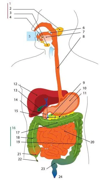
What structure is at 17?
Cecum
Ascending colon
Transverse Colon
Sigmoid Colon
Which is the largest artery in the human body ?
Carotid Artery
Aorta
Subclavin Artery
Femoral Artery
Which vein brings the carbon dioxide rich blood from the upper half of the body to the heart ?
Superior vena cava
Inferior vena cava
Azygos vein
Hemiazygos vein
Which artery carries the oxygen rich blood from the heart to the brain and the head ?
Aorta
Hemiazygos vein
Carotid artery
Subclavin Artery
Vessels that carry high pressure blood
arteries
capillaries
veins

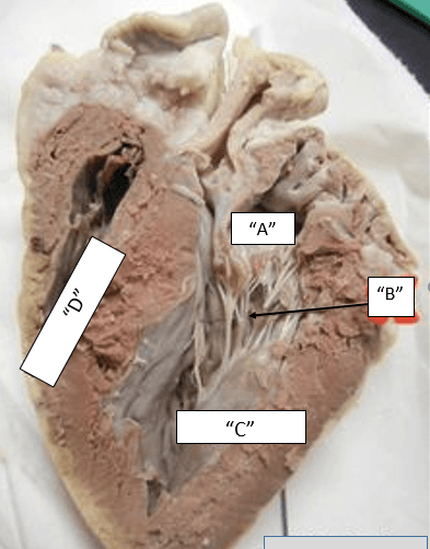
Identify "B" in this diagram.
chordae tendinae
interventricular septum
right ventricle
right atria

What muscle is shown?
Aponeurosis
Internal oblique
Internal intercostals
Diaphragm

What muscle is shown?
Serratus posterior inferior
Erector spinous
Supraspinatus
Serratus posterior superior

What muscle is shown?
Rectus abdominis
Transversus abdominis
External oblique
Internal oblique

What muscle is shown?
External oblique
Transversus abdominis
Internal oblique
Rectus abdominis

What muscle is shown?
Semispinalis capitis
Erector spinous
Trapezius
Latissimus dorsi

What muscle is shown?
External oblique
Rectus abdominis
Internal oblique
Linea alba

What muscle is shown?
Rhomboideus major
Rhomboideus minor
Levator scapulae
Serratus anterior

What muscle is shown?
Serratus posterior inferior
Sternocleidomastoid
Splenius capitis
Semispinalis capitis

What muscle is shown?
Levator scapulae
Teres minor
Teres major
Sternocleidomastoid

What muscle is shown?
Levator scapulae
Teres minor
Teres major
Sternocleidomastoid

What muscle is shown?
Transversus abdominis
Internal oblique
External oblique
Rectus abdominis

What muscle is shown?
Trapezius
Latissimus dorsi
Rhomboideus major
Rhomboideus minor

Identify the muscle
gastrocnemius
semitendinosis
biceps femoris
semimembranosis

name the muscle
Vastus Medialis
Vastus Intermedius
Rectus Femoris
Vastus Lateralis

fibularis longus
soleus
gastrocnemius
tibialis anterior
popliteus

fibularis longus
soleus
gastrocnemius
tibialis anterior
popliteus

fibularis longus
soleus
gastrocnemius
tibialis anterior
popliteus

What is the name of the muscle labeled #1?
temporalis
Masseter
Trapezius
sternocleidomastoid
deltoid
What is the name of the muscle labeled #2?
temporalis
Masseter
Trapezius
sternocleidomastoid
deltoid

What is the name of the muscle labeled #6?
sartorius
gracilis
Gluteus medius
gluteus maximus
Supraspinatus
What is the name of the muscle labeled #4?
Trapezius
deltoid
. latissimus dorsi
infraspinatus
Supraspinatus

Number 30 represents the ____
Gastrocnemius
Tendon of Quadriceps Femoris
Biceps Femoric
Deltoid
Number 12 represents the ____
Tibialis Anterior
Tendons
Soleus
Peroneus

Number 2 represents the ____
Gluteus Maximus
Tensor Fascia Latae
Pectineus
Peroneus
Number 4 represents the ____
Gluteus Maximus
Tensor Fascia Latae
Pectineus
Peroneus
Number 3 represents the ____
Sartorius
Tensor Fascia Latae
Pectineus
Peroneus


What is the name of the small light yellow muscles that run lateral on the upper thighs?
Tensor Fascia Latte
Vastus Lateralis
Vastus Medialis
Rectus Femoris
What is the name of the bright green muscles (part of the Quadriceps)?
Tensor Fascia Latte
Vastus Lateralis
Vastus Medialis
Rectus Femoris
What is the name of the dark blue/purple muscles (part of the Quadriceps)?
Tensor Fascia Latte
Vastus Lateralis
Vastus Medialis
Rectus Femoris
What is the name of the light blue muscles right above the knees (part of the Quadriceps)?
Tensor Fascia Latte
Vastus Lateralis
Vastus Medialis
Rectus Femoris
What is the name of the bright pink muscle?
Sartorius
Gracilis
Vastus Medialis
Rectus Femoris
What is the name of the orange muscle that is lateral to the Gracilis?
Sartorius
Adductor Magnus
Adductor Longus
Rectus Femoris

What is the name of the blue muscles (part of the hamstrings)?
Gluteus Maximus
Gluteus Medius
Semitendinosus
Bicep Femoris
What is the name of the bright green muscles (part of the hamstrings)?
Gluteus Maximus
Semimembranosus
Semitendinosus
Bicep Femoris
What is the name of the neon blue muscles (part of the hamstrings)?
Gluteus Maximus
Semimembranosus
Semitendinosus
Bicep Femoris
What is the name of the orange muscles?
Gastrocnemius
Soleus
Tibialis Anterior
Bicep Femoris

Name this movement
adduction
abduction
inversion
eversion

Name this movement
inversion
eversion
adduction
abduction

