
 Worksheets
AIOC OPTOS
Total questions: 10
Worksheet time: 5mins
What is the core technology for single scan Ultra Wide Field retinal imaging?
Adaptive optics
Elliptical mirror system
Infrared laser diode
Ultra-widefield cameras in a single capture can image what part of the retina
15%
200%
82%
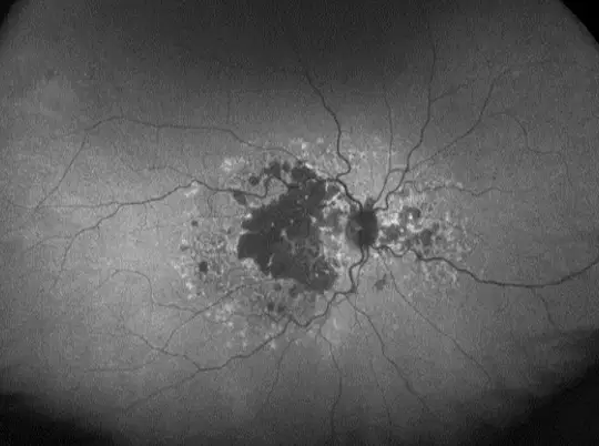
This modality of imaging helps in
Visualization of RPE changes
Inherited retinal diseases
Multifocal central serous retinopathy
All the above
What is the advantage of ultra-widefield retinal imaging over traditional methods?
It provides lower image resolution
It allows for visualization of peripheral retinal pathology
It is invasive in nature
Wide field angiography on OPTOS
Allows for guided treatment
No historical image comparison
Needs full mydriasis
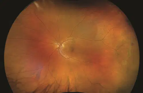
Imaging mode?
FAF
True color
Pseudocolor
Color RGB

A normal appearing macula shows this on the periphery, what is it?
Retinal detachment
Peripheral exudative hemorrhagic chorioretinopathy
Dislocated lens
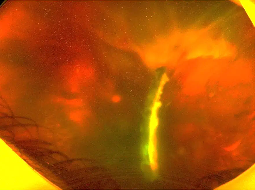
A challenging surgery, what are we looking at?
Intraocular lens
Intraocular cysticercosis
Intraocular foreign body
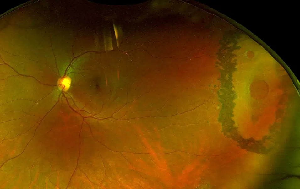
What is this lasered lesion?
Atrophic retinal hole
Horse shoe tear
Lattice degeneration
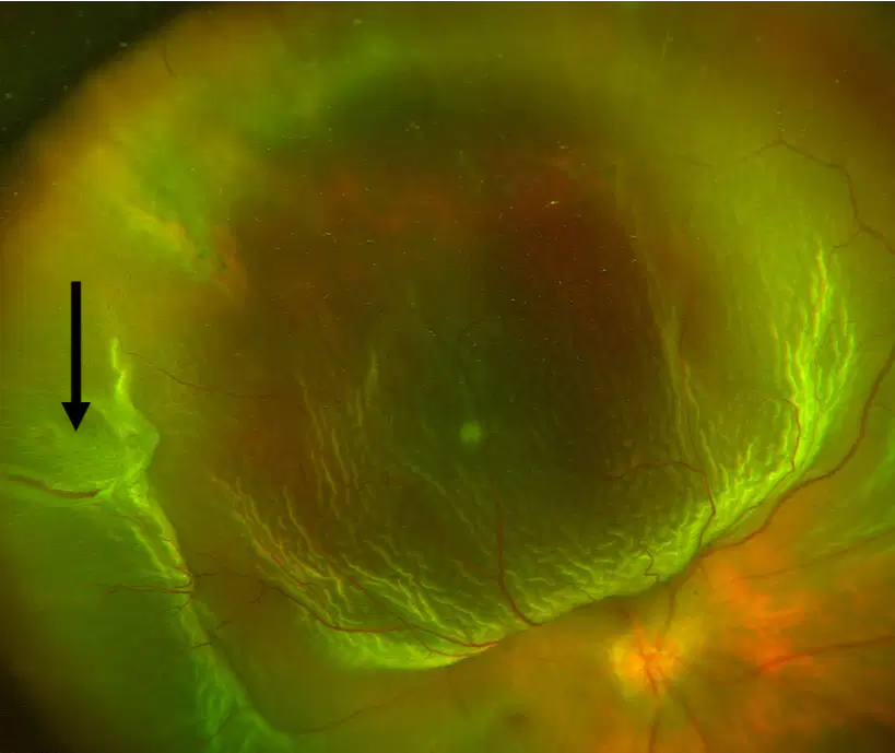
Pay attention to the arrow and diagnose this case
Exudative retinal detachment
Tractional retinal detachment
Rhegmatogenous retinal detachment
Combined retinal detachment

