
 Worksheets
Preoperative Saphenous Vein Mapping Quiz
Total questions: 61
Worksheet time: 31mins
What is the purpose of preoperative saphenous vein mapping?
To create a "map" of the leg veins for the surgeon in preparation for various procedures
To measure the blood pressure in the leg veins
To diagnose varicose veins
To check for blood clots in the leg veins
Which procedure is preoperative saphenous vein mapping commonly used to prepare for?
Bypass graft surgery
Appendectomy
Hip replacement surgery
Cataract surgery
What is one of the main uses of the healthy vein from the leg in preoperative saphenous vein mapping?
Replacing diseased vessels in the body
Measuring blood sugar levels
Treating infections
Monitoring heart rate
Which of the following factors affects vein performance as an arterial conduit?
Vein color
Vein quality
Vein location
Vein temperature
What must be checked during the ultrasound imaging of the saphenous vein?
Vein diameter, vein quality, and vein morphology
Vein color, vein temperature, and vein location
Vein length, vein width, and vein depth
Vein pressure, vein elasticity, and vein flow rate
Which of the following is NOT a part of vein morphology that must be checked?
Presence of recanalization
Sclerotic areas
Thick vein walls
Vein color
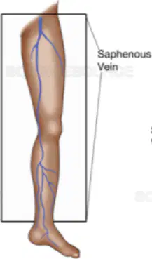
Which vein is most commonly used as a conduit for surgical coronary revascularization?
Long Saphenous Vein (LSV)
Short Saphenous Vein (SSV)
Femoral Vein
Jugular Vein
Which of the following is an advantage of the procedure mentioned in the document?
It is highly invasive.
It is rapidly accomplished.
It requires extensive surgical dissection.
It often leads to wound complications.
What is one of the benefits of the procedure in terms of surgical site selection?
It allows suboptimal surgical site selection.
It does not affect surgical site selection.
It allows optimal surgical site selection.
It complicates surgical site selection.
How does the procedure help in terms of surgical dissection and wound complications?
It increases the need for surgical dissection.
It avoids unnecessary surgical dissection and potential wound complications.
It has no effect on surgical dissection.
It increases potential wound complications.
Which of the following factors is assessed during preoperative saphenous vein mapping?
Blood type
Medical history
Eye color
Height
What is NOT a factor assessed during preoperative saphenous vein mapping?
Condition of skin
Recent or past surgery on the planned donor leg
Current medications
Dietary preferences
During preoperative saphenous vein mapping, which of the following is evaluated regarding the planned donor leg?
Blood pressure
Recent or past surgery
Heart rate
Vision
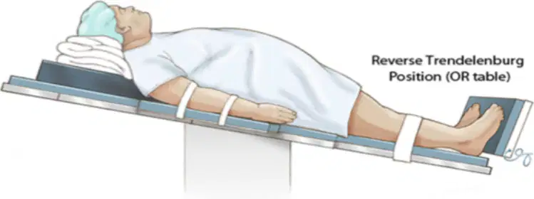
What is the position of the patient as described in the learning material?
Prone
Supine
Lateral
Sitting

Where is the tourniquet placed on the patient?
Around the upper arm
Around the lower leg
Around the upper thigh
Around the neck
What is the purpose of placing the patient in reverse Trendelenburg position?
To aid in arterial filling
To aid in venous filling
To aid in lymphatic drainage
To aid in respiratory function
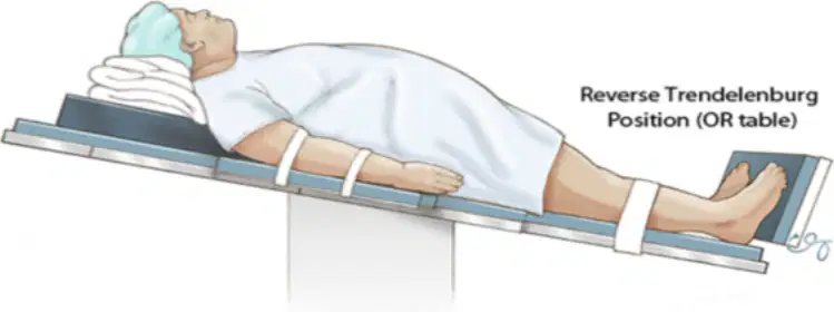
How is the leg positioned to optimize access during the procedure?
Fully extended and internally rotated
Slightly flexed and externally rotated
Fully flexed and internally rotated
Slightly extended and externally rotated
What is the initial step in the ultrasound technique described in the document?
Setting the ultrasound machine to a depth of 2.8 cm
Applying a generous amount of ultrasound gel to the donor leg
Identifying the vein on the ultrasound screen
Adjusting the depth as the thigh is reached
To what depth is the ultrasound machine initially set for scanning below the knee?
1.5 cm
2.8 cm
3.5 cm
4.0 cm
How is the vein identified and confirmed on the ultrasound screen?
As a solid, non-compressible structure
As a tubular, compressible structure
As a flat, non-compressible structure
As a round, non-compressible structure
What is done to the ultrasound machine as the thigh is reached?
The depth is decreased
The depth is increased
The machine is turned off
The gel is reapplied
What does LSV stand for in the context of the document?
Lateral Saphenous Vein
Long Saphenous Vein
Large Saphenous Vein
Lower Saphenous Vein
What must be assessed and measured at multiple sites during the technique?
The length of the vein
The caliber of the vein
The color of the vein
The temperature of the vein
What should be done unless the patient has undergone prior varicose vein stripping of the LSV?
Only one leg is mapped
Both legs are mapped
No legs are mapped
The arms are mapped
Which of the following must be documented during the technique?
Small tributaries
Blood pressure
Large tributaries
Heart rate
Which of the following is NOT required to be documented during the technique?
Large tributaries
Varicosities
Duplicate systems
Blood type
What is the best plane to image a vein for measurements?
Sagittal plane
Transverse plane
Coronal plane
Oblique plane
What should be marked when measuring vein diameter?
Only the initial diameter
Only the final diameter
Any marked changes in vein diameter along the course of the vein
Only the average diameter
At which site is the internal diameter of the vein measured 15 cm distal to the Saphenofemoral junction?
In the distal thigh – 20 cm above the tibial vessels
Just below the knee at the level of the tibial vessels
15 cm distal to the Saphenofemoral junction
Above the ankle 5 cm superior to the medial malleolus
Where is the internal diameter of the vein measured 20 cm above the tibial vessels?
In the distal thigh
Just below the knee at the level of the tibial vessels
At the midcalf 15 cm distal to the tibial vessels
Above the ankle 5 cm superior to the medial malleolus
At which site is the internal diameter of the vein measured just below the knee?
15 cm distal to the Saphenofemoral junction
In the distal thigh – 20 cm above the tibial vessels
Just below the knee at the level of the tibial vessels
At the midcalf 15 cm distal to the tibial vessels
Where is the internal diameter of the vein measured 15 cm distal to the tibial vessels?
At the midcalf
In the distal thigh – 20 cm above the tibial vessels
Just below the knee at the level of the tibial vessels
Above the ankle 5 cm superior to the medial malleolus
At which site is the internal diameter of the vein measured above the ankle 5 cm superior to the medial malleolus?
15 cm distal to the Saphenofemoral junction
In the distal thigh – 20 cm above the tibial vessels
Just below the knee at the level of the tibial vessels
Above the ankle
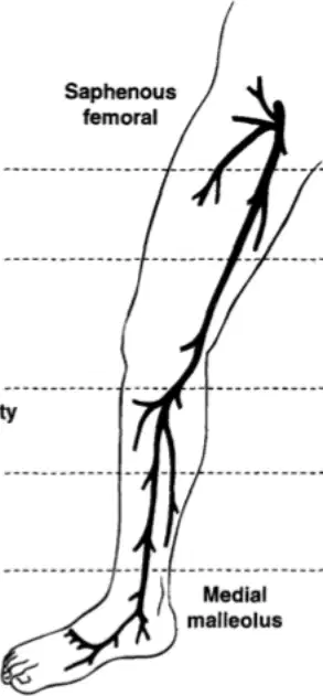
What is the distance of Level 1 from the saphenofemoral junction in the diagram of vein measurements?
5cmdistal
10cmdistal
15cmdistal
20cmdistal
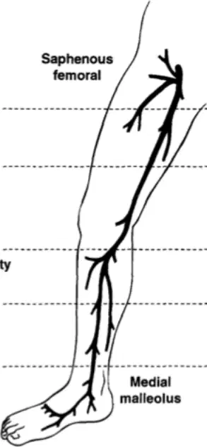
At what level is the tibial tuberosity located according to the vein measurements diagram?
Level 1
Level 2
Level 3
Level 4
How far above the top of the medial malleolus is Level 5 in the vein measurements diagram?
5cm
10cm
15cm
20cm
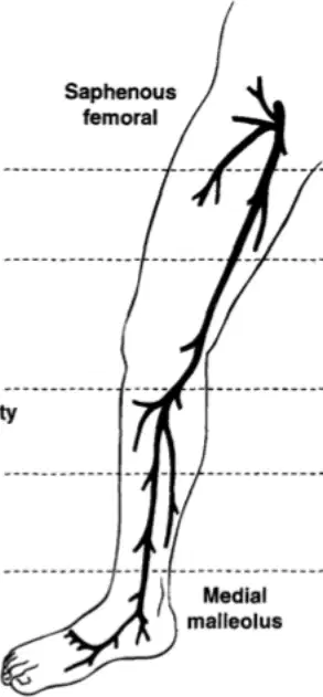
What is the distance of Level 4 from the tibial tuberosity in the vein measurements diagram?
5cmdistal
10cmdistal
15cmdistal
20cmdistal
What frequency range of transducer is generally employed for imaging?
4.5- to 6-MHz
10- to 12-MHz
8- to 10-MHz
12- to 14-MHz
What type of probes are occasionally useful to image deeper veins in extremely obese individuals?
Higher MHz probes
Lower MHz probes
10- to 12-MHz transducer
4.5-MHz pulsed Doppler
What is the primary use of a 4.5-MHz pulsed Doppler in imaging?
To image deeper veins
To check for vein patency
To image superficial veins
To measure blood pressure
What is the importance of preparing the examination area or room for successful mapping?
It helps in minimizing peripheral venoconstriction.
It ensures the patient remains clothed and covered.
It is vital for successful mapping.
It assists with visualization of the ultrasound image.
Why should the room be well heated during the examination?
To keep the patient comfortable.
To minimize peripheral venoconstriction.
To assist with visualization of the ultrasound image.
To ensure the patient remains clothed and covered.
What should be done to the patient during the examination?
The patient should be fully exposed.
The patient should remain clothed and covered, exposing only the necessary limbs.
The patient should be kept in a cold room.
The patient should be kept in a bright room.
Why is it useful to keep the exposed foot covered during the examination?
To minimize peripheral venoconstriction.
To assist with visualization of the ultrasound image.
To keep the patient comfortable.
To ensure the patient remains clothed and covered.
Why is the room generally kept dark during the examination?
To minimize peripheral venoconstriction.
To ensure the patient remains clothed and covered.
To assist with visualization of the ultrasound image on the display.
To keep the patient comfortable.
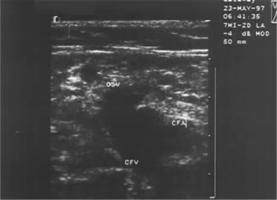
Which of the following is the most common site for imaging the saphenous vein?
A)Theankle
B)Theknee
C)Thegroin
D)Thewrist
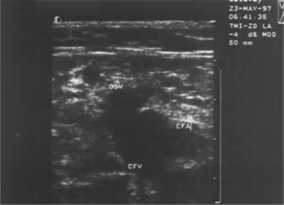
Imaging of the saphenous vein can be started at which of the following sites?
A)Theankle,theknee,thegroin
B)Thewrist,theelbow,theshoulder
C)Theneck,thechest,theabdomen
D)Thehip,thethigh,thecalf
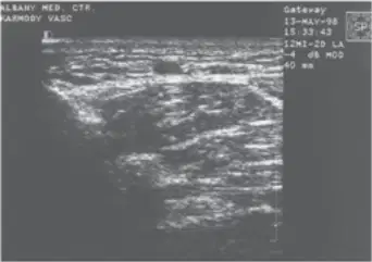
What shape should the vein be in a cross-sectional imaging technique?
Elliptical
Round
Triangular
Square
In the context of imaging techniques, what does an elliptical shape of the vein indicate?
No pressure
Mid pressure
Excess pressure
Normal condition
What is one method to confirm the patency of a vein?
Using a stethoscope
Compressing the vein
Applying heat
Using a thermometer
What technique can be used in conjunction with manual compression of the distal leg if there is doubt about vein patency?
Continuous Doppler
Pulsed Doppler
MRI
X-ray
Why should care be taken to keep the probe as perpendicular to the skin as possible during imaging?
To reduce patient discomfort
To help the surgeon make incisions directly over the vein
To improve image resolution
To prevent infection
What is the next step after identifying the vein in the transverse plane during imaging techniques?
The probe is rotated 180° to insulate the vein in a coronal plane.
The probe is slowly rotated 90° to insolate the vein in a sagittal plane.
The probe is moved distally to mark a new dot every inch.
The probe is rotated 45° to insolate the vein in a transverse plane.
Why is a marker with a chisel tip used in imaging techniques?
Because it will mark through gel and stay wet when left uncapped.
Because it is easier to hold and use.
Because it is more colorful and visible.
Because it is cheaper and more readily available.
What is done as the probe is moved distally during imaging techniques?
The probe is rotated 90°.
A new dot is made every inch.
The marker is changed.
The vein is identified in the transverse plane.
What is done after the remainder of the scan is completed in the imaging technique described?
The operator makes incisions.
The dots are painted over with a continuous line.
The patient is discharged.
The scan is repeated.
What does the continuous line provide the operator with?
A detailed picture of the skin.
A map of the underlying vein.
A list of potential complications.
A guide for medication dosage.
What limitation does the map of the underlying vein have?
It does not show the vein at all.
It does not provide a detailed picture of the vein.
It does not necessarily indicate the best place for incisions.
It does not help in any surgical procedure.
What type of data about the vein wall is considered important but subtle?
Information about the vein wall
Information about the blood flow
Information about the heart rate
Information about the muscle tissue
How does the intimal-medial complex normally appear?
Thick, double, poorly-delineated reflection
Thin, single, well-delineated reflection
Thick, single, well-delineated reflection
Thin, double, poorly-delineated reflection
What should be noted when using the probe in the sagittal plane?
The color of the vein wall
The abnormal appearance of the vein wall
The thickness of the blood
The heart rate

