
 Worksheets
Benign & Malignant CT tumors
Total questions: 20
Worksheet time: 10mins
During a dental check-up, Dr. Rohan encounters a patient with a tumor in the oral cavity. The patient is concerned about the aggressiveness and recurrence of some tumors, even if benign. Dr. Rohan explains that there are different types of tumors and asks which tumor is known for aggressive behavior and frequent recurrence in the oral cavity despite being benign.
Lipoma
Myxoma
Fibroma
Osteoma
During a medical conference, a doctor presented a case study about a patient diagnosed with a malignant tumor arising from cartilage tissue. What type of tumor was the patient diagnosed with?
Chondroma
Osteoma
Chondrosarcoma
Fibroma
Which of the following is NOT a typical feature of a malignant tumor?
Poorly defined margins
Metastasis
Rapid growth
Well-circumscribed lesion
A 50-year-old patient named Aisha has a slow-growing, painless mass in the oral cavity. What is the most likely diagnosis?
Liposarcoma
Fibrosarcoma
Fibroma
Rhabdomyosarcoma
Which of the following is a benign connective tissue tumor of the oral cavity?
Osteosarcoma
Fibroma
Squamous cell carcinoma
Melanoma
During a dental check-up, Dr. Sharma finds a growth in Naira's mouth that he suspects might be a tumor from adipose tissue. Which tumor is he likely referring to?
Lipoma
Osteoma
Myxoma
Fibrosarcoma
What is the source of the tumor neurilemmoma?
Neurites
Axon
Perineural fibroblasts
Schwann cells
Siya has persistent leg pain that improves with aspirin. Which type of bone lesion is classically relieved by aspirin?
Chondroma
Osteoid osteoma
Osteoblastoma
Osteosarcoma
A doctor reviews radiographic images of a patient with unexplained bone pain. The images show multiple punched out lesions. Which condition is most likely associated with these findings?
Paget's Disease
Osteosarcoma
Fibrosarcoma
Multiple myeloma
Which of the following is benign in nature?
Lymphangioma
Lymphoma
Melanoma
Leukemia
Where is Herring bone pattern seen histologically?
Osteosarcoma
Fibrosarcoma
Rhabdomyosarcoma
Lymphoma
Dr. Smith presented a case study about a patient with a rare tumor, mentioning Antoni type A and type B.
Neurofibroma
Neurilemmoma
Neurofibrosarcoma
Traumatic neuroma
What type of tumor has a "honeycomb" or "soap bubble" appearance?
Osteosarcoma
Fibrosarcoma
Central giant cell granuloma
Liposarcoma
Dr. Sharma notices a swelling in Ishaan's mouth and suspects it might be a lymphangioma. He asks Ishaan's parents about the most common location for this condition.
Tongue
Buccal mucosa
Palate
Gingiva
In a medical conference, Dr. Smith presented a case study of a patient with radiographic images showing a 'Sunray' pattern. What type of tumor was most likely diagnosed?
Fibrosarcoma
Chondrosarcoma
Leiomyosarcoma
Osteosarcoma
Dr. Sharma examines a patient named Kiara with persistent left arm pain. An X-ray shows a peculiar radiographic appearance. What is a typical radiographic appearance of an aneurysmal bone cyst?
"Ground-glass" appearance
"Soap bubble" or "blow-out" appearance
"Sunburst" pattern
"Moth-eaten" appearance
During a dental examination, Dr. Naira notices a growth in Aarav's jaw and takes a radiograph to assess it. What is the typical radiographic appearance of an ossifying fibroma?
Well-defined radiolucent area with no calcifications
Mixed radiolucent-radiopaque lesion with well-defined borders
Diffuse radiopacity with ill-defined borders
"Ground-glass" appearance
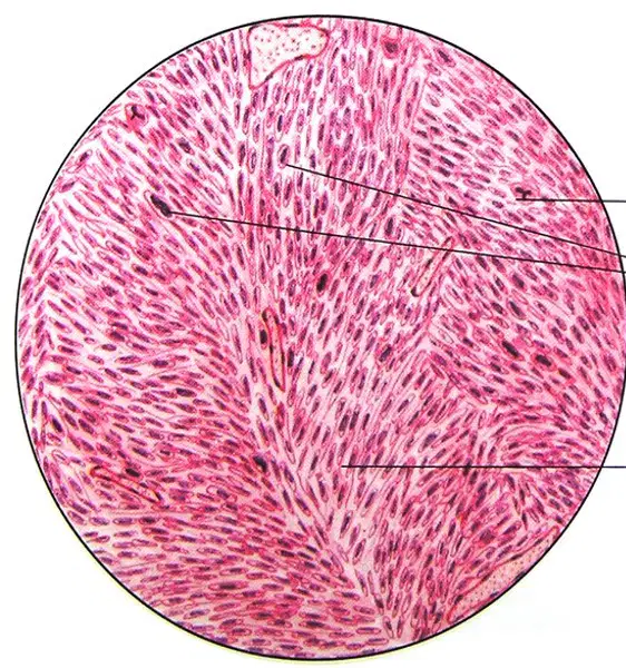
Identify the type of tissue in the histopathology image.
Osteosarcoma
Schannoma
Fibrosarcoma
Traumatic neuroma
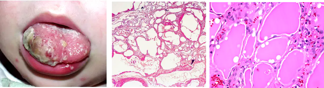
Aarav is a pediatrician examining a child with a soft, raised, reddish lesion on the tongue. He needs to identify the lesion.
Capillary Hemangioma
Cavernous Hemangioma
Lymphangioma
Aneurysmal Bone cyst
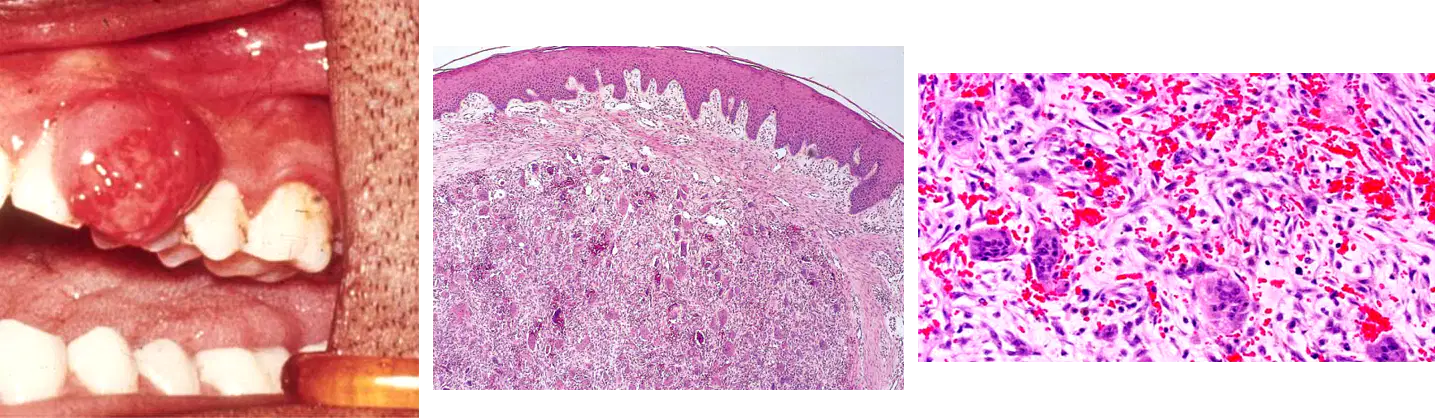
Aashi notices a growth on her gums during a dental check-up. The dentist asks her to identify it from the following options:
Peripheral Ossifying fibroma
Peripheral giant cell granuloma
Aneurysmal bone cyst
Verruciform Xanthoma

