
 Worksheets
Echocardiography
Total questions: 20
Worksheet time: 10mins
What is the likely diagnosis? (movie)
Athlete's heart
Hypertrophic cardiomyopathy
Hypertensive heart disease
Cardiac amyloidosis
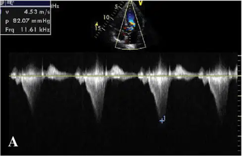
The cause of systolic murmur in this patient is likely to be:
Valvular aortic stenosis
Ventricular septal defect
Mitral valve prolapse
Hypertrophic obstructive cardiomyopathy
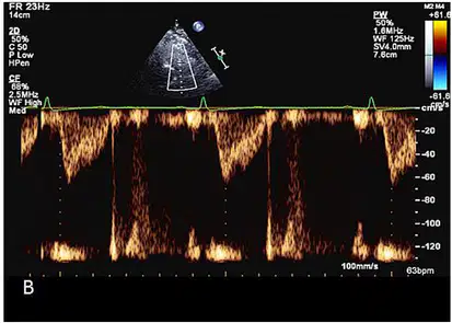
This was a recording of flow across the pulmonary valve using pulsed wave doppler in a patient with severe dyspnea. Likely diagnosis is:
Pulmonary hypertension
Pulmonary stenosis
Pulmonary regurgitation
Tricuspid stenosis
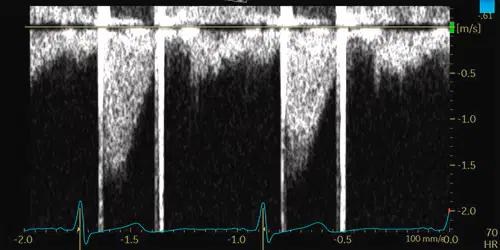
A 68-year-old patient came to the ER with chief complaint of worsening shortness of breath. This patient had been routinely check-up at cardiology outpatient clinic.
This image from TTE evaluation shows continuous wave signal from the apical view. The image is suggestive of:
Moderate aortic stenosis
Severe aortic stenosis
Mitral regurgitation
Prosthetic aortic valve stenosis
The equation for calculating the Relative Wall Thickness (RWT) is:
Notes:
IVST (Interventricular Septal Thickness);
PWT (Posterior Wall Thickness);
LVEDD (Left Ventricular End-Diastolic Diameter)
RWT = (2 x PWT) / LVEDD
RWT = LVEDD / ( 2 x PWT)
RWT = LVEDD / (IVST + PWT)
RWT = (IVST + PWT) / LVEDD
This patient in hospitalized for elective surgery. Blood sample was taken and IV line access was in left brachial. TTE was performed for pre-op evaluation.
What is the most likely condition in this agitated saline testing? (movie)
Coronary Arteriovenous Fistula
Unroofed Coronary Sinus
Persistent Left Superior Vena Cava
Partial Anomalous Pulmonary Venous Drainage
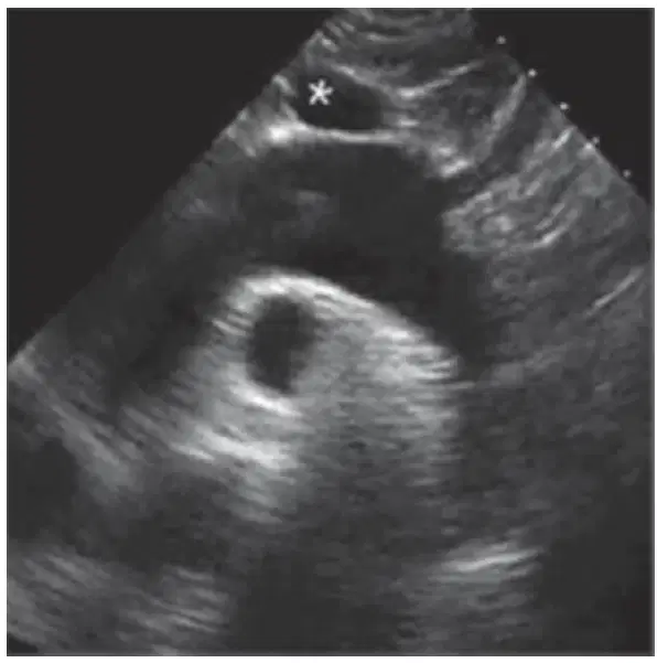
Which vessel is starred (*) in this view?
Right brachiocephalic vein
Right pulmonary artery
Left pulmonary artery
Left brachiocephalic vein
All of the following items are considered in calculating the Wilkins score EXCEPT:
Leflet mobility
Subvalvular calcification
Subvalvular thickening
Leaflet calcification

You are performing a transesophageal echocardiogram for the evaluation of mitral valve pathology.
From a probe position high in the esophagus, you insert the probe deeper while keeping the imaging plane at 0 degrees (horizontal).
The most common MV leaflet segments (scallops) seen initially are:
A1 P1
A2 P2
A3 P3
A3 P1
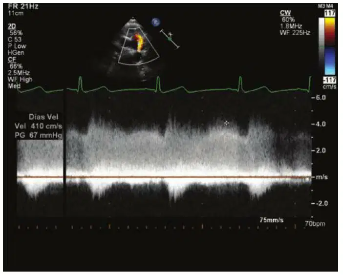
A 20-year-old pregnant female presents for evaluation of a murmur.
The following flow signal is recorded.
The most likely diagnosis is:
Ventricular septal defect
Anomalous pulmonary venous connection
Patent ductus arteriosus
Atrial septal defect
The most likely diagnosis from these TEE findings is: (movie)
Left atrial thrombus
Right atrial myxoma
Left atrial myxoma
Right atrial thrombus
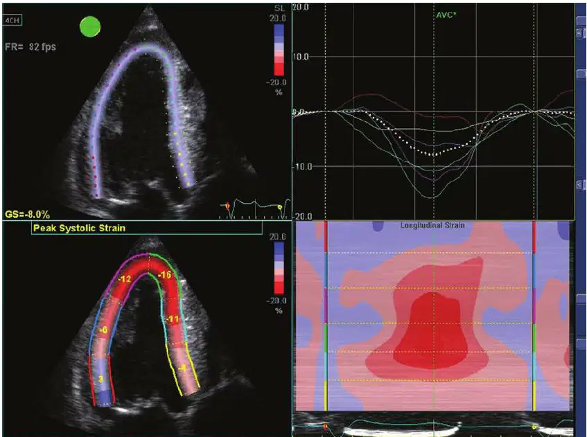
From the analysis provided in the image, what is the likely diagnosis?
Cardiac Sarcoidosis
Cardiac Amyloidosis
Hypertensive Heart Disease
Hypertrophic Cardiomyopathy
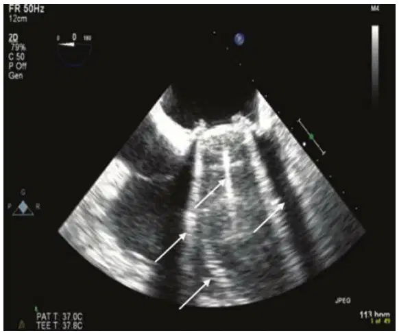
The shown image displays which of the following artifacts?
Reverberation artifact only
Reverberation and speckle artifacts
Reverberation artifact, side lobe artifact, and duplication artifact
Side lobe artifact, reverberation artifact, and attenuation
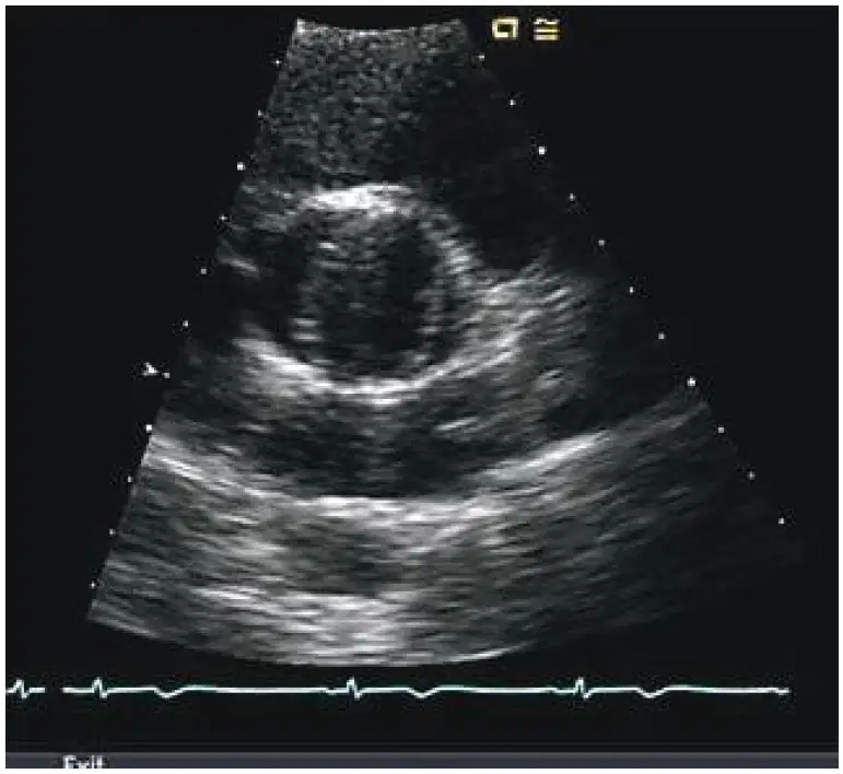
The abnormal finding in this image is:
Aortic dissection flap
Bicuspid aortic valve
Aortic dissection flap
Coarctation of the aorta
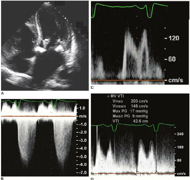
These images are consistent with what diagnosis?
Severe mitral regurgitation
Severe mitral stenosis
Grade 1 diastolic dysfunction
Severe MR and moderate MS
What is the most likely diagnosis? (movie)
RHD Aortic Regurgitation
Bicuspid Aortic Valve
Ascending Aortic Dissection
Aortic Aneurysm
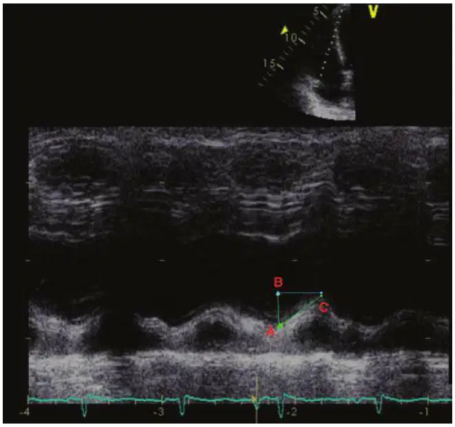
From this image, which one of the following is the accurate value of TAPSE?
Distance between A and B.
Distance between B and C.
Distance between A and C.
Any of the above would be correct.

In this TEE image, number “2” is:
Superior vena cava
Inferior vena cava
Pulmonary artery
Aorta
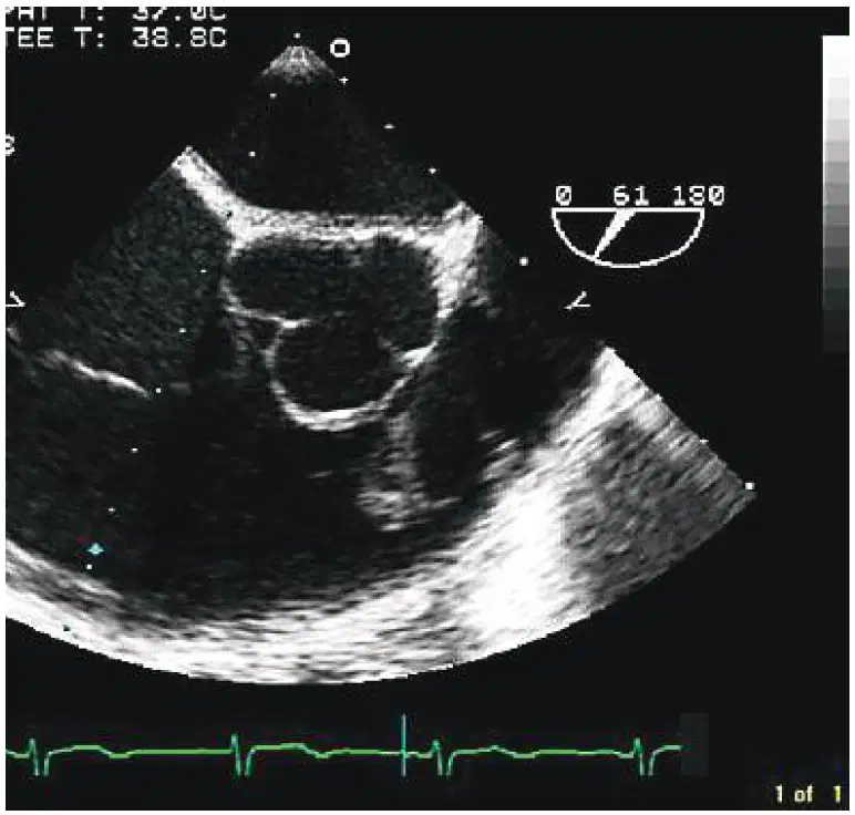
This patient has vegetation on:
Aortic valve
Pulmonary valve
Tricuspid valve
Pacemaker lead
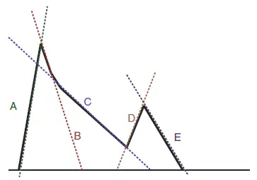
From this schematic mitral inflow, which of the following is the correct slope to measure mitral deceleration time?
A
B
C
D

