
 Worksheets
Echocardiography 2
Total questions: 20
Worksheet time: 10mins

A 56-year-old male is undergoing a mitral valve replacement via thoracotomy. What is shown in the image pointed by arrow?
Crista terminalis
Side-lobe artifact
Chiari network
Eustachian valve

In image here, which Doppler artifact is shown?
Aliasing
Spectral Doppler mirroring
Pseudoflow
Twinkling
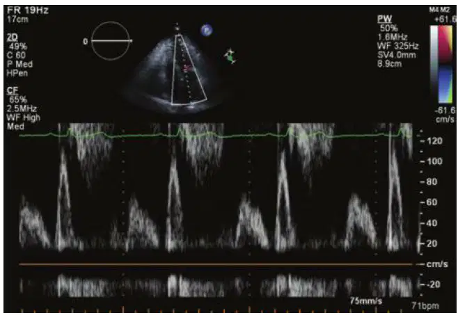
What should be adjusted to improve the Doppler spectral signal in this image?
Increase the wall filter
Reduce the wall filter
Increase the gain
Reduce the gain
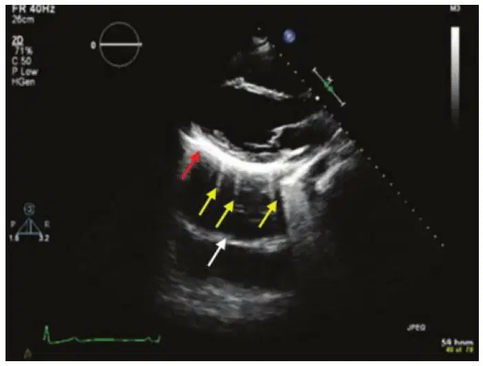
The image shown here demonstrates the following:
Mirror artifact
Mirror artifact and pleural effusion
Mirror artifact, reverberation artifact, and a pericardial
effusion
Mirror artifact, enhancement, and reverberation artifact
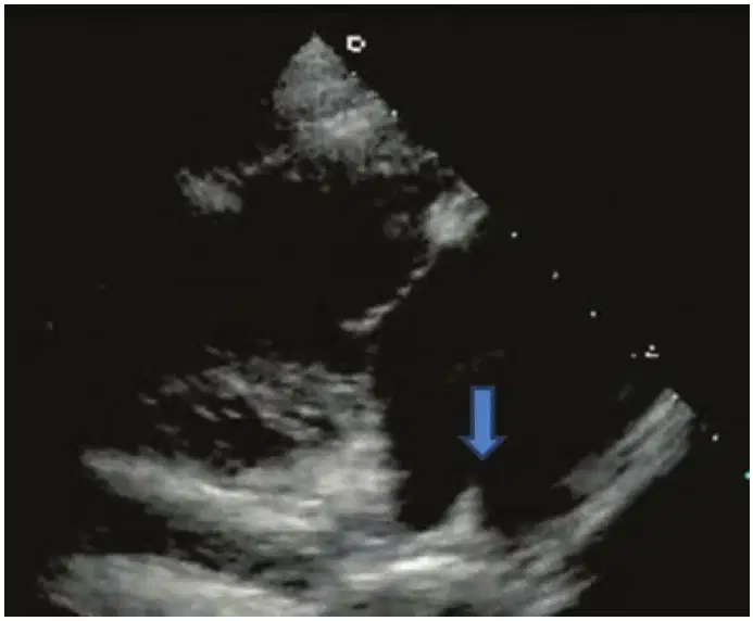
Which structure is highlighted in this TTE view?
Superior vena cava
Inferior vena cava
Eustachian valve
Coronary sinus
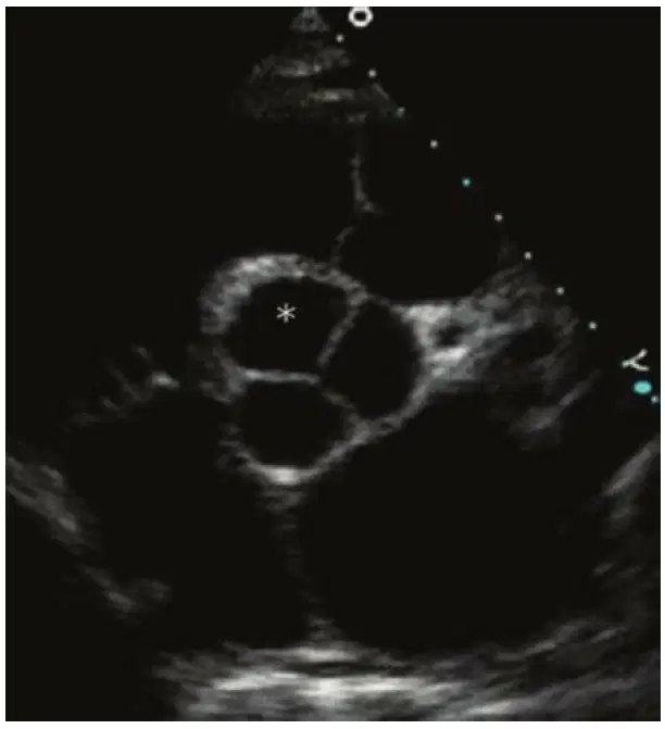
Which aortic valve coronary cusp is starred in this image?
Septal
Right
Left
Noncoronary

A 62-year-old female with a long history of tobacco use and hypertension was referred for an echocardiogram following the detection of atrial fibrillation.
What structure is the arrow pointing to in this image?
Accessory band
Papillary muscle
Moderator band
Chordae tendineae
Which of these M-mode images is suggestive of valvular aortic stenosis?


A 76-year-old male undergoes single-vessel coronary artery bypass grafting with left internal mammary artery (LIMA) to the left anterior descending coronary artery. During weaning from bypass, the surgeon called the echocardiographer to the operating room to review the TEE before closing the chest. The patient’s preoperative echo had mild to moderate (2+) MR with preserved left ventricular ejection fraction. The surgeon reported all his vessels have good flow including the new LIMA graft by Doppler.
What is the etiology of mitral regurgitation in this patient?
Occlusion of the LIMA graft
Stunned myocardium
The preoperative echo was likely misread
Ischemic papillary muscle
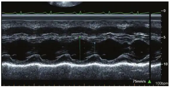
End-diastolic diameter 50 mm
End-systolic diameter 25 mm
Interventricular septum in systole 1.1 cm
Posterior wall in diastole 1.0 cm
What is the fractional shortening?
35%
24%
50%
30%
Which is the correct pair of measures used to calculate an accurate LVEF using the single-plane Simpson method of disks? Consider image quality, image acquisition, and quantitative technique.


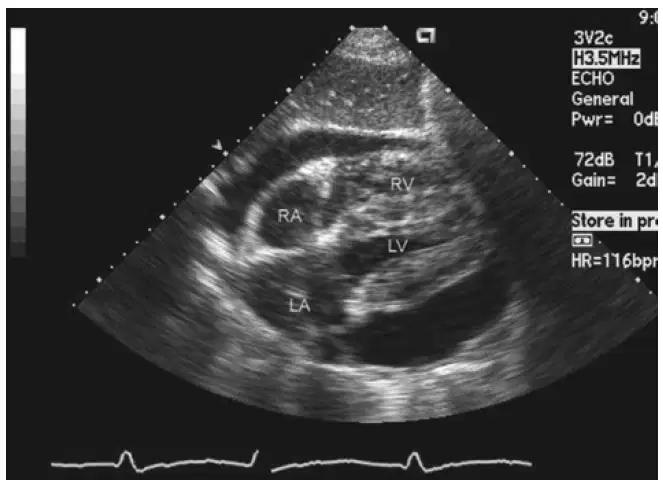
A 72-year-old patient with breast cancer presents to the emergency department with generalized fatigue. A CT scan of the chest reveals a large pericardial effusion, and a stat echocardiogram is performed (shown in the image). The cardiologist concludes that the patient has a large pericardial effusion with 2D echocardiographic features of tamponade.
Which of the following 2D echo findings is most consistent with the cardiogist’s interpretation?
A 1.5-cm pericardial effusion in diastole with late ventricular systolic RVOT collapse
A 2.0-cm pericardial effusion in diastole with early ventricular systolic RA collapse
A 2.1-cm pericardial effusion in diastole with ventricular diastolic LA collapse
A 3.4-cm pericardial effusion noted in ventricular systole

Which of these is the LEAST likely a reason for the pericardial effusion seen in this image?
Uremia from poorly compliant peritoneal dialysis
Bacterial pericarditis
Metastatic melanoma
Pregnancy associated with preeclampsia

In this TEE image, number “2” is:
Superior vena cava
Inferior vena cava
Pulmonary artery
Aorta
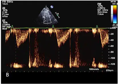
This was a recording of flow across the pulmonary valve using pulsed wave doppler in a patient with severe dyspnea. Likely diagnosis is:
Pulmonary hypertension
Pulmonary stenosis
Pulmonary regurgitation
Tricuspid stenosis
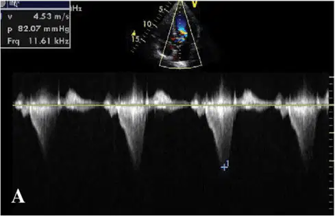
The cause of systolic murmur in this patient is likely to be:
Valvular aortic stenosis
Ventricular septal defect
Mitral valve prolapse
Hypertrophic obstructive cardiomyopathy
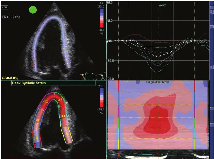
From the analysis provided in the image, what is the likely diagnosis?
Cardiac Sarcoidosis
Cardiac Amyloidosis
Takotsubo Cardiomyopathy
Hypertrophic Cardiomyopathy
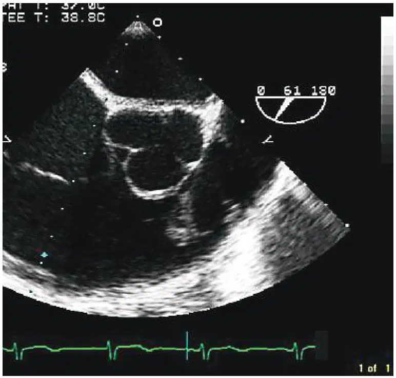
This patient has vegetation on:
Aortic valve
Pulmonary valve
Tricuspid valve
Pacemaker lead
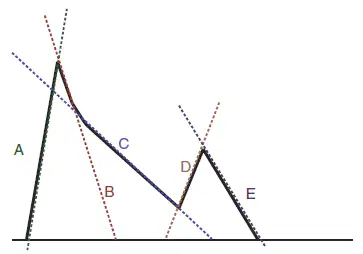
From this schematic mitral inflow, which of the following is the correct slope to measure mitral deceleration time?
A
B
C
D
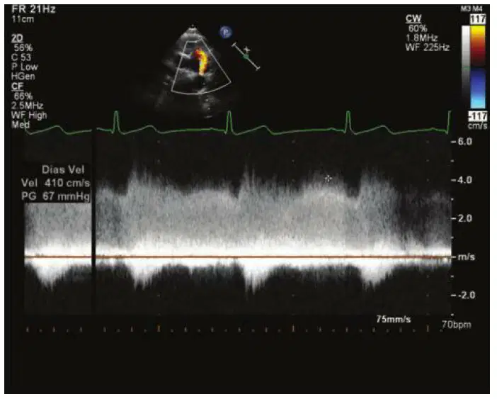
A 20-year-old pregnant female presents for evaluation of a murmur.
The following flow signal is recorded.
The most likely diagnosis is:
Ventricular septal defect
Anomalous pulmonary venous connection
Patent ductus arteriosus
Atrial septal defect

