
 Worksheets
Artifacts
Total questions: 23
Worksheet time: 15mins
An artifact is not:
An incorrect size or shape
Incorrect position
Incorrect brightness
A real structure
What is an assumption of the imaging device that if broken causes an artifact?
Sound travels exactly 1540 m/s
Imaging plane is very thin
Strength if a reflection is related to the characteristics of the tissue
Sound travels directly to a reflector and back
All of the above
Which artifact is primarily produced by refraction?
Posterior enhancement
Shadowing
Comet tail
Refraction

Which adjustment will reduce the artifact in the cystic lesion in image A resulting in image B?
Turn on harmonics
Change to a 1.5 D linear transducer
Decrease gain
Change imaging plane

What is the best way to fix this image?
Leave it- it helps characterize the tissue
TGC adjustment
Overall gain
Change angle

How to change the image artifact shown by B?
Use a 1 D linear transducer
Decrease output power
Move focus
Decrease image depth

What is the artifact shown by the white arrow?
Speed error
Mirror image artifact
Comet tail
Focal enhancement

How would you adjust this image?
Use spatial compounding
Decrease depth
Use temporal compounding
Change focal zone

What are the artifacts seen in this image?
Edge shadow
Posterior shadowing
Temporal artifact
Posterior enhancement
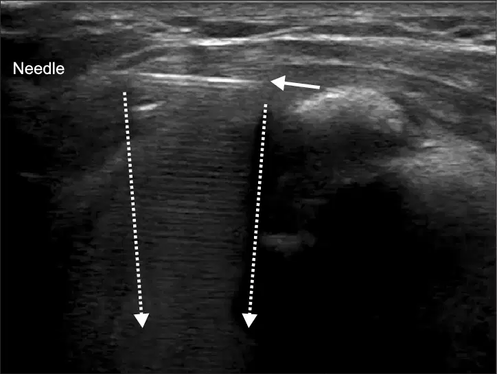
How would you correct this image?
Change depth
Decrease gain
Decrease output power
Harmonics

What causing the shadowing in this image?
A highly attenuating structure
Too little attenuation
A gallstone
Two closely placed reflectors
A speed error artifact caused by a faster speed of sound in the tissue causing a step ___.
Down
Up
There is no way to tell
Correcting for aliasing causes what artifact?
Speed
Refraction
Range ambiguity
Enhancement
What is the most common artifact of Doppler?
Cross talk
Aliasing
Flow reversal
Ghosting
Circle the enhancement artifact on the image.
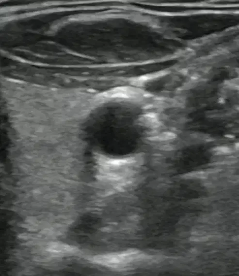
What is the artifact created by the contamination of doppler signals by the heart walls?
Clutter
Acoustic speckle
Ghosting
Cross talk
What is the artifact best fixed by using multi-focusing?
Shadowing
Enhancement
Focal banding
Incorrect TGC usage
What is the artifact characterized by an artifact side by side with the true anatomy?
Lobe
Refraction
Slice thickness
Lateral resolution
A curved or oblique reflector cause sound to bounce away from the transducer this can cause-
Edge shadow
An absent reflector
Reflectors appear too weak
All of the above
The clinic gets a new monitor with a pixel density of 200 per square inch, when the previous monitor had a density of 70 per square inch. What happens to the spatial resolution of the images?
Increases
Decreases
Stays the same
Noise is caused by:
Movement of the heart
Signal Processes
Electrical interference
Constructive and destructive interference of wavelets
All of the above
What artifacts can cause true anatomy to not be seen on the image?
Shadowing
Speed error
Mirror
All of the above
What determines spatial resolution?
Line density
Axial resolution
Lateral resolution
Depth

