
 Worksheets
DS 3262 Ch 41- Normal anatomy & physiology of the females pelvis
Total questions: 281
Worksheet time: 2hrs 21mins
Which of the following statements about ultrasound approaches for evaluating the female pelvis is True?
transabdominal ultrasound provides a more detailed study than endovaginal ultrasound
endovaginal ultrasound requires a full bladder for optimal imaging
transabdominal ultrasound offers a wider field of view but less detail
a complete pelvic examination consists only of endovaginal ultrasound
What is the recommended bladder status for a transabdominal ultrasound of the female pelvis?
empty bladder
full bladder
bladder status does not matter
catheterized bladder
Which ultrasound approach provides a more detailed study of the female pelvic structures?
transabdominal ultrasound
endovaginal ultrasound
both provide equal detail
neither approach provides detailed images
Why is endovaginal ultrasound limited in its imaging capabilities?
it requires a full bladder
it has a lower image resoluation
it has limited filed of view and depth
it cannot image the uterus
Which of the following is a characteristic of transabdominal ultrasound?
empty bladder
more detailed images
limited depth
wider field of view
At what stage of life do ALL ova begin their development?
during puberty
at birth
during embryonic life
at menarche
What constitutes a complete pelvic ultrasound examination?
transabdominal ultrasound only
endovaginal ultrasound only
both transabdominal and endovaginal ultrasound
only if abnormalities are found
How many bones make up the bony pelvis?
2
3
4
5
Which bones form the anterior and lateral margins of the bony pelvis?
sacrum and coccyx
ilium and ischium
two innominate bones
pubic bones
The sacrum and coccyx together form which part of the bony pelvis?
anterior wall
lateral margins
posterior wall
pelvic floor
What is the role of the sacrum in the pelvic structure?
supports the bladder
forms the anterior wall
forms the posterior wall
connects the femur
Which of the following is NOT one of the bones that make up the bony pelvis?
Innominate bone
Sacrum
Coccyx
Femur
What are the two main compartments of the pelvis?
upper pelvis and lower pelvis
anterior pelvis and posterior pelvis
true pelvis and false pelvis
medial pelvis and lateral pelvis
which anatomical marks the division between the true and false pelvis?
the ischial tuberosity
the pelvic brim
the sacroiliac joint
the acetabulum
The plane that divides the true and false pelvis passes through which of the following?
Inferior border of the sacrum to the inferior margin of the pubic symphysis
Iliac crest to the obturator foramen
superior border of the sacrum to the superior margin of the pubic symphysis
Ischial spine to the sacrospinous ligament
The iliopectineal line is associated with which pelvic structure?
pelvic outlet
ischial spine
division between true and false pelvis
attachment of the gluteus medius
Where is the true pelvis located?
above the pelvic brim
below the pelvic brim
at the level of the iliac crest
b/w the coccyx and sacrum
Which of the following best describes the false pelvis?
it is located below the pelvic brim and houses pelvic organs
it is the space b/w the sacrum and the pubic symphysis
it is located above the pelvic brim and communicates with the abdominal cavity superiorly
it is a separate compartment that does not connect with the abdominal or pelvic cavity
The false pelvis communicates with which of the following?
the vertebral column and the gluteal region
the perineum and the inguinal canal
the abdominal cavity superiorly and the pelvic cavity inferiorly
the urinary bladder and rectum
which of the following statements is FALSE?
the true pelvis lies below the pelvic brim
the false pelvis is part of the abdominal cavity
the false pelvis contains most of reproductive organs
the false pelvis is continuous with the abdominal cavity
Which muscles form the pelvic floor?
piriformis and obturator internus
coccygeus and piriformis
levator ani and coccygeus
iliopsoas and obturator internus
Which of the following organs is located in the ANTERIOR pelvic cavity?
rectum
colon
ovary
ileum
how do pelvic muscles appear on ultrasound viewed in their long axis?
hyperechoic with anechoic striations
isoechoic with no striations
hypoechoic with hyperechoic striations
hyperechoic with shadowing
which muscles combine to form the iliopsoas muscle?
psoas major and obturator internus
psoas major and iliacus
iliacus and coccygeus
levator ani and psoas
the right and left iliopsoas muscles are located:
posterior to the uterus
anterior to the vagina
lateral to the urinary bladder
medial to the rectum
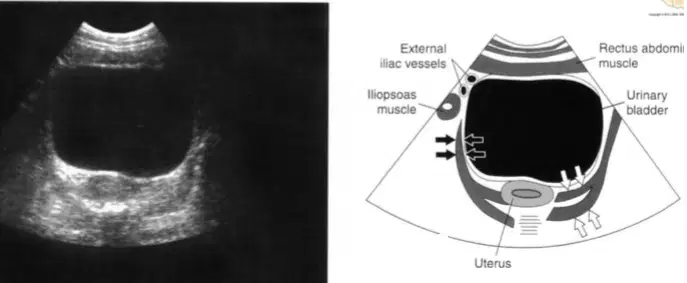
on ultrasound, which muscle is identified with black arrows?
ilipsoas
piriformis
obturator internus
levator ani
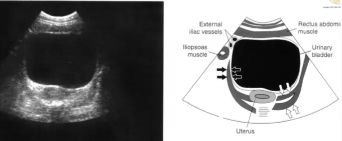
on ultrasound, which muscle is identified with the white arrows?
iliopsoas
piriformis
obturator internus
levator ani
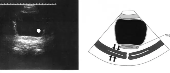
Where are the curved levator ani muscles located relative to the vagina?
anterior and lateral
posterior and lateral
medial and anterior
superior and posterior
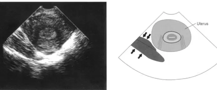
Which muscle is indicated by black arrows ?
obturator internus
iliopsoas
piriform muscle
levator ani
When empty, the bladder remains:
in the false pelvis
above the symphysis pubis
behind the anterior abdominal wall
entirely within the true pelvis
Where is the bladder located within the pelvic cavity?
Posterior portion of the pelvic cavity
Anterior portion of the pelvic cavity
Superior portion of the abdominal cavityLateral portion of the pelvic cavity
What anatomical structure lies directly anterior to the bladder?
Sacrum
Uterus
Pubic symphysis
Rectum
What happens when the bladder becomes distended?
It shifts into the retroperitoneal space
It descends into the perineum
It rises behind the anterior abdominal wall
It compresses the spinal cord
Ureters enter the bladder at which location?
ureteropelvic junction
fundus
trigone
apex
From where do the ureters descend?
liver
bladder
kidneys
urethra
What is the general direction of the ureters as they descend?
superior and lateral
inferior and posterior
anterior and medially
posterior and laterally
Which muscle do the ureters pass anterior to?
Gluteus maximus
Rectus abdominis
Psoas muscle
Obturator internus
How can the ureters be identified sonographically?
By detecting peristalsis in the urethra
By observing urine pooling in the renal cortex
By locating the ureteral jets in the posteroinferior bladder
By measuring bladder wall thickness
The vagina lies:
anterior to the urethra and bladder
posterior to the bladder and anterior to the rectum
between the rectum and color
posterior to the rectum and anterior to the urethra
What structure is described as a collapsed muscular tube?
urethra
fallopian tube
vagina
ureter
The vagina extends from the external genitalia to what structure?
urethra
cervix of the uterus
bladder
rectum
Which structure lies directly anterior to the vagina?
rectum
cervix
urethra and urinary bladder
sigmoid colon
Which structure is located posterior to the vagina?
ovaries
bladder
pubic symphysis
rectum
The uterus develops from:
endodermal folds
embryonic mullerian ducts
wolffian bodies
mesonephric tubules
The uterus is derived from which embryonic structure?
wolffian ducts
mesonephric ducts
mullerian ducts
cloacal membrane
When do the mullerian ducts fuse to form the uterine lumen?
Between the 4th and 6th weeks of development
Between the 7th and 12th weeks of development
at birth
after puberty
Which of the following best describes the shape of a normal uterus?
cylindrical
oval
pear-shaped
round
Which are the main anatomical divisions of the uterus?
Fundus, body, and ovary
Fundus, body, cervix
Isthmus, ovary, and cervix
Corpus, ovary, and infundibulum
What is the average length of a menarcheal uterus?
3-4 cm
4-6 cm
6-8 cm
8-10 cm
What are the typical anterior-posterior and transverse dimensions of a menarcheal uterus?
2-3 cm
3-5 cm
5-7 cm
7-9 cm
Which of the following factors influence the size of the uterus?
BMI and blood pressure
Age and parity
Hormone levels only
Ethnicity and weight
The fallopian tubes enter the uterus at the:
isthmus
cornua
endometrium
cervix
What is the widest and most superior portion of the uterus called?
cervix
body
fundus
cornua
What structure is located at the lateral borders of the fundus where the fallopian tubes enter the uterus?
cervical os
cornua
isthmus
infundibulum
What is the largest portion of the uterus?
fundus
body
cornua
cervix
Where is the body of the uterus located?
Between the cervix and vagina
Between the ovaries and fallopian tubes
Between the fundus and the cervix
Between the bladder and rectum
The internal and external os are part of the:
fundus
myometrium
endocervix
ectocervix
What is the cervix?
The upper dome-shaped portion of the uterus
The widest part of the uterus
The lower cylindrical portion of the uterus
The muscular layer of the uterus
What is the endocervix?
The external surface of the cervix
The area where the fallopian tubes enter
The canal within the cervix
The outer muscular layer of the cervix
The endocervix is constricted at its upper and lower ends by which two structures?
Internal os and fundus
External os and isthmus
Internal os and external os
Cornua and vagina
What is the ectocervix?
The opening into the uterine body
The portion of the cervix continuous with the vagina
The internal lining of the endometrium
The muscular layer of the uterus
Which layer of the uterus is muscular and the thickest?
endometrium
myometrium
serosa
peritoneum
What is the outermost layer of the uterine wall called?
endometrium
myometrium
serosa
peritoneum
Which layer of the uterus is the thickest and composed of smooth muscle?
endometrium
serosa
cervical canal
myometrium
What type of muscle is found in the myometrium?
Skeletal muscle
Cardiac muscle
Smooth muscle
Voluntary muscle
The muscle fibers in the myometrium are arranged in which patterns?
Spiral and oblique
Longitudinal and circular
Horizontal and diagonal
Vertical and transverse
What is the innermost layer of the uterus called?
myometrium
endometrium
perimetrim
cervix
What type of tissue is the endometrium composed of?
cartilage
connective tissue
mucous membrane
adipose tissue
Which ligament supports the uterus in its midline position?
broad ligament
round ligament
uterosacral ligament
all of the above
How many layers does the endometrium have?
one
two
three
four
What is the name of the superficial layer of the endometrium that sheds during menstruation?
Zona basalis
Zona functionalis
Myometrium
Serosa
What is the role of the zona basalis?
It stores menstrual blood
It supports the myometrium
It regenerates the new endometrium
It initiates ovulation
Which layer of the endometrium contains blind ends of endometrial glands?
Zona functionalis
Zona basalis
Myometrium
Serosa
The appearance of the endometrium on ultrasound varies based on what factor?
Patient’s age only
Diet and exercise
Hormone levels and menstrual status
Size of the uterus
What causes dynamic changes in the endometrium?
Progesterone only
Uterine contractions
Cyclic hormonal flux from ovulation
Temperature changes in the uterus
the mesosalpinx encloses the:
ovary
fallopian tube
uterine artery
cervix
What are the broad ligaments composed of?
Smooth muscle
Connective tissue
Double fold of peritoneum
Adipose tissue
Where do the broad ligaments extend?
From the ovaries to the cervix
From the uterus to the bladder
From the lateral aspect of the uterus to the pelvic sidewall
From the rectum to the sacrum
What structures are contained within the broad ligaments?
Only lymph nodes
Fallopian tubes and ovaries
Uterine blood vessels and nerves
Ligaments and cartilage
What is the name of the upper fold of the broad ligament?
Mesometrium
Mesosalpinx
Mesovarium
Mesocervix
What structure does the mesosalpinx enclose?
Ovary
Uterus
Fallopian tube
Bladder
What is the name of the posterior fold of the broad ligament?
Mesometrium
Mesocervix
Mesovarium
Mesosalpinx
What does the mesovarium enclose?
uterine artery
ovary
cervix
urethra
What is the function of the round ligaments?
Support the ovaries
Suspend the fallopian tubes
Help hold the uterine fundus and body in a forward position
Attach the uterus to the pelvic floor
Which ligaments help maintain the forward position of the uterus?
Uterosacral ligaments
Broad ligaments
Round ligaments
Ovarian ligaments
What is the role of the cardinal and uterosacral ligaments?
Keep the fallopian tubes in place
Hold the cervix in a fixed position
Support the bladder
Suspend the ovaries
Which of the following ligaments does not contribute to holding the cervix fixed?
Cardinal ligament
Uterosacral ligament
Round ligament
None of the above
Which ligaments help hold the cervix in a fixed position?
Round ligaments
Broad ligaments
Cardinal and uterosacral ligaments
Ovarian ligaments
What is the average position of the uterus ?
Retroverted and retroflexed
Anterverted and anteflexed
Midline and upright
Inverted and rotated
What does anteverted mean?
The uterus is tilted backward
The fundus is aligned with the cervix
The Fundus and body are bent forward, forming a 90 degree angle with the vaginal canal
The cervix points straight down

What does anteflexed mean?
The uterine body curves backward over the cervix
The uterine body is straight and rigid
The body and Fundus curve forward over the cervix
The cervix curves toward the sacrum
In a nulliparous female, what helps maintain the anteverted, anteflxed position of the uterus?
Broad ligaments
Uterosacral ligaments
Round ligaments
Ovarian ligaments

What term describes the position when the entire uterus is tilted posteriorly, and the cervical canal forms an angle greater than 90 degrees with the vaginal canal?
anteverted
retroflexed
retroverted
midposition

In which uterine position does the uterine fundus & body curve backward upon the cervix?
retroverted
anteflexed
retroflexed
dextroverted

A uterus that is retroflexed differs from one that is retroverted because it:
is tilted entirely forward
has the cervix forming a 90-degree angle with the vaginal canal
bends backward at the junction of the body and cervix
is only found in nulliparous females
In multiparous females, what change in uterine position is commonly observed?
The uterus moves anteriorly and becomes anteverted
The uterus often remains in the same position ad before pregnancy
The entire uterus may tip, altering its normal orientation
The cervix becomes fixed and immobile
What term describes a uterus that is titled to the right of midline?
Levoverted
Retroverted
Dextroverted
Anterflexed
When the uterus is titled to the left of midline, it is referred to as:
Dextrovertedq
Levoverted
Anteverted
Retroflexed
Uterine prolapse occurs when:
The uterus is retroverted and retroflexed
The bladder is too full
The uterine ligaments and pelvic floor muscles are weak
The cervix becomes more rigid
What is a consequence of uterine prolapse?
The uterus rotates laterally
The uterus protrudes into the vaginal canal
The cervix shortens
The uterine fundus enlarges
How does a full bladder typically affect the position of an anteverted and anteflexed uterus?
it tips the uterus forward
it causes the uterus to rise superiorly
It pushes the uterus laterally
It tips the uterus backward
What is the typical length of the fallopian tubes?
4-8 cm
6-8 cm
10-12 cm
14-16 cm
What is the approximate diameter range of the fallopian tubes?
0.5 to 1mm
1 to 4mm
4 to 6mm
5 to 8 mm
How many anatomic portions in each fallopian tube divided into?
2
3
4
5
Which of the following is NOT an anatomical portion of the fallopian tube?
Infundibulum
Ampulla
Corpus
Interstitial
The fallopian tubes open into which of the following at the lateral end?
Uterine cavity
Cervical canal
Vagina
Peritoneal cavity
What is the most lateral portion of the fallopian tube?
Ampulla
Isthmus
Infundibulum
Interstitial
What is the shape of the opening of the infundibulum?
Round
Trumpet-shaped
Funnel-shaped
Oval
What are the finger-like projections on the infundibulum called?
Villi
Papillae
Fimbriae
Cilia
What is the approximate length of the infundibulum?
1 cm
2 cm
3 cm
4 cm
Which part of the fallopian tube is the WIDEST and LONGEST?
Infundibulum
Isthmus
Interstitial
Ampulla
What is the approximate length of the ampulla?
2 cm
3 cm
4 cm
5 cm
Which characteristic best describes the shape and structure of the ampulla?
straight and rigid
narrow and flat
convoluted and curving over the ovary
spiral and fixed
What is a key functional feature of the ampulla?
It houses the fimbrae
It connects directly to the uterus
It is the most common site for fertilization
It is where implantation occurs
The ampulla is described as having which of the following wall characteristics?
Thick and stiff
Thin and dilatable
Muscular and rigid
Spongy and immobile
Which portion of the fallopian tube is located medially, closest the uterus?
Infundibulum
Ampulla
Isthmus
Interstitial
What is the approximate length of the isthmus?
1 cm
2 cm
3 cm
5 cm
What is the approximate diameter of the isthmus>
1 mm
2 mm
3 mm
4 mm
Which portion of the fallopian tube passes through the uterine cornu?
Infundibulum
Ampulla
Isthmus
Interstitial
What is the approx. length of the interstitial portion of the fallopian tube?
2 cm
3 cm
1 cm
4 cm
The interstitial portion of the fallopian tube is located:
Near the ovary
Outside the uterus
Within the uterine wall
B/w the ampulla and fimbriae
Why are fallopian tubes difficult to distinguish on US?
They are always filled with fluid
They are the same echogenicity as bone
They resemble surrounding ligaments & vessels
They are NOT located in the pelvis
What imaging technique may help differentiate fallopian tubes from prominent blood vessels?
M-mode
3D reconstruction
doppler interrogation
harmonic imaging
On ultrasound. fallopian tubes are most often mistaken for:
muscles
ovaries
ligaments and vessels
bladder wall
What is the typical shape of the ovaries?
round
pear-shaped
almond-shaped
triangular
What is the approx. length of the ovary in a menarcheal female?
1 cm
2 cm
3 cm
4 cm
The ovaries are suspended from which part of the broad ligament?
anterior aspect
lateral edge
posterior aspect
medial base
Where are the ovaries usually located in relation to the external iliac vessels?
Lateral
Posterior
Medial
Superior
In relation to the internal iliac vessels and ureter, the ovaries are typically located?
Posterior
Inferior
Anterior
Lateral
Ovaries receive a dual blood supply from the ovarian artery and the:
Internal iliac artery
uterine artery
renal artery
external iliac artery
The ovarian arteries arise directly from which vessel?
IVC
internal iliac artery
renal artery
aorta, below the renal arteries
On the right side, the ovarian vein drains directly into the:
left renal vein
IVC
aorta
uterine vein
On the left side, the ovarian drains into the
IVC
Aorta
Left renal vein
Common iliac vein
Which of the following is TRUE about ovarian venous drainage?
both sides drain directly into the IVC
The left ovarian vein drains into the uterine vein
The right ovarian vein drains into the renal vein
The left ovarian vein drains into the renal vein and the right into the IVC
What part of the ovary contains follicles in various stages of development?
medulla
tunica albuginea
cortex
germinal epithelium
What is the cortex in relation to the medulla?
It lies deep to the medulla
It surrounds the central medulla
It is located in the ovarian hilum
It is the central core of the ovary
What type of tissue makes up the tunica albuginea?
smooth muscle
epithelial tissue
dense connective tissue
cartilage
What layer surrounds the tunica albuginea?
medulla
germinal epithelium
peritoneum
ovarian capsule
The central medulla of the ovary contains all of the following except:
blood vessels
lymphatics
follicles
nerves
What is the reproductive cell produced by the ovaries called?
zygote
ovum
embryo
follicle
Which hormone is secreted by ovarian follicles?
progesterone
estrogen
testosterone
prolactin
What is one function of progesterone?
stimulating ovulation
increasing bone density
preparing the uterus for implantation
suppressing estrogen production
Progesterone contributes to which of the following?
milk ejection reflex
muscle development in the uterus
maintenance of secondary gender characteristics
breakdown of the uterine lining
Which hormone is involved in the development of the mammary glands in females?
estrogen
progesterone
FSH
oxytocin
Which ligament supports the ovaries medially?
broad ligament
suspensory ligament
ovarian ligament
round ligament
Which ligament supports the ovaries laterally?
round ligament
broad ligament
ovarian ligament
suspensory ligament
The ovaries are attached to the posterior aspect of the broad ligament via the:
round ligament
mesosalpinx
mesovarium
mesometrium
The broad ligament primarily functions to:
anchor the ovaries to the uterine fundus
support the uterus only
provide lateral support to the female reproductive organs
connect the ovaries the fallopian tubes
The common iliac arteries course in which direction relative to the psoas muscles?
posterior and lateral
anterior and medial
superior and medial
inferior and lateral
The common iliac arteries supply blood to the:
abdominal organs only
uterus and ovaries only
pelvic cavity and lower extremities
kidneys and adrenal glands
Into which two branches do the common iliac arteries bifurcate?
femoral and profunda femories arteries
aorta and renal arteries
external and internal iliac arteries
inferior and superior epigastric arteries
what is another name for the internal iliac artery?
iliolumbar artery
hypogastric artery
median sacral artery
gonadal artery
The external iliac arteries continue as:
internal iliac arteries
femoral arteries
common iliac arteries
inferior epigastric arteries
The internal iliac arteries are primarily responsible for perfusing which region?
lower limbs
abdominal wall
pelvic structures
thoracic cavity
Which of the following structures is NOT typically supplied by branches of the internal iliac artery?
Urinary bladder
Uterus
Rectum
Small intestine
Which pelvic organ receives blood supply from the internal iliac artery?
Kidney
Ovary
Uterus
Liver
The vagina receives arterial blood supply primarily from branches of the:
External iliac artery
Renal artery
Internal iliac artery
Abdominal Aorta
The uterine artery arises from which artery?
External iliac artery
Ovarian artery
Anterior branch of the internal iliac artery
Renal artery
At what level does the uterine artery enter the uterus?
fundus
body
cornua
cervix
The uterine artery travels within which structure to reach the uterine cornua?
round ligament
suspensory ligament
broad ligament
uterosacral ligament
Which uterine layer receives the blood supply from the uterine artery?
endometrium
serosa
myometrium
perimetrium
What is a notable characteristic of the uterine artery's course?
it runs straight up the uterus
it forms a circular loop around the uterus
it is tortuous and spirals along the uterine sides
it descends from the fundus to the cervix
Which vessels encircle the uterus and are formed by the uterine artery?
Radial arteries
Spiral arteries
Arcuate vessels
Straight arteries
The radial arteries extend through which uterine layer?
Perimetrium
Endometrium
Myometrium
Cervical canal
Which arteries branch from the radial arteries to supply the zona basalis of the endometrium?
Spiral arteries
Uterine arteries
Arcuate arteries
Straight arteries
Spiral arteries supply which part of the endometrium?
Zona basalis
Zona functionalis
Myometrium
Cervical canal
What happens to blood from the spiral arteries during menstruation?
It is absorbed back into the uterus
It nourishes the ovum
It is shed
It supplies the fallopian tubes
Which 3 endocrine structures are essential for the normal physiology of the menstrual cycle? **
Thyroid, pituitary and adrenal glands
Hypothalamus, pituitary, and ovarian axis
Pineal gland, uterus and ovary
Pancreas, ovary and hypothalamus
The reproductive years typically begin at what age range ?
8-10 years
11-13 years
14-16 years
17-19 years
The reproductive years generally end around what age?
40
45
50
55
When does the menstrual cycle begin?
After ovulation
With implantation
With the first day of bleeding
With hormone secretion
What is the average length of a typical menstrual cycle?
21 days
25 days
28 days
35 days
What term describes menstrual cycles with intervals shorter than 21 days?
Amenorrhea
Polymenorrhagia
Oligomenorrhagia
Menometrorrhagia
What terms describes menstrual cycles that occur more than 35 days apart?
amenorrhea
polymenorrhagia
oligomenorrhagia
dysmenorrhea
What terms refers to the physiological state before the onset of menstruation?
menopause
menarche
premenarche
oligomenorrhea
Menarche is defines as:
The first ovulation
The time when menstruation permanently stops
The onset of regular menstrual cycles after puberty
Irregular menstrual bleeding
What terms describes the cessation (brought to an end) of menstruation?
Menarche
Premenarche
Polymenorrhagia
Menopause
Which part of the brain regulates the menstrual cycle?
cerebellum
pituitary gland
hypothalamus
hippocampus
The menstrual cycle is primary dependent on the cyclic release of which two ovarian hormones?
LH and FSH
Estrogen and progesterone
Testosterone and prolactin
Oxytocin and cortisol
When does ovulation typically occur in a 28 day menstrual cycle?
day 7
day 10
day 14
day 21
How often is an ovum released from the ovary under normal conditions?
weekly
twice a month
once a month by one of the two ovaries
every 3 months
It is speculated that ovum release alternates b/w:
fallopian tubes
hormonal cycles
menstrual cycles
the two ovaries
In what state do immature oocytes remain until menarche?
primary follicles
secondary follicles
suspended animation within preantral follicles
atretic bodies
approx. how many oocytes does each ovary contain at birth?
50,000
100,000
200,000
500,000
What happens to the majority of oocytes during a woman's reproductive years?
they are fertilized
they remain dormant
they degenerate
they all undergo ovulation
Ovulation is primarily regulated by which structure in the brain?
cerebrum
cerebellum
pituitary gland
hypothalamus
The hypothalamus begins the pulsatile release of which hormone to initiate ovulation
estrogen
progesterone
gonadotropin- releasing hormone (GnRH)
luteinizing hormone (LH)
What does GnRH (gonadotropin releasing hormone) stimulate?
ovaries to release progesterone
posterior pituitary gland to release oxytocin
anterior pituitary gland to release gonadotropins
hypothalamus to release estrogen
Which two gonadotropins are secreted by the anterior pituitary in response to GnRH (gonadotropin releasing hormone)?
estrogen and progesterone
LH and prolactin
FSH and LH
GnRH and oxytocin
What gland secretes FSH (Follicle Stimulating Hormone)?
Hypothalamus
Adrenal Gland
Pituitary Gland
Thyroid Gland
FSH is responsible for stimulating:
endometrial shedding
growth of the uterine lining
ovarian follicle development
progesterone release
The follicular phase begins on:
the day of ovulation
the first day of bleeding
the last day of menstruation
day 21 of the cycle
How long does the follicular phase typically last in a 28-day cycle?
7 days
10 days
14 days
28 days
As ovarian follicles develop, what to they fill with and secrete?
blood and LH
fluid and estrogen
mucus and progesterone
GnRH and estrogen
How many preantral follicles typically begin to develop during each cycle?
2-3
4-6
5-8
9-12
What is the name of the dominant follicle that matures?
primary follicle
antral follicle
graafian follicle
luteinized follicle
approx. how large is the graafian follicle right before ovulation?
1 cm
1.5 cm
2 cm
3 cm
What happens as estrogen levels rise in the blood?
FSH levels increase
The pituitary gland stops producing LH
The pituitary is inhibited from producing more FSH
ovulation is suppressed
What hormone does the pituitary begin to secrete in response to rising estrogen?
FSH
Progesterone
GnRH
LH (luteinizing hormone)
LH is secreted by which gland?
hypothalamus
thyroid gland
pituitary gland
adrenal gland
The LH surge typically occurs how long before ovulation?
48-72 hours
24-36 hours
12-18 hours
6-10 hours
What is the LH surge commonly used to predict?
menopause
fertility decline
timing of ovulation for conception
onset of menstruation
LH levels peak approx. how many hours before ovulation?
24 hours
6 hours
10 to 12 hours
48 hours
What does the LH surge trigger?
menstruation
ovulation
implantation
follicular development
on what day of the menstrual cycle does ovulation typically occur?
day 7
day 10
day 14
day 28
what phase follows ovulation?
menstrual phase
follicular phase
luteal phase
ovulatory phase
How long does the luteal phase typically last?
7 days
10 days
14 days
21 days
What structure is formed from the ruptured follicle after ovulation?
graafian follicle
primordial follicle
corpus albicans
corpus luteum cyst
what hormone is secreted by the corpus luteum?
estrogen
FSH
progesterone
LH
what happens to the corpus luteum 9 to 11 days after ovulation if there is NO pregnancy?
it enlarges
it secretes estrogen
it degenerates
it implants in the uterus
What hormone's decline leads to menstruation?
estrogen
LH
progesterone
FSH
what event is triggered by the decline in progesterone levels?
ovulation
fertilization
menstruation
implantation
if PREGNANCY occurs, what hormone is produced by the zygote?
FSH
LH
Estrogen
hCG (human chorionic gonadotropin)
What is the function of hCG during early pregnancy?
inhibits estrogen
triggers menstruation
causes corpus luteum to persist
stimulates ovulation
how long does the corpus luteum continue to secrete progesterone during early pregnancy?
1 month
until the end of the menstrual cycle
3 months
6 months
what structure eventually takes over progesterone production during pregnancy?
corpus albicans
hypothalamus
ovary
placenta
ovulation is the release of an ovum from which structure?
corpus luteum
antral follicle
graafian follicle
primordial follicle
what is commonly seen sonographically mid-cycle as a result of ovulation?
thickened endometrium
enlarged uterus
small amounts of fluid in the posterior cul-de-sac
follicle rupture on both ovaries
some women can detect ovulation due to:
increased urination
abdominal bloating
dull ache pain in the lower abdomen
sharp chest pain
the term "mittelschmerz" refers to:
the first menstrual period
ovulation-related pain
postmenopausal bleeding
pain from implantation
mittelschmerz typically occurs:
during menstruation
after menopause
at ovulation, mid-cycle
during implanation
What is one of the main roles of progesterone in the menstrual cycle?
Stimulates the release of LH during the mid-cycle surge
Induces secretory activity in the endometrial glands primed by estrogen
Inhibits endometrial development after ovulation
Increases estrogen levels directly
What is one of the main roles of progesterone in the menstrual cycle?
Stimulates the release of LH during the mid-cycle surge
Induces secretory activity in the endometrial glands primed by estrogen
Inhibits endometrial development after ovulation
Increases estrogen levels directly
What is one of the main roles of progesterone in the menstrual cycle?
Stimulates the release of LH during the mid-cycle surge
Induces secretory activity in the endometrial glands primed by estrogen
Inhibits endometrial development after ovulation
Increases estrogen levels directly
How does progesterone help prepare the uterus for a fertilized ovum?
By promoting the thickening of the myometrium
By increasing LH levels to maintain ovulation
By inducing secretory changes in the endometrium
By stimulating follicle growth in the ovary
What is one of the primary roles of progesterone in the endometrium?
To stimulate follicle development
To increase LH secretion
To trigger menstruation
To induce secretory activity in endometrial glands
How does progesterone affect basal body temperature?
lowers it
keeps it stable
has no effect
increases it
What is the effect of progesterone on LH secretion?
Stimulates a mid-cycle surge
Inhibits LH to prevent ovulation
Has no effect on LH
Increases LH to maintain the corpus luteum
Which of the following are main functions of progesterone?
Induces secretory changes in the endometrium primed by estrogen and to prepare a site for the fertilized ovum
Increases basal body temperature
Inhibits LH secretion to prevent the LH surge and ovulation
All of the above
How many phases are typically recognized in the endometrial cycle?
2
3
4
5
What are the three main phases of endometrial changes?
Follicular, Ovulatory, Luteal
Menstrual, Proliferative, Endometrial
Menstrual, Proliferative, Secretory
Early, Mid, Late
How long does the menstrual phase typically last?
1-3 days
1-5 days
5-10 days
7-14 days
What hormonal change triggers the start of the menstrual phase?
Rising estrogen levels
Declining estrogen levels
Declining estrogen levels
Declining progesterone levels
What part of the endometrium is shed during the menstrual phase?
Zona basalis
Zona functionalis
Entire endometrium
Myometrium
The menstrual phase of the uterine cycle coincides with which phase of the ovarian cycle?
Luteal phase
Ovulatory phase
Follicular phase
Secretory phase
What hormone stimulates regeneration of the superficial endometrial layer during the menstrual phase?
progesterone
estrogen
LH
FSH
Which hormone primarily influences the proliferative phase?
Progesterone
Estrogen
LH
FSH
When does the proliferative phase typically end?
At the start of menstruation
Around day 10 of the cycle
At the luteinization of the Graafian follicle, around ovulation (day 14)
At the implantation of the fertilized ovum
The proliferative phase of the endometrial cycle corresponds to which phase of the ovarian cycle?
Luteal phase
Menstrual phase
Ovulatory phase
Follicular phase
What is the primary function of estrogen during the proliferative phase?
To maintain the corpus luteum
To trigger ovulation
To stimulate endometrial growth
To inhibit LH secretion
Which hormone primarily controls the secretory phase?
estrogen
LH
progesterone
FSH
What happens to the endometrial glands during the secretory phase?
They shrink and degenerate
They become fibrotic
They enlarge and prepare for implantation
They release eggs
When does the secretory phase occur in the menstrual cycle?
Day 1 to 5
Day 6 to 14
Day 15 until the onset of menses
Throughout the entire cycle
The secretory phase of the endometrial cycle corresponds to which phase of the ovarian cycle?
Follicular phase
Menstrual phase
Ovulatory phase
Luteal phase
What is the main purpose of the endometrial changes during the secretory phase?
To support ovulation
To regenerate the zona basalis
To prepare the uterus for implantation
To shed the uterine lining
How is the early proliferative endometrium typically described on imaging?
Thick and echogenic
Normal and thin
Irregular and heterogeneous
Not visualized
What is a characteristic ultrasound appearance of the late proliferative endometrium?
Homogeneous and hyperechoic
Thick and irregular
Triple layer appearance
Poorly defined margins
What is the typical ultrasound appearance of the early secretory endometrium?
Anechoic
Isoechoic
Hypoechoic
Hyperechoic
How does the late secretory endometrium typically appear on ultrasound?
Thin and hypoechoic
Thick and hyperechoic
Thin and hyperechoic
Thick and hypoechoic
What is the typical ultrasound appearance of the postmenstrual endometrium?
Thick and hyperechoic
Normal and thin
Triple layer pattern
Irregular and heterogeneous
How does the thickness of the endometrium change during menstruation?
It increases
It decreases
It remains the same
It becomes irregular
During the early proliferative phase, the endometrium typically appears as:
A thick, hyperechoic line
A thin, echogenic line
A triple layer pattern
A thick, hypoechoic structure
What is the “three-line sign” in the proliferative phase referring to?
Zona basalis anteriorly and posteriorly, and a central line representing the uterine cavity
Three separate layers of the myometrium
Three blood vessels within the endometrium
Triple layering of the zona functionalis
During the proliferative phase, the endometrium thickens to an average of:
1 to 3 mm
4 to 8 mm
9 to 12 mm
12 to 15 mm
How is the endometrial thickness measured on ultrasound?
As a single layer thickness from anterior to posterior
As a double layer thickness from anterior to posterior
From the fundus to the cervix
Only on the posterior wall
During which phase does the endometrium reach its thickest dimension?
Menstrual phase
Early proliferative phase
Secretory phase (after ovulation)
Follicular phase
What is the typical thickness range of the endometrium during the secretory phase?
2 – 5 mm
4 – 8 mm
7 – 14 mm
15 – 20 mm
How does the endometrium appear on ultrasound during the secretory phase?
Thin and hypoechoic
Echogenic with blurring of the 3-line appearance
Triple-layered and hypoechoic
Anechoic and thin
How does the endometrium typically appear in anovulatory patients?
Thick and hyperechoic
Thin and echogenic line
Thick with a triple-layer pattern
Irregular and hypoechoic
What is the normal endometrial thickness limit for postmenopausal patients not on hormone replacement therapy (HRT)?
Less than 3 mm
Less than 5 mm
Less than 8 mm
Less than 10 mm
Postmenopausal patients on hormone replacement therapy (HRT) or taking Tamoxifen may have a normal endometrial thickness of up to:
5 mm
6 mm
8 mm
10 mm
What is menorrhagia?
Short or light periods
Heavy or long periods
Absence of menstruation
Irregular bleeding between cycles
Menorrhagia is often associated with all of the following EXCEPT:
Uterine fibroids
Intrauterine devices (IUDs)
Hormonal imbalance
Polycystic ovary syndrome (PCOS)
What does polymenorrhagia refer to?
Heavy bleeding with normal cycle length
Shortening of the menstrual cycle to less than 21 days
. Long menstrual cycles over 35 days
Complete absence of menstruation
Oligomenorrhagia means:
Heavy or prolonged periods
Shortening of the menstrual cycle
Short or light periods occurring more than 35 days apart
Bleeding between cycles
Oligomenorrhagia can be associated with all of the following EXCEPT:
Polycystic ovary syndrome (PCOS)
Emotional and physical stress
Chronic illnesses
Uterine fibroids
What is amenorrhea?
Heavy menstrual bleeding
Absence of menstruation
Painful periods
Irregular menstruation
Primary amenorrhea is defined as:
Absence of menstruation after age 18 without previous cycles
Cessation of menstruation after having regular cycles
Painful menstruation caused by endometriosis
Heavy or prolonged menstrual bleeding
Secondary amenorrhea refers to:
Absence of menstruation by age 18
Cessation of menses in women who previously had menstrual cycles
Painful menstruation
Shortening of the menstrual cycle
Dysmenorrhea means:
Absence of menstruation
Heavy menstrual bleeding
Painful periods
Irregular menstrual cycles
Dysmenorrhea is commonly associated with which condition?
Polycystic ovary syndrome (PCOS)
Endometriosis
Uterine fibroids
Hormonal imbalance
Where is the vesicouterine recess located?
Between the uterus and rectum
Between the bladder and uterus, anterior to the uterine fundus
Between the bladder wall and the pubic symphysis
Behind the bladder
The rectouterine recess (Pouch of Douglas) is located:
Anterior to the uterus
Posterior to the uterus between the uterus and rectum
Between the bladder and pubic symphysis
Inside the urinary bladder
Which of the following is the most inferior and posterior region of the peritoneal cavity?
Vesicouterine recess
Rectouterine recess (Pouch of Douglas)
Retropubic space
Subcutaneous fat space
The retropubic space is located:
Between the uterus and bladder
Between the bladder wall and the pubic symphysis
Between the uterus and rectum
Behind the rectum
What does the retropubic space normally contain?
Fluid
Muscle tissue
Subcutaneous fat
Bone
What might cause displacement of the urinary bladder posteriorly in the retropubic space?
Pregnancy
Hematoma or abscess
Uterine fibroids
Menstrual bleeding
Where is it normal to observe a small accumulation of free fluid during the menstrual cycle?
Anterior cul-de-sac
Posterior cul-de-sac (Pouch of Douglas)
Retropubic space
Vesicouterine recess
Which of the following conditions can cause hemorrhage or infection within the free fluid in the posterior cul-de-sac?
Ruptured cyst
Ascites
Ruptured corpus luteum cyst
All of the above
Which of the following is NOT typically associated with free fluid accumulation in the posterior cul-de-sac?
Ectopic pregnancy
Pelvic inflammatory disease (PID)
Uterine fibroids
Ruptured corpus luteum cyst
True or False: Some women may have a small amount of fluid in the posterior cul-de-sac continuously.
True
False

