
 Worksheets
Biliary System and Pancreas
Total questions: 118
Worksheet time: 2hrs 52mins
RT and LT hepatic ducts
(a)
Common hepatic duct
(a)
Common bile duct
(a)
Gallbladder
(a)
Cystic duct
(a)
Each hepatic duct is formed by the union of (a) from the liver lobules.
RT & LT hepatic ducts unite at the Porta hepatis to form the (a)
CHD then joins with the Cystic duct (CD) from GB to form the (a) (CBD), which runs parallel with Portal vein.
CBD is joined by the (a)
The CBD and MPD open through a small ampulla, (a) , into the duodenal wall
The end parts of both ducts (CBD and MPD) and the ampulla are surrounded by circular muscle fibers known as the (a)
Measurement of the gallbladder
3 mm
8-12cm long, less than 4cm diameter
0.5-2.5 cm long
0.1-0.7cm diameter
Measurement of the Gallbladder wall
8-12cm long
3 mm thick
0.1-0.2 diameter
0.5-0.8 diameter
Measurement of right and left hepatic ducts
3 mm thick
0.5-2.5cm long
0.1-0.2cm diameter
2.0-6.5 cm long
0.1-0.2 cm diameter
0.5-0.8 cm long
0.1-0.4 cm diameter
Measurement of the common hepatic duct
0.5-2.5 cm long (Lt > Rt)
0.1-0.2 cm diameter
2.0-6.5 cm long
0.1-0.2 cm diameter
0.5-0.8 cm long
0.1-0.4 cm diameter
2.0-6.5 cm long
0.1-0.2 cm diameter
Measurement of the common bile duct
8-12 cm long
Less than 4 cm diameter
2.0-6.5 cm long
0.1-0.2 cm diameter
5-15 cm long
0.1-0.7 cm diameter
0.5-0.8 cm long 0.1-0.4 cm diameter
Measurement of the cystic duct
0.5-0.8 cm long
0.1-0.4 cm diameter
5-15 cm long
0.1-0.7 cm diameter
2.0-6.5 cm long
0.1-0.2 cm diameter
0.5-2.5 cm long (Lt > Rt)
0.1-0.2 cm diameter
A short, narrow tube that carries bile from GB
Joins the CHD to form the CBD
(a)
AKA spiral mucosal folds
(a)
located on the inner surface of the Cystic duct (CD)
(a)
help maintain the patency of the CD, which can be narrow and tortuous
(a)
play a role in facilitating gallbladder emptying
(a)
Posteriorinferior
Related to main lobar fissure
Describes the location of the: (a)
Posteriorinferior (Rt lobe)
Related to main lobar fissure
Position changes with patient position
All describes the location of the: (a)
Right and left (a) merge at the liver hilum (aorta hepatic)
Proximal portion after merging pf the right and left hepatic ducts
Describes the location of (a)
2-4cm long, 1-5mm diameter
(a)
Distal portion in the continuation with the CHD
Describes the location of the (a)
The (a) is divided into the fundus, body, and neck
The rounded fundus usually projects below the inferior margin of the liver,
(a)
The portal triad looks like (a)
Encounters the anterior abdominal wall at the level of the ninth right costal cartilage.
(a)
The gallbladder (a) generally lies in contact with the visceral surface of the liver and is directed upward, backward, and to the left.
The gallbladder (a) becomes continuous with the cystic duct, which turns into the lesser omentum to join the right side of the common hepatic duct to form the common bile duct
The portal vein is (a) in images with the portal triad
The CBD lies __ and __ in sonographic images
(a)
The hepatic artery lies __ and __ in sonographic images
(a)
Right ear- CBD
Left ear- HA
Face- PV
(a)
Majority of the (a) lies in the retroperitoneal cavity, except for a small portion of the head.
Connective prevertebral tissues are ___ ___ in pancreatic anatomy
(a)
Portal-splenic confluence is a (a) structure in pancreatic anatomy
superior mesenteric vessels are (a) structures in pancreatic anatomy
Aorta and IVC are (a) structures in pancreatic anatomy
lower border of the diaphragm is a (a) structure of the pancreatic anatomy
The stomach, duodenum, and transverse colon are __ and __ structures in pancreatic anatomy
(a)
Epigastrium and left hypochondrium; retroperitoneal
(a)
Anterior to IVC, Aorta, diaphragm
(a)
Posterior to stomach and transverse colon
(a)
Level of L1–L2
(a)
From C- duodenal loop to splenic hilum
(a)
Across/anterior to aorta
(a)
U-shaped / dumbbell /comma-shaped/ sausage
(a)
Measurement of the pancreas length
1-2cm
2-3 cm
12-18 cm
1.5-2.5 cm
Measurement of Pancreas head (larger segment)
2-3 cm
12-13 cm
1.5-2.5 cm
1-2cm
Pancreas AP Neck measurement
12-18cm
1.5-2.5cm
2-3cm
1-2cm
AP Pancreas body length
1-2cm
2-3cm
12-18cm
1.5-2.5cm
AP Pancreas tail length
1-2cm
2-3cm
1.5-2.5cm
12-18cm
Pancreas is a (a) organ. 90%
Pancreas is a (a) organ. 10%
Pancreatic juice component Amylase acts on (a)
Pancreatic juice component lipase acts upon (a)
Pancreatic juice components Trypsin, chymotrypsin, carboxypeptidase acts on (a)
Pancreatic juice component Nucleases acts upon (a)
Pancreatic juice component Sodium bicarbonate
(a)
Islets of Langerhans 60%-70%
(a)
Pancreatic juice component Alpha (15%-20%)
(a)
Pancreatic juice component beta acts on:
(a)
Pancreatic juice component Delta (5%) acts on
(a)
Pancreatic juice component acts on
(a)
Pancreatic juice component Epsilon (<1%)
(a)
(merging of superior mesenteric vein and splenic vein) – Posterior to neck of pancreas.
(a)
branch of the CHA) Inferior and lateral to neck of pancreas
(a)
Anterior and posterior superior pancreaticoduodenal branches. Supply head of pancreas and duodenum.
(a)
crosses anterior to the uncinate process of the head of the gland and posterior to the neck and body of the pancreas.
(a)
in the superior border of the pancreas. Supplies body and tail
(a)
Venous drainage is through (a) of the splenic and superior mesenteric veins.
Which of the following are characteristics of the pancreas?
Homogenous
Smooth to lightly lobular (islets of langerhans)
> liver, </> spleen (depends on fatty/fibrous texture)
heterogeneous
Celiac axis and branches
Splenic artery
Common hepatic artery
Gastroduodenal artery
Superior mesenteric artery
Portal vein and tributaries
Common bile duct
(a)
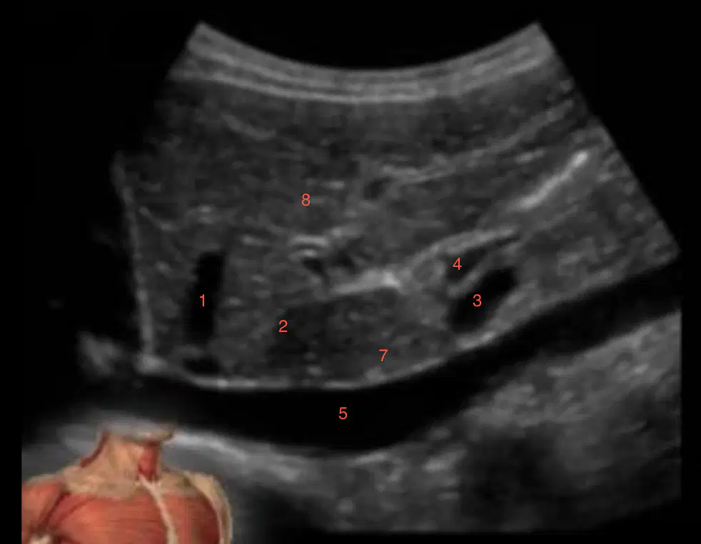
What is labeled 1?
(a)
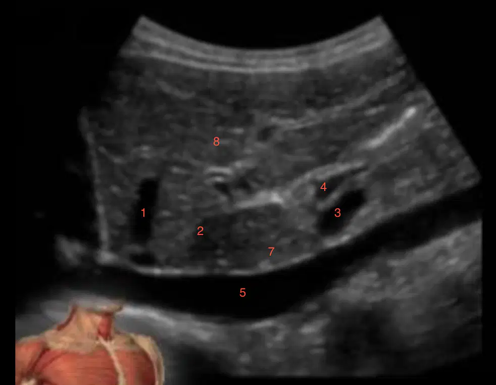
What is labeled 2?
(a)

What is labeled 3?
(a)
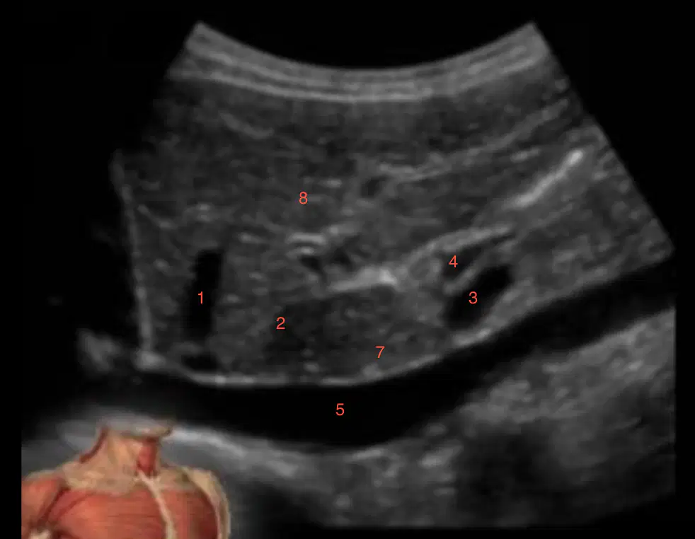
What is labeled 4?
(a)
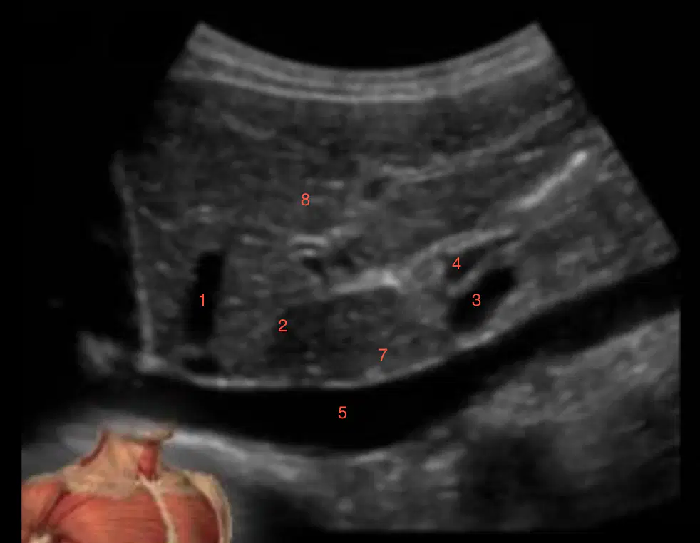
What is labeled 5?
(a)
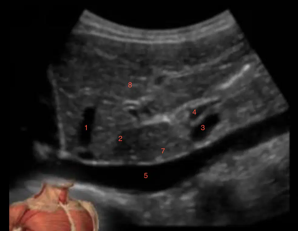
What is labeled 7?
(a)
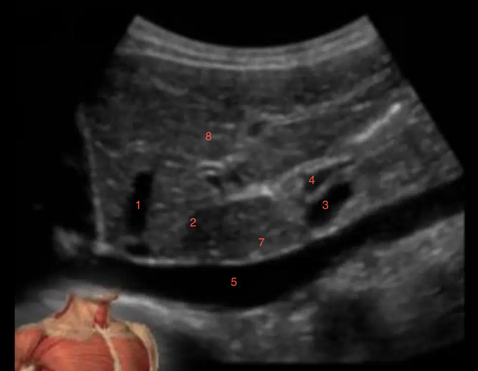
What is labeled 8?
(a)
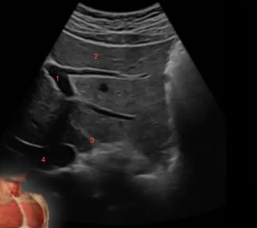
What is labeled 1?
(a)
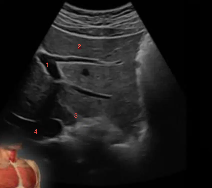
What is labeled 2?
(a)
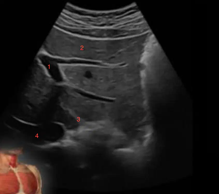
What is labeled 3?
(a)
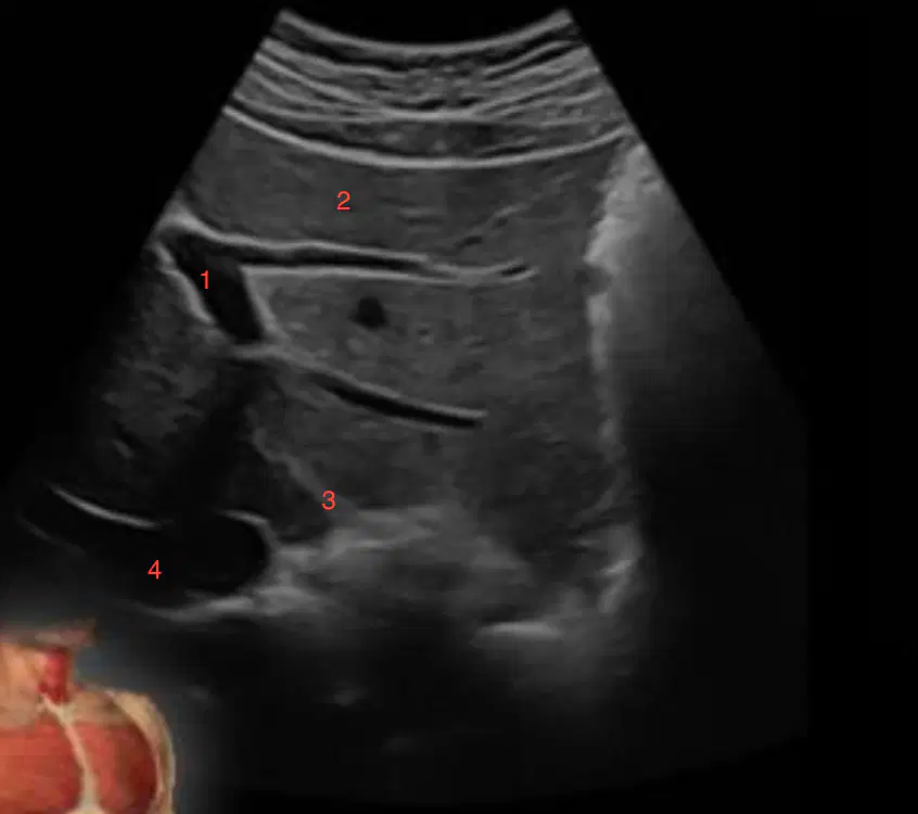
What is labeled 4?
(a)
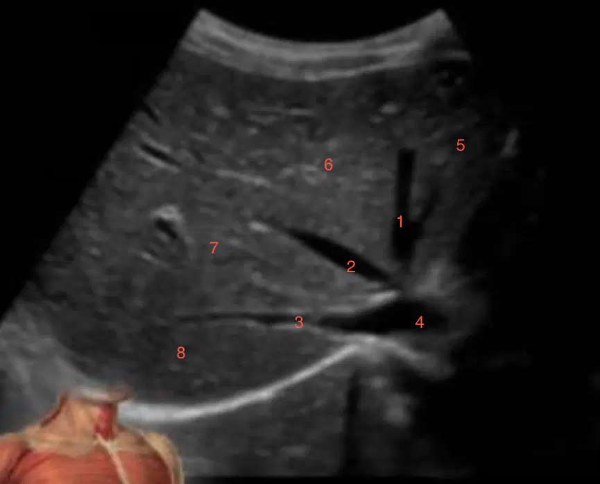
What is labeled 1
(a)
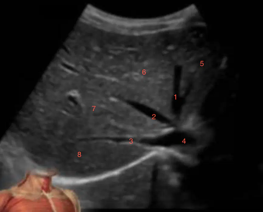
what is labeled 2
(a)
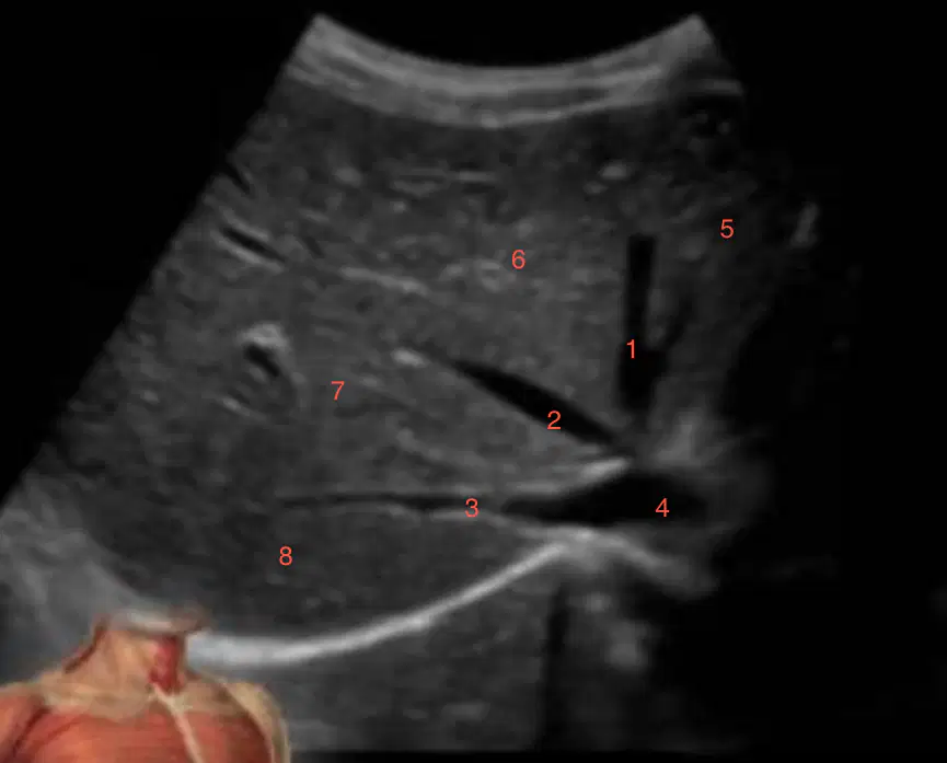
What is labeled 3
(a)
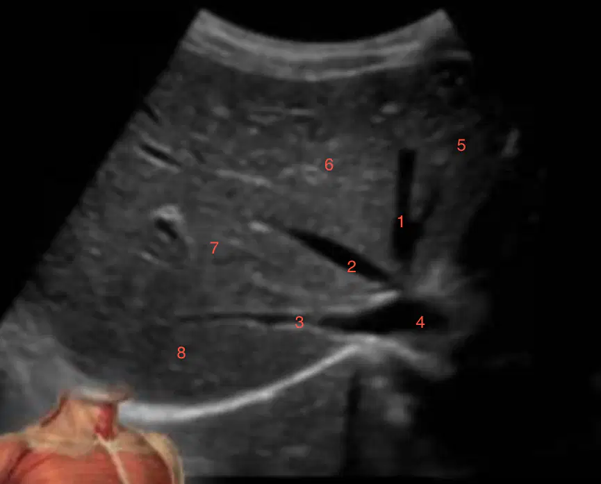
What is labeled 5
(a)
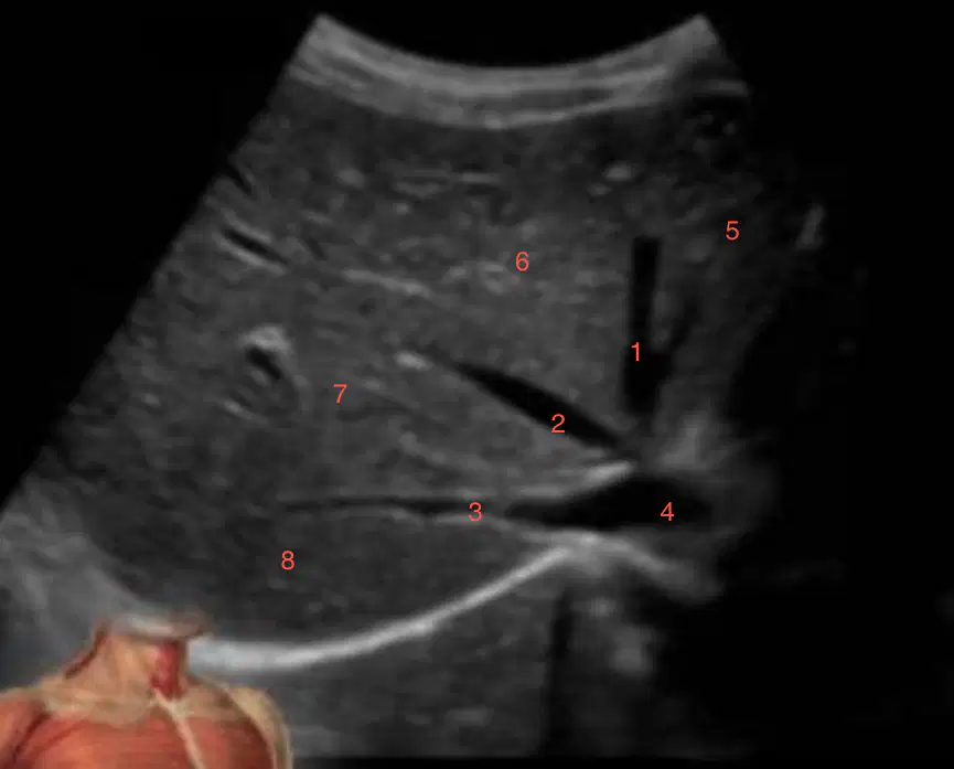
What is labeled 6
(a)
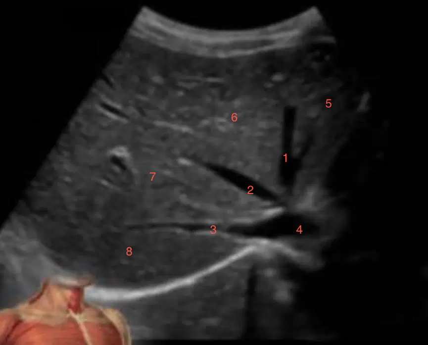
What is labeled 7
(a)
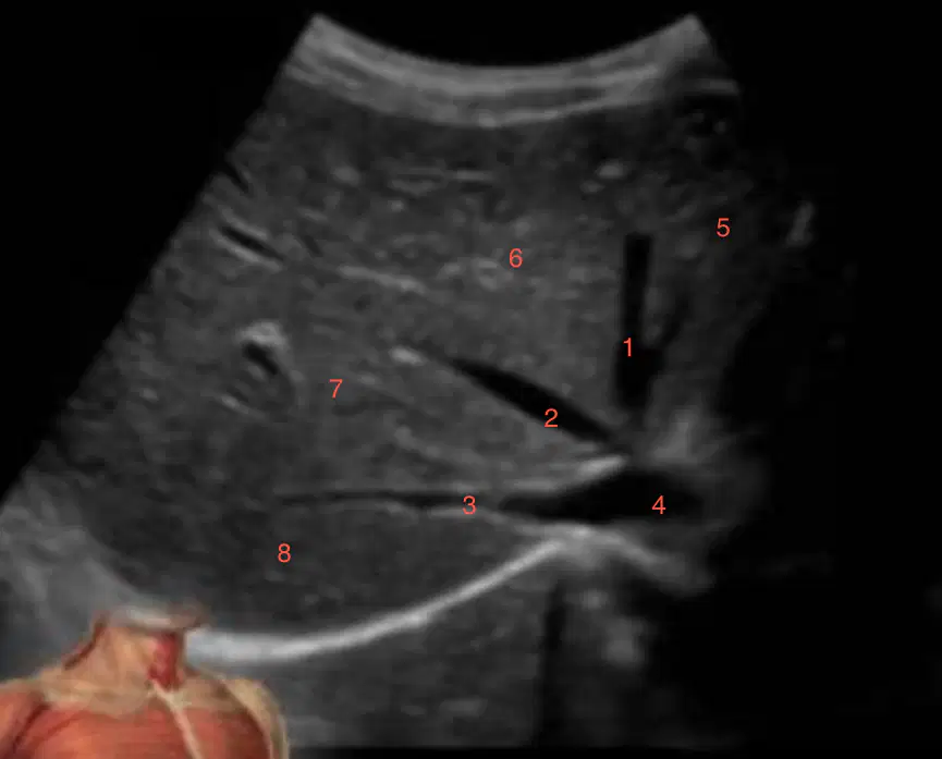
What is labeled 8
(a)
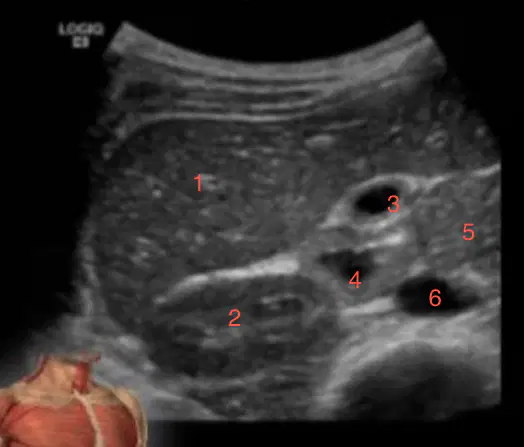
What is labeled 1
(a)

What is labeled 2
(a)
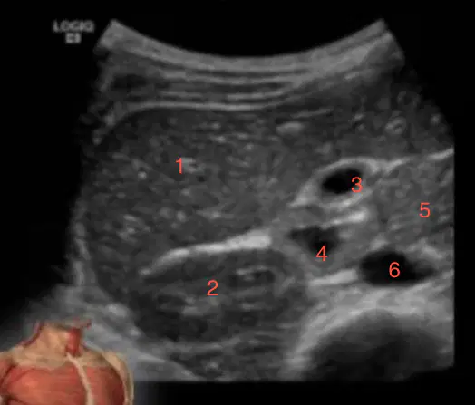
What is labeled 3
(a)
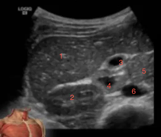
What is labeled 4
(a)
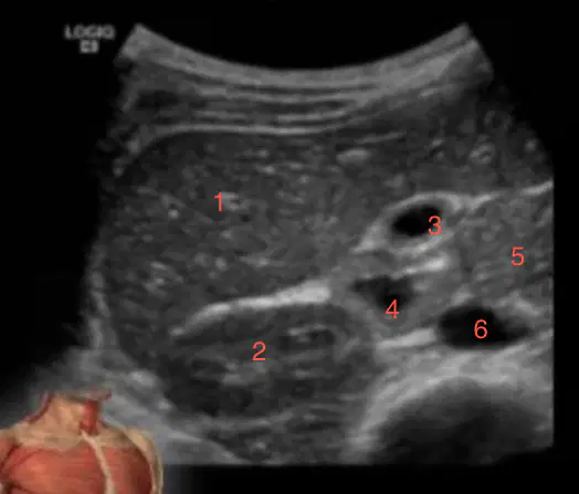
What is labeled 5
(a)
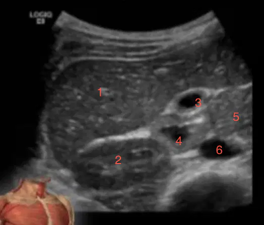
What is labeled 6
(a)

What is labeled 1
(a)
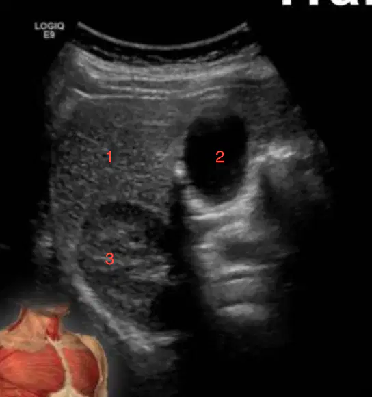
What is labeled 2
(a)
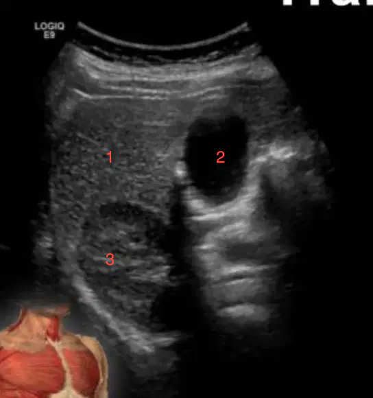
What is labeled 3?
(a)
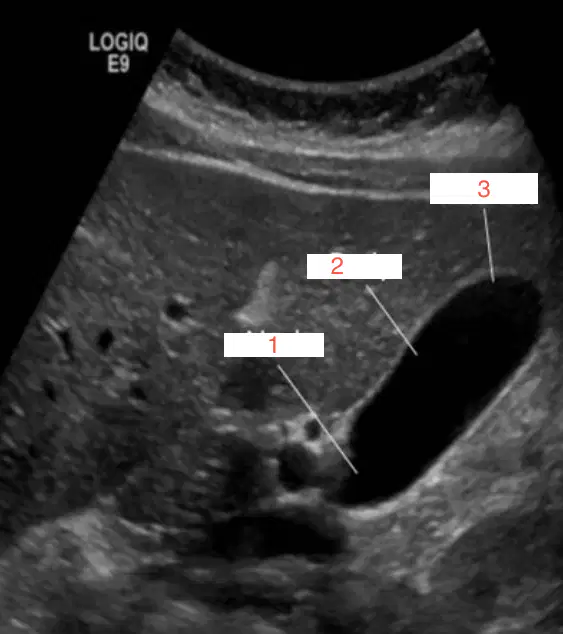
What is labeled 1
(a)
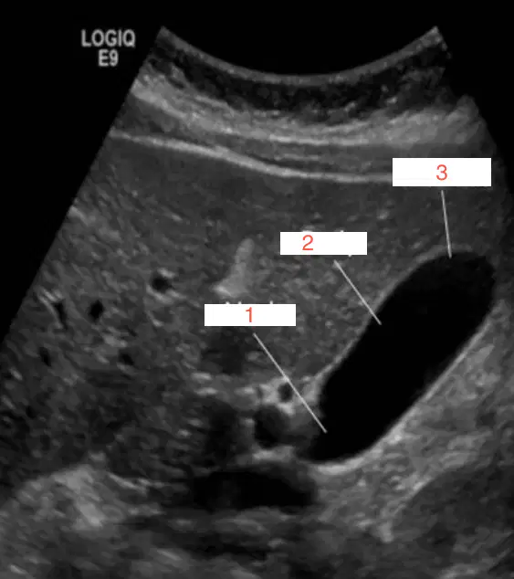
What is labeled 2
(a)
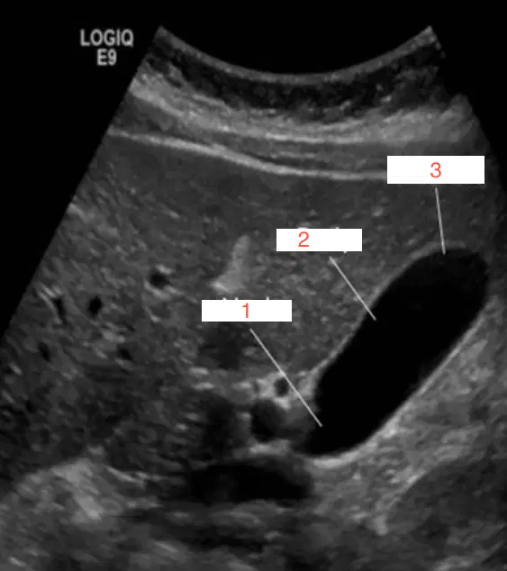
What is labeled 3
(a)
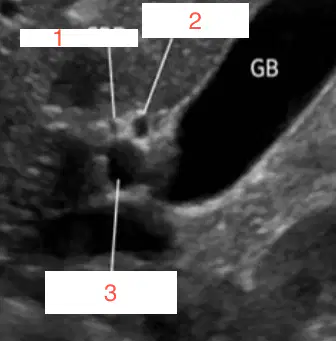
What is labeled 1
(a)
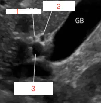
What is labeled 2
(a)
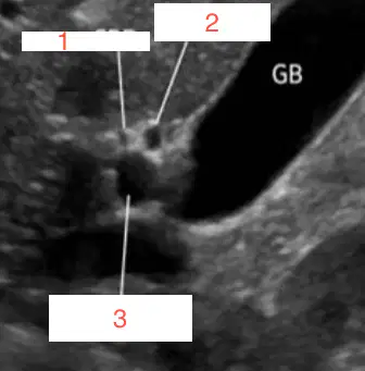
What is labeled 3
(a)
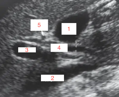
What is labeled 1
(a)
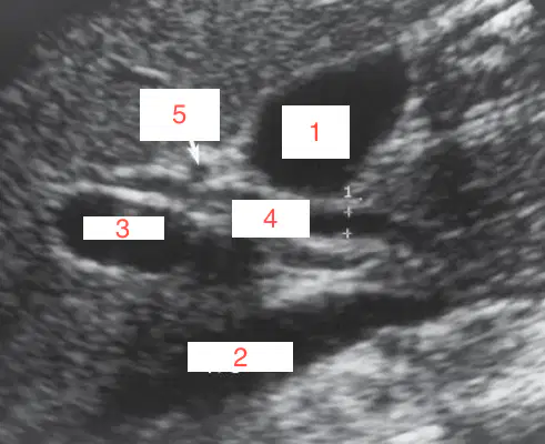
What is labeled 2
(a)
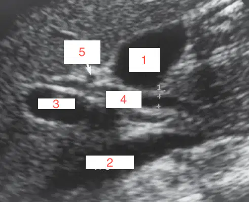
What is labeled 3
(a)
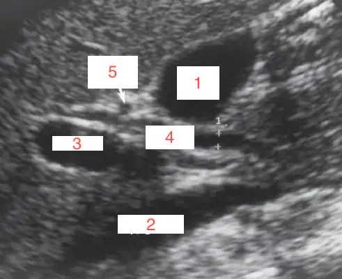
What is labeled 4
(a)
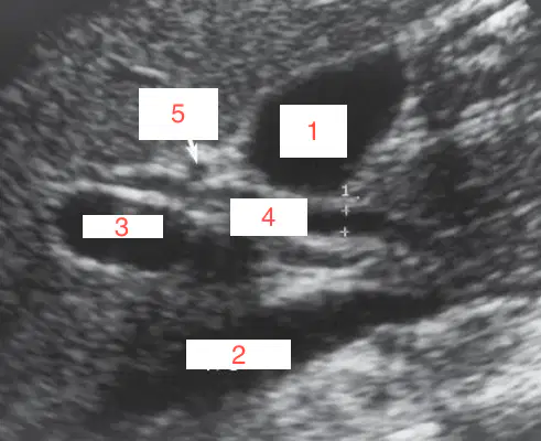
What is labeled 5?
(a)
The portal system supplies (a) % of total blood flow to the liver
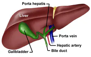
The main portal vein enters the (a) and divides into left and right branches

