
 Worksheets
Amyloid interpretation
Total questions: 10
Worksheet time: 7mins
What defines a positive Florbetaben PET scan?
Two or more brain areas (each larger than a single cortical gyrus) in which there is reduced or absent gray-white contrast.
Area(s) of tracer uptake equal to or higher than that present in white matter extending beyond the white matter rim to the outer cortical margin involving the majority of the slices within at least one of the four brain regions.
One or more cortical gyri as bright or brighter than the middle cerebellar peduncle.
Three or more brain areas (each larger than a single cortical gyrus) in which there is reduced or absent gray-white contrast.
The criteria for determining amyloid PET scan positivity are identical across all radiotracers.
True
False
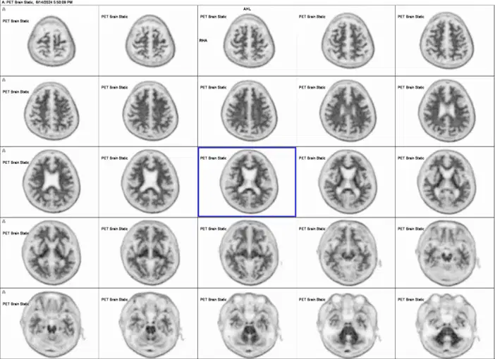
Positive or Negative
Positive
Negative
Which four brain regions are assessed when interpreting a Florbetaben PET scan?
Hippocampus, precuneus, parietal lobes, and lateral temporal lobes.
Lateral temporal lobes, precuneus/posterior cingulate, frontal lobes, and parietal lobes.
Precuneus, anterior cingulate, inferior frontal gyri, hippocampus.
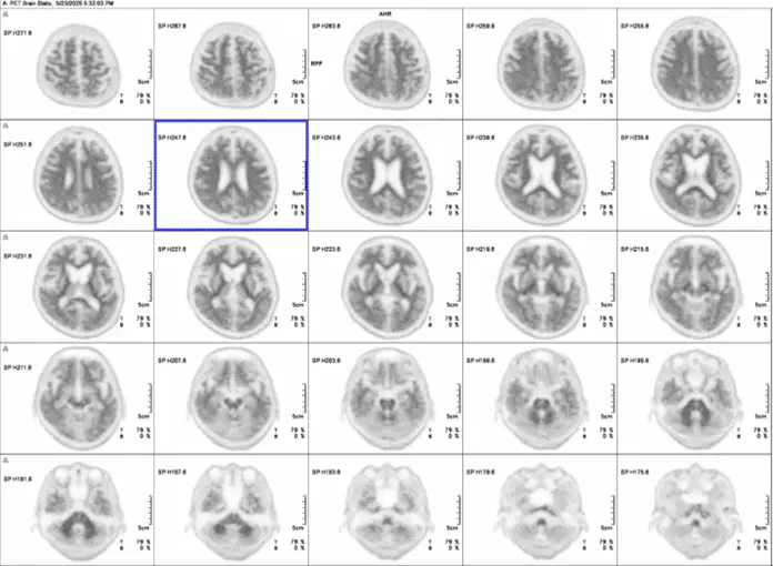
Positive or Negative
Positive
Negative
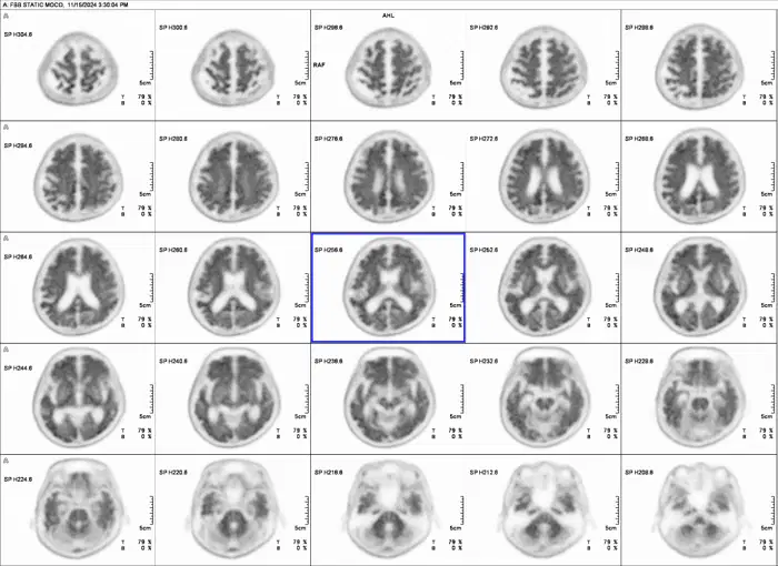
Positive or Negative
Positive
Negative
At this time, it is important to distinguish "moderate" and "pronounced" ß-amyloid deposition for anti-amyloid eligibility.
True
False
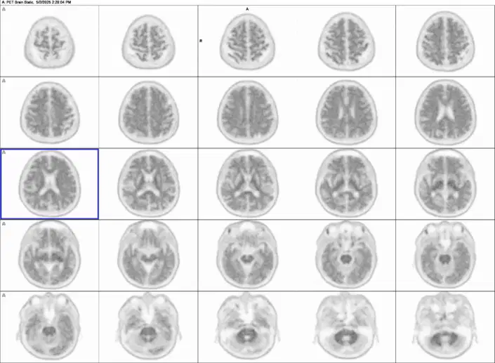
Positive or Negative
Positive
Negative
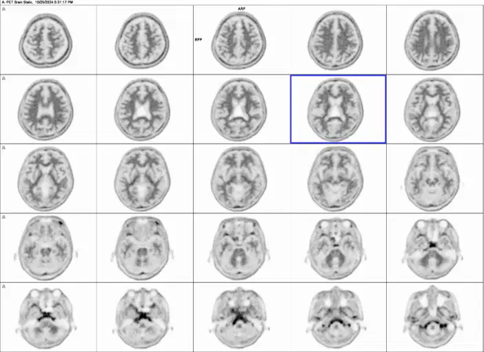
Positive
Negative
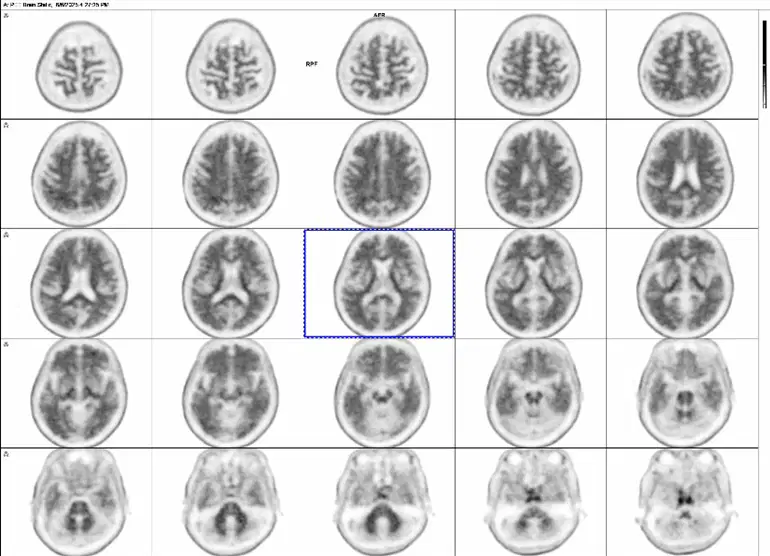
From the picture ,estimate the range of CL for this patient
<10
10-15
20-30
30-50
>50

