
 Worksheets
GI System
Total questions: 128
Worksheet time: 2hrs 57mins
The (a) system is responsible for breaking down food, absorbing nutrients, and eliminating waste.
The GI system involves both __ and __ organs
(a)
Which of the following are considered hollow organs?
Stomach
Liver
Pancreas
Intestines
Which of the following are considered accessory organs?
Stomach
Pancreas
Liver
Intestines
The primary functions of the digestive system include:
Ingestion
Digestion (mechanical and chemical)
Excretion of waste products
Absorption of nutrients
Destruction of toxins
The (a) is a continuous tube that begins at the mouth and ends at the anus
The GI tract is also called the __ and __
Digestive tract
Esophageal junction
Alimentary canal
Mouth tract
The mouth, pharynx (throat), salivary glands, and esophagus are digestive tract structures located (a) the diaphragm
The stomach, small intestines, and large intestines are gastrointestinal tract structures located (a) the diaphragm
(primarily in the abdomen)- These organs support digestion but are NOT part of the GI Tract itself
(a)
The liver, gallbladder, and pancreas are all (a) organs of the GI tract.
True or false: The GI tract accounts for a major portion of the digestive system.
true
false
Food is ingested through the (a) and chewed
Glands in the mouth that release enzymes that initiate the breakdown of food particles into small, digestible molecules.
(a)
Extends from the undersurface of the skull to the level of the cricoid cartilage in front and to the intervertebral disk between the fifth and sixth cervical vertebrae behind
(a)
The (a) is a continuation of the pharynx and ends at the stomach, after passing through the left dome of the diaphragm at the tenth thoracic vertebra level
Food is mixed and the principal chemical changes occur => food is reduced and converted to chyme
(a)
The (a) is the first part of the small intestine
Most of the digestive processes take place in the (a) bowel
Digests carbohydrates, proteins, fats, vitamins, and some fluids including water and electrolytes are digested and absorbed.
(a)
The __ __ absorbs much of the remaining fluid and finally eliminates the undigested products
(a)
Which of the following are salivary glands?
Sublingual
Submandibular
Parotid
Carotid
How many parts is the esophagus divided into?
1
2
3
4
The (a) part of the esophagus travels through the neck
The (a) part of the esophagus is located in the thorax, more specifically in the mediastinum
The (a) part of the esophagus travels past the diaphragm into the abdomen, reaching the stomach
Which part of the esophagus lies in a groove on the posterior aspect of the LT lobe of liver; connects with the cardiac region of the stomach?
(a)
The entrance of the esophagus into the stomach occurs through at the (a)
Visualized on a sagittal ultrasound as a "bulls-eye" or target pattern anterior to the aorta and posterior to the LT lobe of the liver.
(a)
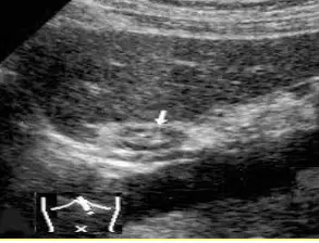
What is the arrow in this image pointing to?
Aorta
SMA
Gastroesophageal junction
LT lobe of liver
Muscular sac, that accepts the digested food passing through the esophagus.
(a)
How many portions does the stomach have?
3
4
2
1
Which of the following is NOT a portion of the stomach?
Fundus
Body
Neck
Pylorus
The stomach includes the __ and __ curvatures
(a)
The (a) are small projections on the internal surface of the stomach
How many parts does the pylorus have?
1
2
4
3

The antrum, pyloric canal, and sphincter are parts of the (a)
Which structure starts immediately after the pyloric sphincter?
Antrum
Duodenum
Large intestine
Pancreas tail
The muscles of the pyloric canal and sphincter can be affected by (a) , hence become stenotic in infants
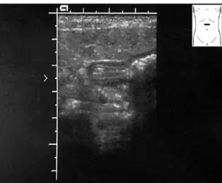
This image shows the (a)
It is the longest segment of the digestive tract, extending from the stomach to the large intestine
(a)
22 feet (6.7 meters) long.
(a)
How long is the duodenum?
25 cm (10 inches)
22 cm
35 cm
67 cm
How many parts of the duodenum are there?
1
2
3
4
The (a) part of the duodenum is a short part that terminates at the level of GB
The (a) part of the duodenum is C-shaped portion outlined by the head of the pancreas
The (a) part of the duodenum is horizontal right to left crossing anterior to IVC, Aorta, and posterior to the SMA
The (a) part of the duodenum is a short part that is not seen with imaging
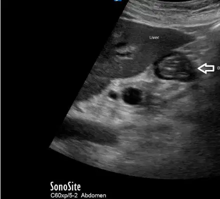
What is the arrow pointing to in this image?
(a)
The (a) is 2.5 meters (8.2 ft) long
The (a) is 3.5 meters (11.5 feet) long
The (a) is the last portion of the small intestine, it extends from the jejunum
The (a) terminates at the ileocecal valve at the cecum of the large intestine
__ and __ projections of small intestines are helping with the absorption of the nutrients from the chyme
(a)
Mucosal and submucosal projections of small intestines are helping with the absorption of the nutrients from the chyme
(a)
The (a) are larger folds giving the specific appearance to small intestines; these help with slowing down chymes movement for better absorption
The large intestines are the (a) part of the GI tract
The large intestines start with the (a)
Ileum connected to Cecum with ____ ____
(a)
Another cecal landmark is the (a)
The (a) is the rudimental intestinal blind-ended extension (vermiform process)
How many parts does the large intestines have?
1
2
5
6
The (a) runs from the cecal portion cephalic towards the livers inferior margin; makes a left turn as hepatic flexure and continues on as the transverse colon.
The (a) runs horizontally right to left, makes downturn at Splenic flexure and continues on as a the descending colon.
The (a) is located at the LLQ, becomes the sigmoid colona
The (a) is the terminal section of the large intestine that turns into the rectum
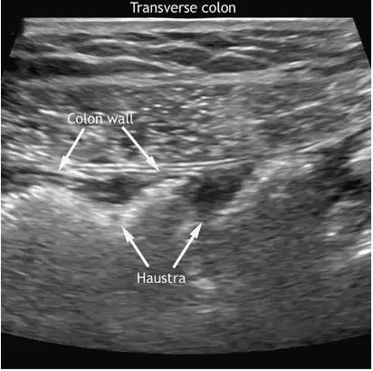
The (a) refers to the small segmented pouches of the bowel separated by pastoral folds.
The (a) are formed by circumferential contraction of the inner muscular layer of the colon
Haustral contractions are slow segmenting, uncoordinated movements that occur approximately every ___ minutes
10
20
25
30
One haustsrum distends as it fills with chyme, which stimulates muscles to (a) , pushing the contents to the next haustrum
The stomach, first part of duodenum, jejunum, ileum, and cecum are all (a) organs
The appendix, transverse colon, sigmoid colom, upper third of rectum, liver, and spleen are considered (a) organs
The second and third parts of the duodenum, ascending column, pancreas, and kidneys are considered (a) organs
The descending colon, middle third of rectum, adrenal glands, proximal ureters and gonadal blood vessels are considered (a) organs
The renal vessels, IVC, and AO are considered (a) organs
The (a) is supplied by the descending aorta, left gastric artery, and left phrenic artery.
The (a) is supplied by the RT and LT gastric arteries, gastroepiploic artery, and gastric vasa brevia.
The (a) are supplied by the SMA, GDA, superior pancreaticoduodenal artery, and inferior pancreaticoduodenal artery
The (a) are supplied by the inferior and superior mesenteric arteries
The venous system parallels the arterial system and empties in the (a)
Evaluation of the bowel with US begins with the patient history, followed by US assessment of the wall thickness, gut signature, and motility
(a)
Which of the following are indications for an ultrasound of the GI tract?
Pyloric stenosis in pediatric patients
Stomach pains
Intussusception (inversion)
Bowel obstruction/ Inflammatory process
Neoplasms
Blood in stool, leukocytosis, and carcinoembryonic marker are all relevant (a) data for GI tract examinations
Other imaging modalities for GI tract include:
Rectal Inversions
X-ray
CT scans
Endoscopy
Colonoscopy
Intraluminal gas obscuring the posterior structures is a (a) of ultrasound imaging
Reverberation artifact, comet-tail artifact, dirty shadow artifact is a (a) of ultrasound imaging
Regardless of its caliber, the bowel shows a stratified morphology consisting of five concentric rings of alternating echogenicity referred to as the (a)
The normal bowel from the stomach to the colon has an average mural thickness of __ to __ mm, depending on the degree of distention
(a)
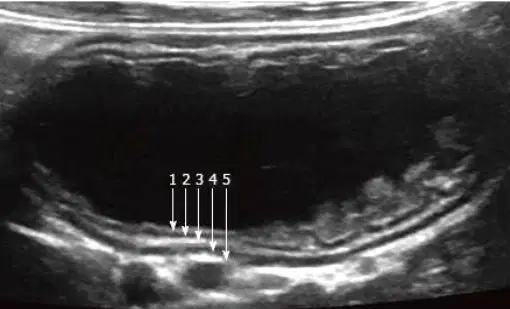
What is labeled 1 in this image?
(a)
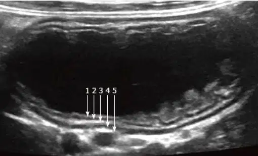
What is labeled 2 in this image?
(a)
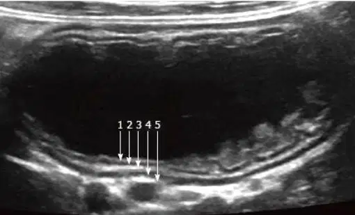
What is labeled 3 in this image?
(a)
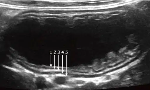
What is labeled 4 in this image?
(a)
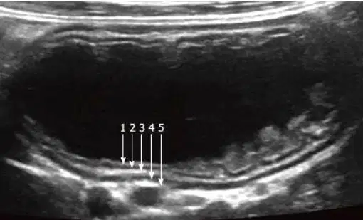
What is labeled 5 in this image?
(a)
The bowel has how many layers sonographically?
1
2
4
3
The (a) layer of the bowel directly contacts the intraluminal contents; lined with epithelial folds; echogenic
The (a) layer of the bowel is hypoechoic.
The (a) layer of the bowel is echogenic
The (a) contains circular and longitudinal bands of fiber; hypoechoic
The (a) is a thin, loose layer of connective tissue; echogenic
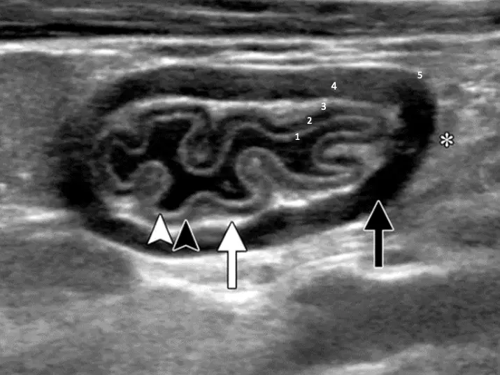
What is labeled 1?
(a)
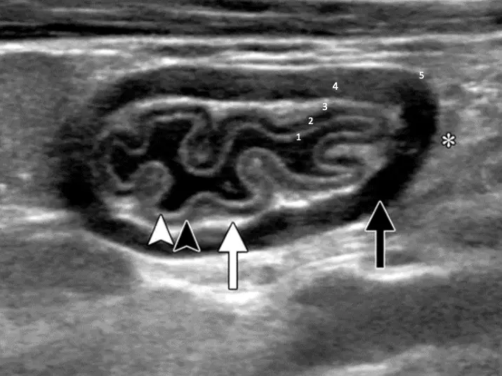
What is labeled 2?
(a)

What is labeled 3?
(a)
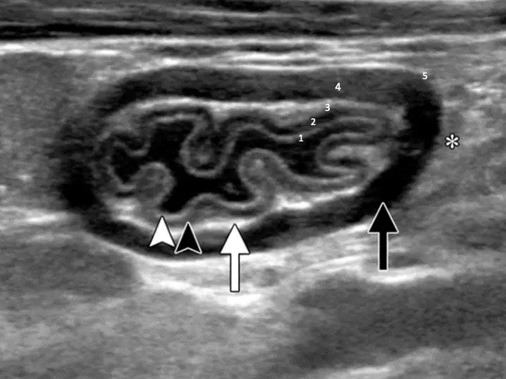
What is labeled 4?
(a)
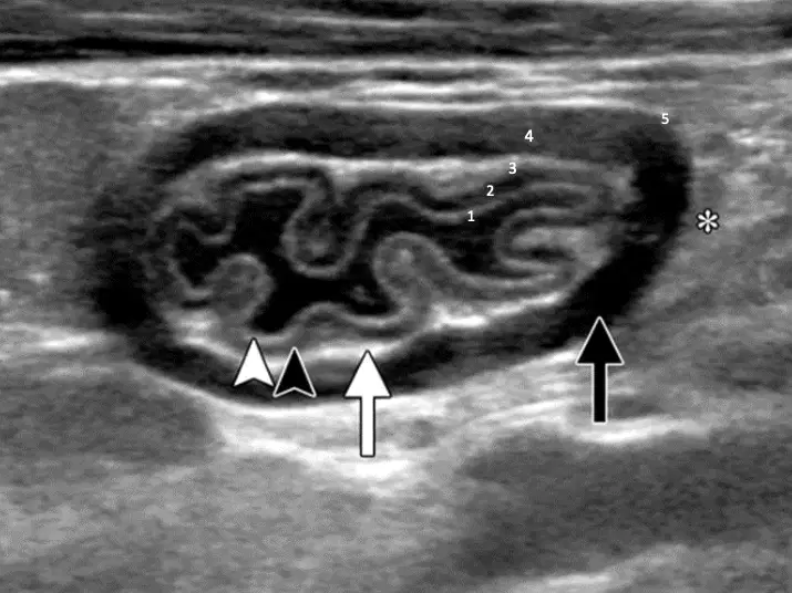
What is labeled 5?
(a)
The odd numbered walls (first, third, and fifth) are (a)
The even numbered walls are (second and fourth) (a)
The upper GI tract (stomach and duodenum) can be scanned by standard longitudinal and transverse sections seen in the picture
True
False
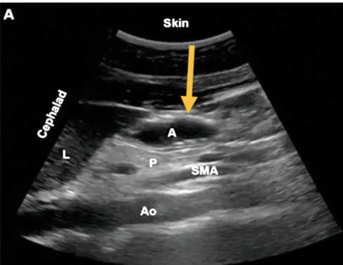
What is labeled A in this image?
(a)
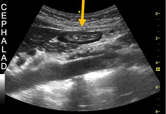
The arrow in this image is pointing to the (a)
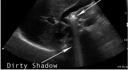
The arrow in this image is pointing to the (a)
Examination of the small bowel performed by systematic parallel and overlapping scanning of the abdomen
True
False
(a) can be visualized on its course over Iliopsoas muscle and Iliac vessels
the small bowel wall is less than (a) mm thick
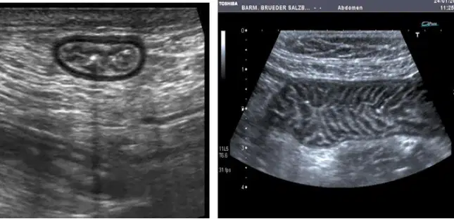
These images show the (a)
(a) , air movement, or movement of intraluminal fluid contents is easily seen
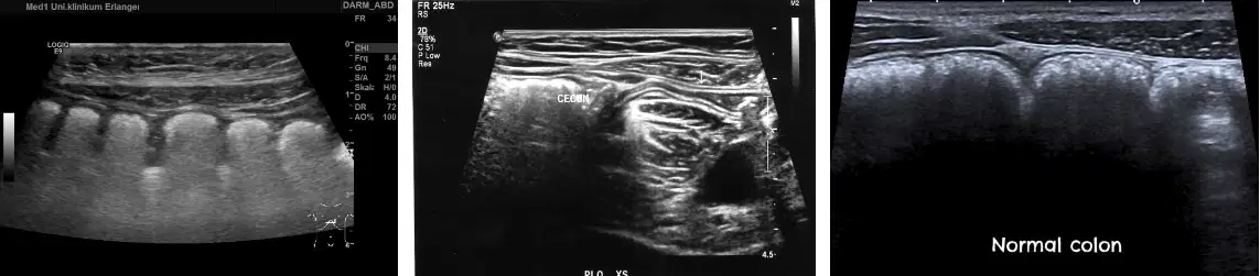
These images show the (a)
The normal thickness of the colon measures at ___ mm
1
4
2
3
The entire halo of the colon should measure less than ___ cm (target sign)
2
3
4
5
If the colon is dilated, the sonographer should measure from the fluid to the (a) of the wall
When is distention considered significant?
When the colon is larger than 3cm in diameter
When the colon is larger than 5cm in diameter
When the colon is larger than 4cm in diameter
When. the colon is larger than 6 cm in diameter
(a) is located on the abdominal wall under McBurney’s point
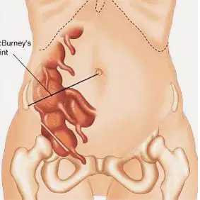
(a) point is located by drawing a line from the right anterosuperior iliac spine to the umbilicus
The appendix varies from which of the following in length?
10 to 30cm
2 to 20cm
3 to 80 cm
70 to 80 cm
The average length of the colon is __ cm
10
11
9
12
The outer layer of the appendix should be less than (a) mm
Position of the (a) varies; among the known configurations – retrocecal variant –
challenging to visualize
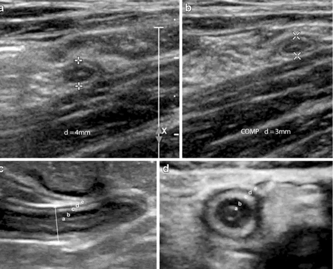
Target-shaped appearance
(a)
the appendix is located immediately (a) to abdominal wall muscle and adipose tissue, at the depth of 2 cm in that patient (with pressure applied)

