
 Worksheets
Microbiology Ch. 3/4
Total questions: 83
Worksheet time: 42mins
Which microscope is used to see detail of a 300-nm virus?
Electron microscope
Fluorescence microscope
DIC microscope
Phase-contrast microscope
Darkfield microscope
Bacterial smears are fixed before staining to
Kill the bacteria.
Affix the cells to the slide.
Make their walls permeable.
Kill the bacteria and Affix the cells to the slide.
Accept stain.
The counterstain in the acid-fast stain is
A negative stain.
A basic dye.
A mordant.
Necessary to determine acid-fast cells.
An acid dye.
The signal molecule produced in quorum sensing is
Light.
An inducer.
An endospore.
A counterstain.
A simple stain.
The appearance of gram-negative bacteria after addition of the mordant in the Gram stain.
Brown
Colorless
Purple
Red
Which of the following pairs is mismatched?
Safranin - acid dye
Iodine - mordant
Alcohol-acetone - decolorizer
Crystal violet - basic dye
Carbolfuchsin - basic dye
Which microscope takes advantage of differences in the refractive indexes of cell structures?
Fluorescence microscope
Darkfield microscope
Electron microscope
Compound light microscope
Phase-contrast microscope
Which microscope is most useful for visualizing a biofilm?
Transmission electron microscope
Compound light microscope
Phase-contrast microscope
Atomic force microscope
Scanning acoustic microscope
What structure does light pass through after leaving the condenser in a compound light microscope?
Ocular lens
Illuminator
Specimen
Objective lens
The best use of a negative stain is
To see endospores.
To determine cell shape.
To determine cell size and To determine cell shape
To determine cell size.
To determine Gram reaction.
Which of the following is NOT correct?
1 µm = 10^3 nm
1 nm = 10-9 m
1 µm = 10-6 m
1 nm = 10-6 μm
1 µm = 10-3 mm
The appearance of gram-positive bacteria after adding the counterstain in the Gram stain.
Purple
Brown
Red
Colorless
Van Leeuwenhoek's microscope magnified up to 300x. This was a(n)
Compound microscope.
Electron microscope.
Simple microscope.
Confocal microscope.
Phase-contrast microscope.
What is the total magnification of a chloroplast viewed with a 10x ocular lens and a 45x objective lens?
450x
10x
100x
45x
4.5x
Place the steps of the Gram stain in the correct order: 1-Alcohol-acetone; 2-Crystal violet; 3-Safranin; 4-Iodine.
1-3-2-4
2-4-1-3
2-1-4-3
4-3-2-1
1-2-3-4
Which of the following microscopes uses visible light?
DIC
Confocal microscope
Fluorescence microscope
Scanning acoustic microscope
Scanning electron microscope
In this microscope, the observer does NOT look at an image through a lens.
Fluorescence microscope
Phase-contrast microscope
Compound light microscope
Electron microscope
Darkfield microscope
What Gram reaction do you expect from acid-fast bacteria?
Gram-negative
Can't tell
Both gram-positive and gram-negative
Gram-positive
The resolution of a microscope can be improved by changing the
Wavelength of light.
Coarse adjustment.
Diaphragm.
Condenser.
Fine adjustment.
Which microscope is used to see intracellular detail in a living cell?
Atomic force microscope
Brightfield microscope
Transmission electron microscope
Two-photon microscope
Fluorescence microscope
The purpose of the ocular lens is to
Increase the light.
Magnify the image from the objective lens.
Decrease the refractive index.
Improve resolution.
Decrease the light.
The purpose of a mordant in the Gram stain is
To make gram-negative cells visible.
To prevent the crystal violet from leaving the cells.
To remove the simple stain.
To make the bacterial cells larger.
To make the flagella visible.
Which microscope is used to observe a specimen that emits light when illuminated with an ultraviolet light?
Compound light microscope
Electron microscope
Fluorescence microscope
Darkfield microscope
Phase-contrast microscope
Which microscope uses two beams of light to produce a three-dimensional, color image?
Electron microscope
Fluorescence microscope
Phase-contrast microscope
DIC microscope
Darkfield microscope
Where are phospholipids most likely found in a eukaryotic cell?
Ribosomes
Around organelles and Plasma membrane
Plasma membrane
Around organelles
Flagella
Which of the following organelles most closely resembles a prokaryotic cell?
Mitochondrion
Nucleus
Golgi complex
Vacuole
Cell wall
Which of the following statements about the glycocalyx is false?
It may be composed of polysaccharide.
It is used to adhere to surfaces.
It protects from osmotic lysis.
It may be responsible for virulence.
It may be composed of polypeptide.
Where are phospholipids most likely found in a prokaryotic cell?
Plasma membrane
Around organelles and Plasma membrane
Ribosomes
Around organelles
Flagella
Which of the following statements about gram-negative cell walls is false?
They are sensitive to penicillin.
Their Gram reaction is due to the outer membrane.
They are toxic to humans.
They have an extra outer layer composed of lipoproteins, lipopolysaccharides, and phospholipids.
They protect the cell in a hypotonic environment.
By which of the following mechanisms can a cell transport a substance from a lower to a higher concentration?
Active transport
Extracellular enzymes
Aquaporins
Facilitated diffusion
Simple diffusion
You have isolated a motile, gram-positive cell with no visible nucleus. You can safely assume that the cell
Has a nucleus.
Has a mitochondrion.
Has 9 pairs + 2 flagella.
Has a cell wall.
Lives in an extreme environment.
What will happen if a bacterial cell is placed in 10% NaCl with penicillin?
Penicillin will diffuse into the cell.
Water will enter the cell.
The cell will plasmolyze.
No change will result; the solution is isotonic.
The cell will undergo osmotic lysis.
Which of the following is NOT a possible function of magnetosomes?
Store iron
Get cells to the North Pole
Locate suitable environments
Protect cells from hydrogen peroxide accumulation
The difference between simple diffusion and facilitated diffusion is that facilitated diffusion
Can move materials from a lower to a higher concentration.
Requires ATP.
Requires transporter proteins.
Doesn't require ATP.
Can move materials from a higher to a lower concentration.
What will happen if a bacterial cell is placed in distilled water with lysozyme?
Lysozyme will diffuse into the cell.
The cell will plasmolyze.
Water will leave the cell.
No change will result; the solution is isotonic.
The cell will undergo osmotic lysis.
Which of the following are NOT energy reserves?
Lipid inclusions.
Polysaccharide granules.
Metachromatic granules.
Sulfur granules.
Carboxysomes.
In bacteria, photosynthetic pigments are found in
Ribosomes.
Cytoplasm.
Chloroplasts.
Chromatophores.
Mesosomes.
Which of the following statements best describes what happens when a bacterial cell is placed in a solution containing 5% NaCl?
Sucrose will move into the cell from a higher to a lower concentration.
No change will result; the solution is isotonic.
Water will move into the cell.
The cell will undergo osmotic lysis.
Water will move out of the cell.
Which of the following is NOT a distinguishing characteristic of prokaryotic cells?
They have cell walls containing peptidoglycan.
They lack membrane-enclosed organelles.
They lack a plasma membrane.
Their DNA is not associated with histones.
Their DNA is not enclosed within a membrane.
Which of the following pairs is mismatched?
Endoplasmic reticulum - internal transport
Golgi complex - secretion
Lysosome - digestive enzymes
Mitochondria - ATP production
Centrosome - food storage
Which of the following statements is true?
A cell can produce many endospores.
Endospores are for reproduction.
A cell produces one endospore and keeps growing.
Endospores are easily stained in a Gram stain.
Endospores allow a cell to survive environmental changes.
Place the following steps in the correct sequence: 1-Staining; 2-Making a smear; 3-Fixing.
2-3-1
3-2-1
The order doesn't matter
1-3-2
1-2-3
In which microscope does the image look like a negative stain?
Phase-contrast microscope
Two-photon microscope
Darkfield microscope
Scanning acoustic microscope
Fluorescence microscope
The appearance of gram-negative bacteria after addition of the decolorizing agent in the Gram stain.
Brown
Purple
Red
Colorless
The appearance of gram-positive bacteria after addition of the first dye in the Gram stain.
Purple
Red
Colorless
Brown
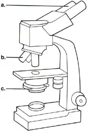
In Figure 3.1, line "b." points to the microscope's
Condenser.
Illuminator.
Objective lens.
Ocular lens.
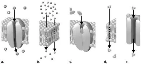
Figure 4.4 shows movement across a plasma membrane. Which figure best represents simple diffusion?
a
b
c
d
e
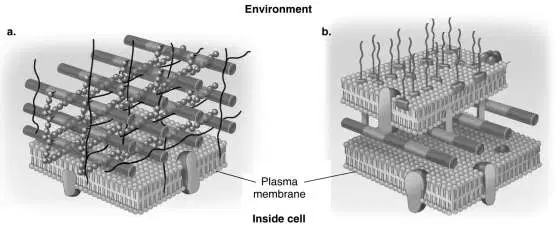
In Figure 4.3, which diagram of a cell wall is decolorized by acetone-alcohol?
b
Both a and b
Can't tell
Neither a nor b
a
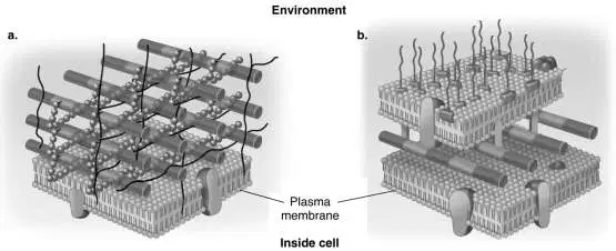
In Figure 4.3, which diagram of a cell wall is a toxic cell wall?
Neither a nor b
Can't tell
Both a and b
a
b
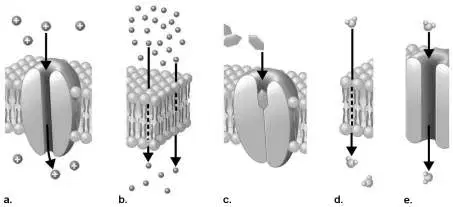
Figure 4.4 shows movement across a plasma membrane. Which figure best represents a specific transporter protein?
a
b
c
d
e
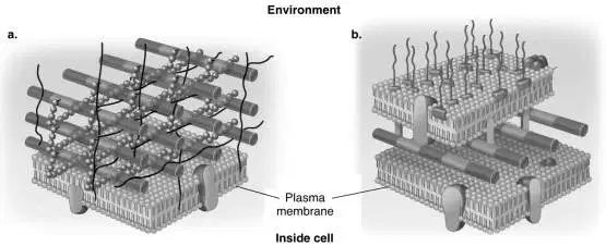
In Figure 4.3, which diagram of a cell wall is a gram-negative cell wall?
Can't tell
Both a and b
a
Neither a nor b
b
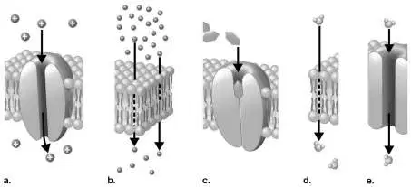
Figure 4.4 shows movement across a plasma membrane. Which figure best represents an aquaporin?
a
b
c
d
e
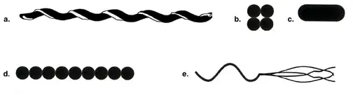
Which drawing in Figure 4.1 possesses an axial filament?
a
b
c
d
e
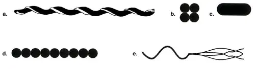
Which drawing in Figure 4.1 is a tetrad?
a
b
c
d
e
You find colorless areas in cells in a Gram-stained smear. What should you do next?
A capsule stain
A simple stain
An endospore stain
A flagella stain
An acid-fast stain
Simple staining is often necessary to improve contrast in this microscope.
Darkfield microscope
Compound light microscope
Electron microscope
Phase-contrast microscope
Fluorescence microscope
Which microscope is used to see internal structures of cells in a natural state?
Darkfield microscope
Electron microscope
Fluorescence microscope
Compound light microscope
Phase-contrast microscope
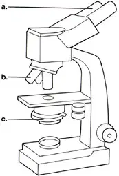
In Figure 3.1, line "c." points to the microscope's
Condenser.
Illuminator.
Objective lens.
Ocular lens.
Which of the following pairs is mismatched?
Alcohol-acetone - decolorizer
Crystal violet - simple stain
Iodine - mordant
Acid-alcohol - decolorizer
Gram-negative bacteria - negative stain
Which of the following is NOT equal to 1 m?
109 nm
106 µm
0.001 km
10 dm
100 mm
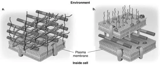
In Figure 4.3, which diagram of a cell wall has a wall that protects against osmotic lysis?
Neither a nor b
Both a and b
Can't tell
a
b
Which of the following statements about a gram-positive cell wall is false?
It is sensitive to lysozyme.
It maintains the shape of the cell.
It protects the cell in a hypertonic environment.
It contains teichoic acids.
It is sensitive to penicillin.
Which of the following structures is NOT found in prokaryotic cells?
Peritrichous flagella
Axial filament
Flagellum
Cilium
Pilus
The best definition of osmotic pressure is
The movement of solute molecules from a higher to a lower concentration.
The active transport of a substance out of a cell to maintain equilibrium.
The movement of solute molecules from a lower to a higher concentration across a semipermeable membrane.
The force with which a solvent moves across a semipermeable membrane from a higher to a lower concentration.
The movement of a substance across a semipermeable membrane from a higher to a lower concentration.
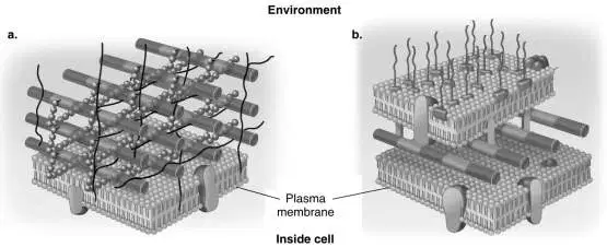
In Figure 4.3, which diagram of a cell wall contains porins?
Can't tell
a
b
Neither a nor b
Both a and b
Which of the following have a cell wall?
Protoplasts
Animal cells
Fungi
L forms
Mycoplasmas
Which of the following is NOT found in mitochondria and prokaryotes?
Cell wall
ATP-generating mechanism
70S ribosomes
Circular chromosome
Binary fission
Which of the following is NOT a chemical component of a bacterial cell wall?
Cellulose
Peptide chains
Peptidoglycan
Teichoic acids
N-acetylmuramic acid
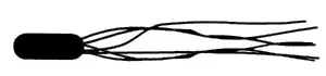
Which of the following terms best describes the cell in Figure 4.2?
Axial filament
Amphitrichous flagella
Monotrichous flagella
Peritrichous flagella
Lophotrichous flagella
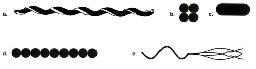
Which drawing in Figure 4.1 is streptococci?
a
b
c
d
e
Which of the following pairs is NOT a functionally analogous pair?
Nucleus - nuclear region
Mitochondria - prokaryotic plasma membrane
9+2 flagella - bacterial flagella
Cilia - pili
Chloroplasts - thylakoids
Which microscope can be used to visualize DNA or botulinum toxin?
Confocal microscope
Scanning tunneling microscope
Phase-contrast microscope
Compound light microscope
Scanning electron microscope
Which microscope achieves the highest magnification and greatest resolution?
Phase-contrast microscope
Fluorescence microscope
Darkfield microscope
Compound light microscope
Electron microscope
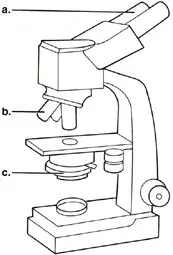
In Figure 3.1, line "a." points to the microscope's
Objective lens.
Ocular lens.
Condenser.
Illuminator.
This microscope produces an image of a light cell against a dark background; internal structures are NOT visible.
Darkfield microscope
Compound light microscope
Fluorescence microscope
Phase-contrast microscope
Electron microscope
The counterstain in the Gram stain is
Necessary to determine the Gram reaction.
A mordant.
A basic dye.
A negative stain.
An acid dye.
The appearance of gram-negative bacteria after completing the Gram stain.
Purple
Red
Colorless
Brown
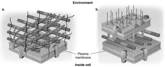
In Figure 4.3, which diagram of a cell wall contains teichoic acids?
Neither a nor b
Both a and b
Can't tell
b
a
Which one of the following pairs is mismatched?
Metachromatic granules - phosphate storage
Lipid inclusions - energy reserve
Ribosomes - protein storage
Gas vacuoles - flotation
Sulfur granules - energy reserve
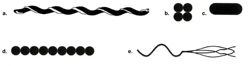
Which drawing in Figure 4.1 is a bacillius?
a
b
c
d
e
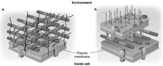
In Figure 4.3, which diagram of a cell wall is resistant to many antibiotics (e.g., penicillin)?
Can't tell
Both a and b
a
Neither a nor b
b
Cells are differentiated after which step in the Gram stain?
Safranin
Alcohol-acetone
Iodine
Crystal violet
Which of the following statements about prokaryotic cells is generally false?
They have a semirigid cell wall
They are motile by means of flagella
They possess 80S ribosomes
They reproduce by binary fission
They lack membrane-bound nuclei

