
 Worksheets
Cerebrovascular Circulation and Arteries
Total questions: 135
Worksheet time: 2hrs 0mins
Cerebrovascular circulation consists of
Extracranial
Willy
Outercranial
Intracranial
Carotid arteries and branches
(a)
Circle of Willis and Ophthalmic arteries
(a)
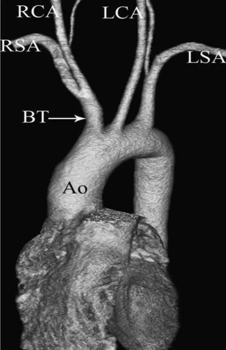
Brachiocephalic artery, Left Carotid artery, Left Subclavian artery
(a)
Right Subclavian artery and Right Carotid artery
(a)
BRACHIOCEPHALIC artery is also called:
(a)
Both Vertebral Arteries are branching from (a)
main branch of cerebrovascular circulation
(a)
CCA branches into ICA ad ECA at the level of the (a) cartilage
The common carotid artery Courses in the (a) aspect of neck, lateral to trachea, esophagus, larynx, and pharynx
The common carotid artery bifurcates into __ and __
(a)
What is the size of the internal carotid artery?
5-6mm
4-5mm
3-4mm
1-2mm
(a) supplies Circle of Willis and an eye via Ophthalmic artery
Posterolateral branch of CCA
(a)
80% of the carotid flow flows through __
ECA
ICA
CCA
RCA
Does the internal carotid artery (ICA) have cervical branches?
Yes
No
What is the first branch of the ICA?
Left common carotid
Extracranial opthalmic artery
Intracranial ophthalmic artery
Right common carotid
Ophthalmic artery is an important (a) pathway in cases of ICA occlusion
ICA terminates at (a) of MCA, ACA and PCA of the Circle of Willis (lateral aspect of the COW)
How many segments is the ICA divided into?
1
2
3
4
Which of the following is NOT major segments of the ICA?
Cervical
Subclavian
Cavernous
Petrous
Cerebral
Which of the following correctly describes the course of the cervical segment of the ICA?
runs from the base of the skull through the petrous bone;
runs through the cavernous sinus
runs from above the carotid bulb through the neck to the base of the skull
runs above the clinoid process through the dura into the subarachnoid space
Which of the following correctly describes the course of the petrous segment of the ICA?
runs from the base of the skull through the petrous bone;
runs through the cavernous sinus
runs from above the carotid bulb through the neck to the base of the skull
runs above the clinoid process through the dura into the subarachnoid space
Which of the following correctly describes the course of the Cavernous segment of the ICA?
runs from the base of the skull through the petrous bone;
runs from above the carotid bulb through the neck to the base of the skull;
runs through the cavernous sinus
runs above the clinoid process through the dura into the subarachnoid space
segment C1 - runs from above the carotid bulb through the neck to the base of the skull;
(a)
segment C2 - runs within the carotid canal in the petrous portion of the temporal bone
(a)
segment C3 - runs through the cavernous sinus
(a)
segment C4 - runs above the clinoid process through the dura into the subarachnoid space
(a)
branch of the CCA
(a)
20% of the carotid's blood supplies the face and neck via the:
(a)
Supplies blood to the tissues of the face and scalp
(a)
What is the size of the external carotid artery?
4-5mm
3-4mm
2-3mm
6-7mm
is the first branch of the ECA
(a)
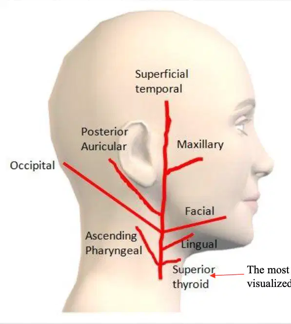
Ascending pharyngeal artery is a cervical branch of the (a)

S
(a)

L
(a)
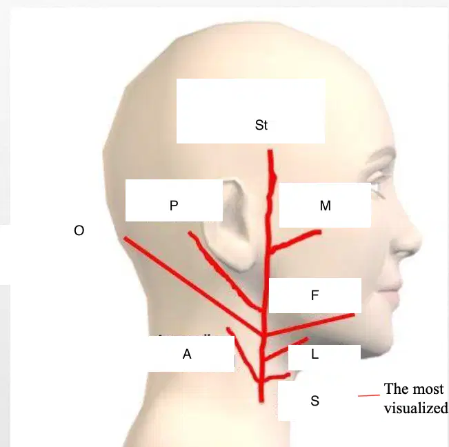
F
(a)
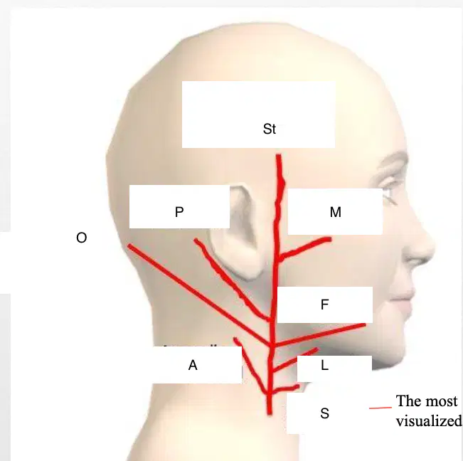
M
(a)
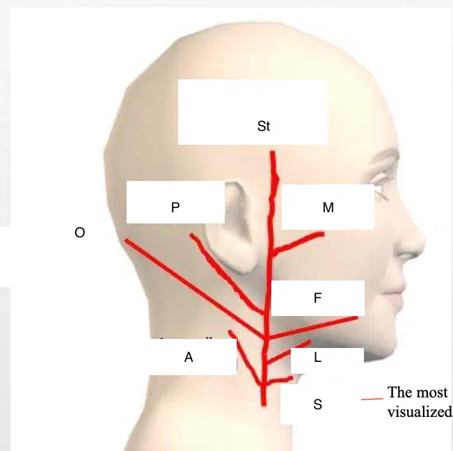
ST
(a)
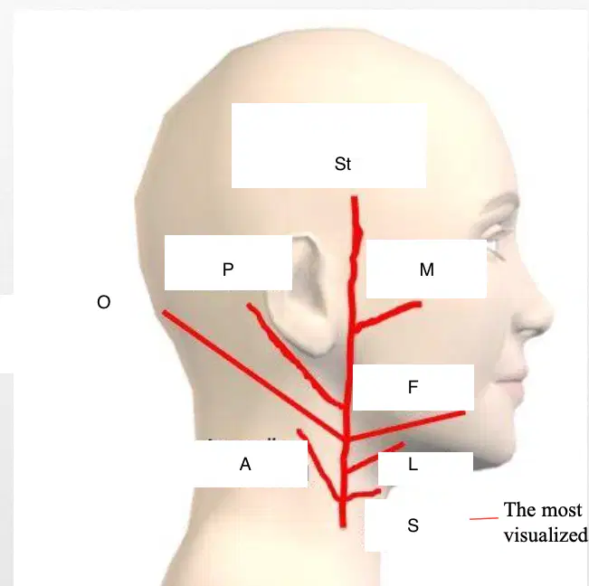
P
(a)
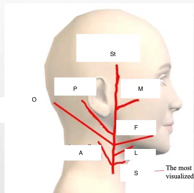
O
(a)
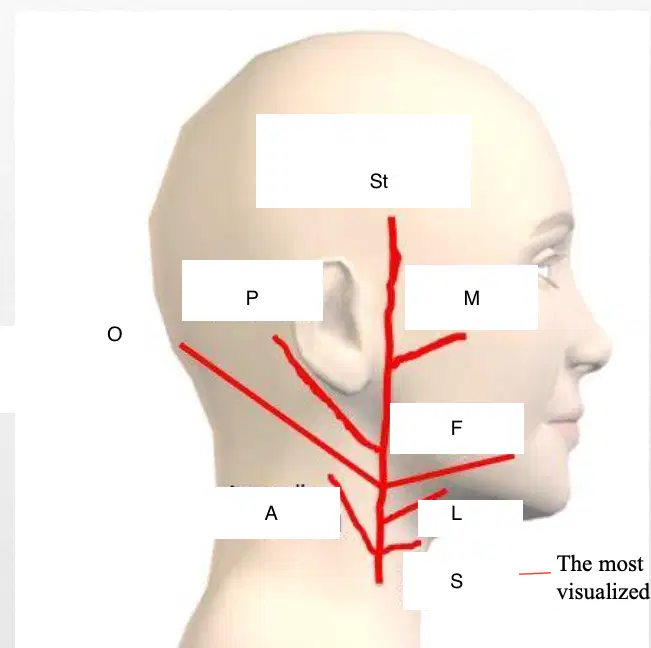
A
(a)
Ascending pharyngeal artery, Lingual artery, Facial artery, Occipital artery, Posterior auricular artery, Maxillary artery (External & Internal), Superficial temporal artery- continuation of the ECA
(a)
The ICA, the ECA, and some of their main branches
(a)
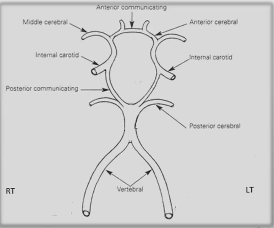
The RT and LT vertebral arteries => the Basilar artery =>
the Posterior Cerebral arteries forming part of the (a)
the first branch of the Subclavian artery
(a)
Courses superiorly via the spinal column; runs through Foramen Magnum to form Basilar Artery (vertebrobasilar system, posterior portion of the COW)
(a)
Which artery is 2-3mm in diameter
vertebral artery
internal carotid
middle cerebral
posterior cerebral
(a) portion of the vertebral artery begins as it pierces dura and arachnoid immediately below base of skull at Foramen Magnum
Continues anterior and medial to anterior surface of medulla and unites with contralateral vertebral artery to form basilar artery
(a)
formed by union of two Vertebral arteries; is evaluated during TCD imaging
(a)
Variable in pathway, size, length
(a)
Usually originates at lower border of pons, extends anteriorly and superiorly, and terminates by dividing into paired posterior cerebral arteries
(a)
Gives off several branches, including anterior inferior cerebellar arteries, internal auditory (labyrinthine) arteries, pontine branches, and superior cerebellar arteries just proximal to posterior cerebral arteries
(a)
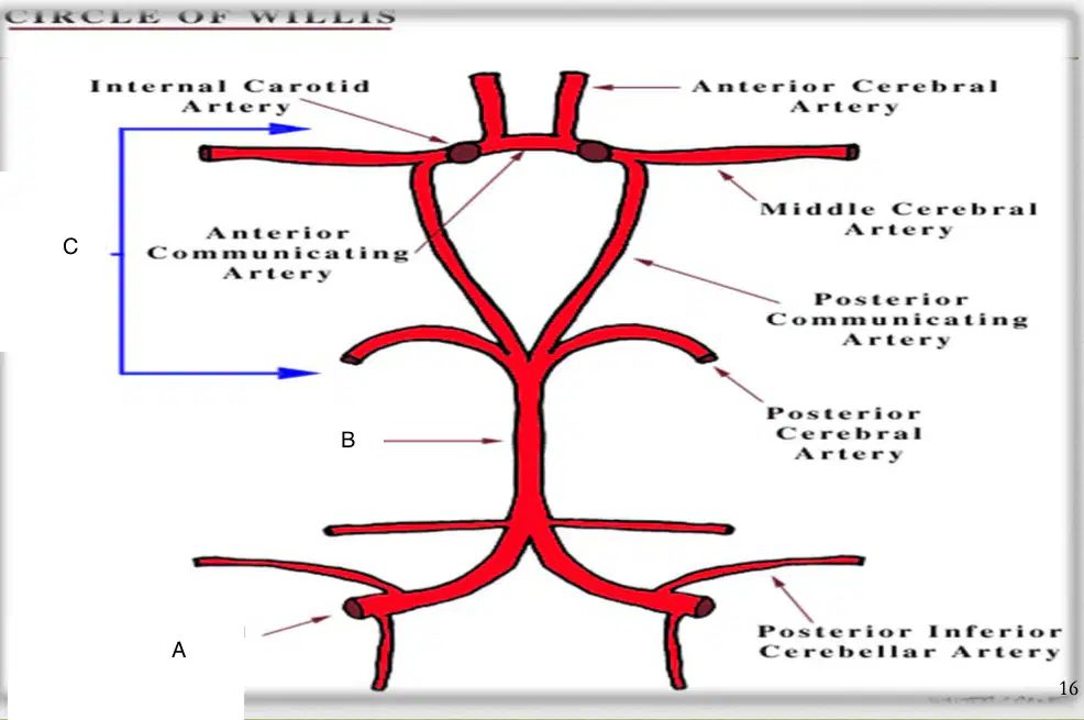
A
Circle of wills
Basilar artery
Vertebral artery

B
Circle of willis
Basilar artery
Vertebral artery
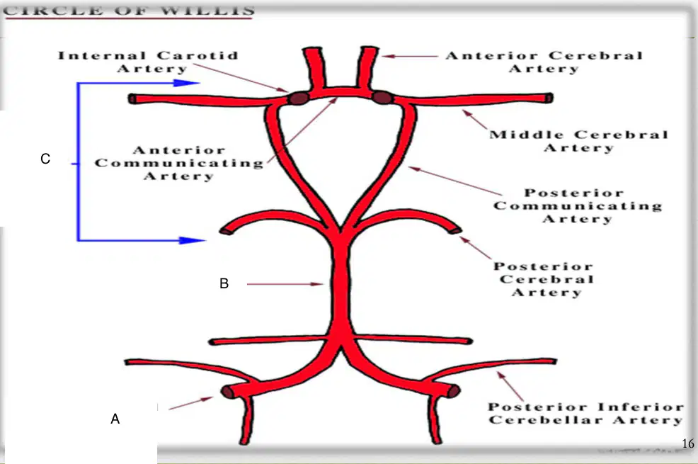
C
Circle of willis
Basilar artery
Vertebral artery
Does the ECA go to the brain?
yes
no
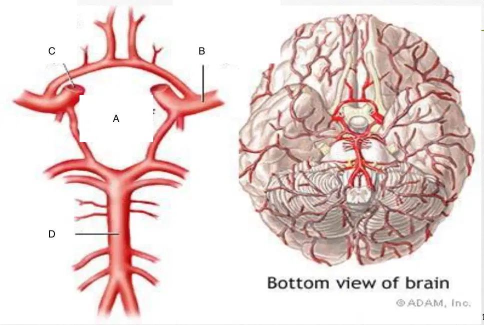
A
(a)
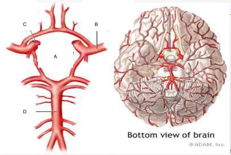
B
(a)

C
(a)

D
(a)
What is the first branch of the AORTA?
Brachiocephalic or Innominate
ICA
ECA
Aorta
Anechoic lumen
Thin, hyperechoic walls
(a)
(a) arteries course through fossae of transverse vertebral processes
What is the 2nd branch of the aortic arch?
RCA
LCA
CCA
What is the 3rd branch of the aortic arch
LCA
Right subclavian artery
Left subclavian artery
RCA
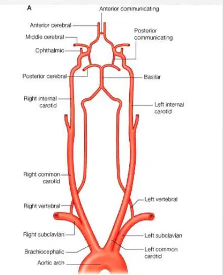
Basilar artery is the union of two arteries together
True
False
Where is the circle of willis located?
Neck
Brain
Ear
Eye

A
(a)
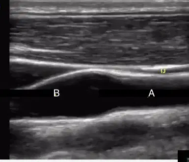
B
(a)
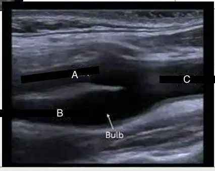
A
(a)

B
(a)
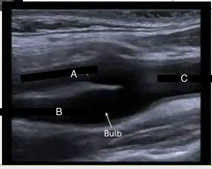
C
(a)
Origin of the ICA is slightly dilated; hence, the term the (a) or carotid sinus
Bulb may encompass the most (a) segment of the CC
Most common site for the plaque formation
(a)
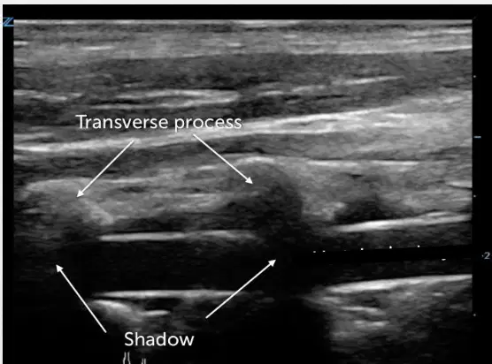
This image shows the:
ICA
ECA
Vertebral artery
Basilar artery
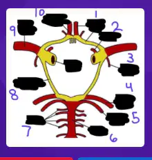
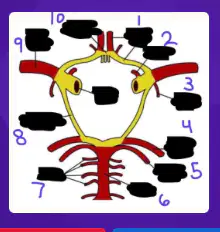
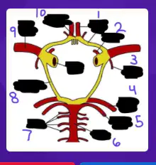
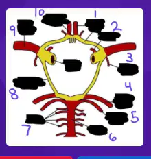
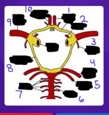
A circulatory anastomosis of arteries(polygon vascular ring)at the base of the brain
(a)
Provides a redundant blood supply to the brain
(a)
The circle of willis connects the __ and __systems, ensuring continuous blood flow even if one artery is blocked.
(a)
Permits communication between right and left cerebral hemispheres and between anterior and posterior systems
(a)
These communications are important when there is significant disease or occlusion of one of major cervical arteries
(a)
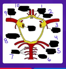
1
(a)

2
(a)

3
(a)
4
(a)
5
(a)
6
(a)
7
(a)
8
(a)
9
(a)

10
(a)
Comprises the arteries of Circle of Willis
(a)
Anterior Communicating (ACoA)
Anterior Cerebral (ACA) x 2
Supraclinoid segment of ICA x 2
Posterior communicating (PCoA) x 2
Posterior cerebral arteries (PCA) x 2
(a)
MCA is the (a) terminal branch of ICA.
From its origin, the MCA extends __ and __ in lateral cerebral fissure
(a)
What is the first branch of the ECA?
Superior thyroid
Occipital
Maxillary
Facial
Horizontal segment may course downward or upward
(a)
It should be noted that although the MCA is always included in a noninvasive transcranial evaluation (TCD imaging), anatomically it (a) lie within the circle of Willis
Is the smaller of two terminal branches of ICA
(a)
Is midline a marker
(a)
From its origin, ACA courses (a) over optic chiasm and optic nerve to interhemispheric fissure.
is short vessel that connects Anterior Cerebral arteries (segment A1) at interhemispheric fissure.
(a)
May be absent or may be single, duplicated, or multichanneled system
(a)
originate from the terminal bifurcation of the Basilar artery and as they emerge, they course anteriorly and then laterally around the midbrain.
(a)
Many perforating branches that supply brainstem and thalamus originate from this segment
(a)
Courses posteriorly and medially from ICA to join Posterior Cerebral artery.
(a)
Variable in size and may angle upward or downward • Hypoplasia common PCoA anomaly
(a)
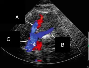
A
(a)
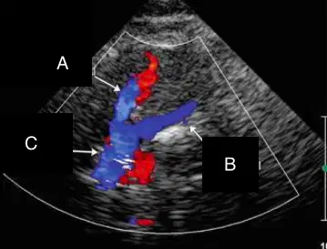
B
(a)

C
(a)

A
(a)

B
(a)

C
(a)
The innominate artery divides into the ___________ and ___________ arteries.
right common carotid; right subclavian
left common carotid; left subclavian
right subclavian; right vertebral
left subclavian; left vertebral
Which one of the following branches of the ECA is the most visualized
Facial
Superior thyroid
Lingual
Superficial temporal
The union of the two Vertebral arteries forms which one of the following?
Basilar artery
Posterior cerebral artery
Posterior communicating artery
Anteroinferior cerebellar artery
The large terminal branch of the Internal carotid artery is which one of the following?
Anterior cerebral artery
Middle cerebral artery
Posterior cerebral artery
Posterior communicating artery
The ________________ artery wraps around the cerebral peduncle
Posterior cerebral
Anterior cerebral
Basilar
Superior thyroid
Which branch of the ECA is the most commonly visualized?
Vertebral
Facial
Ophthalmic
Superior Thyroid
the largest terminal branch of the internal carotid artery is the
MCA
ICA
ECA
CCA
The second main branch of the aortic arch is the
CCA
Left Subclavian
LCA
ICA
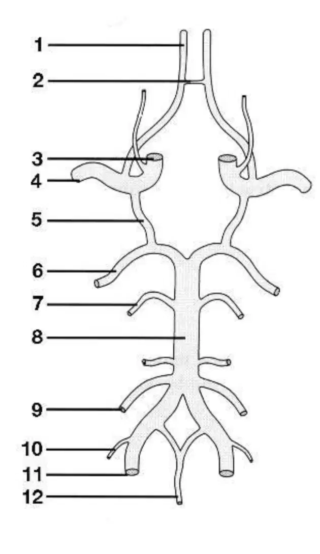
1
(a)
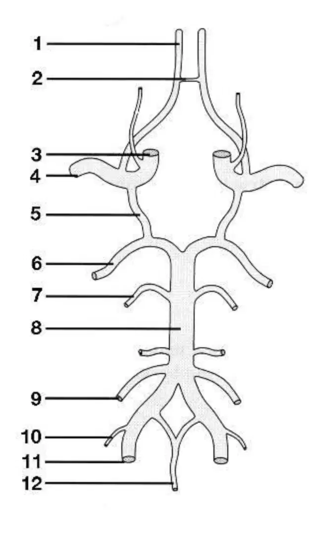
2
(a)
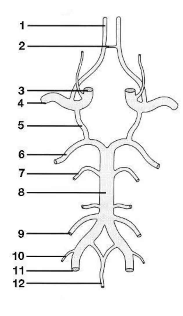
3
(a)

4
(a)
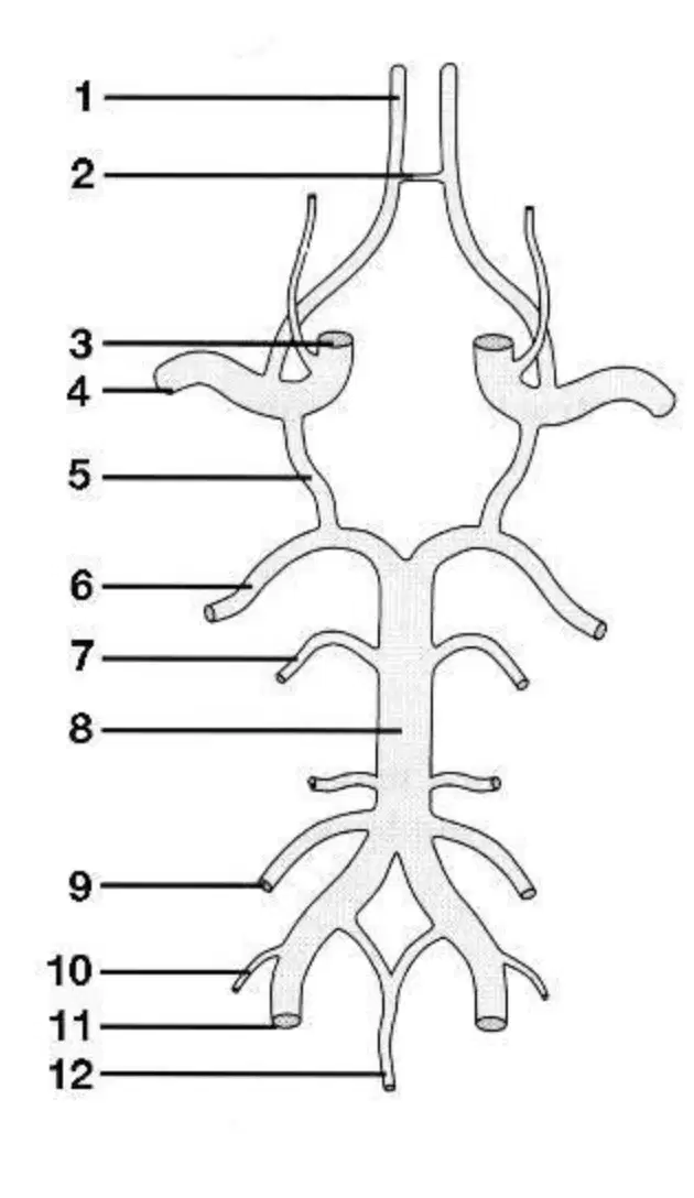
5
(a)
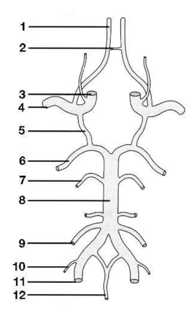
6
(a)
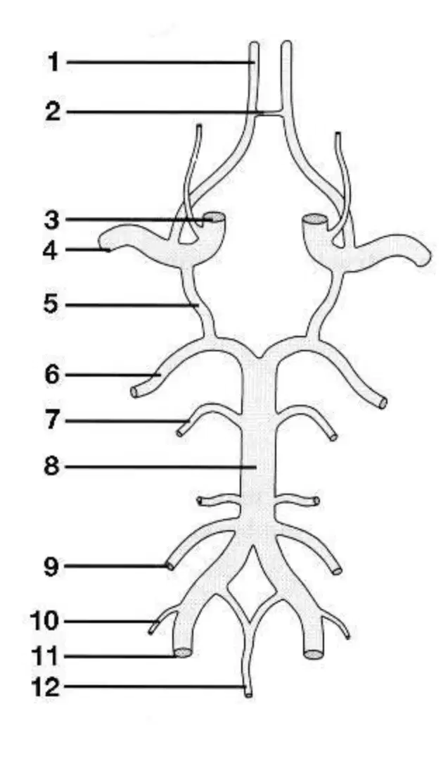
7
(a)
8
(a)
9
(a)

10
(a)

11
(a)

12
(a)

