
 Worksheets
Panoramic Radiography
Total questions: 37
Worksheet time: 1hrs 4mins

Arrow #1 is pointing to:
Stylohyoid ligament
Hyoid Bone
External auditory meatus

Arrow #2 is pointing to:
Hyoid bone
External auditory meatus
Articular eminence

Arrow #3 is pointing to:
External auditory meatus
Articular eminence
Head of the mandibular condyle

Arrow #4 is pointing to:
Articular eminence
Head of the mandibular condyle
Lower border of the zygomatic arch

Arrow #5 is pointing to:
Head of the mandibular condyle
Lower border of the zygomatic arch
Zygomatic process of the maxilla process

Arrow #6 is pointing to:
Lower border of the zygomatic arch
Zygomatic process of the maxilla process
Inferior concha/ turbinate

Arrow #7 is pointing to:
Zygomatic process of the maxilla
Inferior concha/ turbinate
Nasal septum

Arrow #8 is pointing to:
Inferior concha/ turbinate
Nasal septum
Inferior orbital rim

Arrow #9 is pointing to:
Nasal septum
Inferior orbital rim
Maxillary sinus

Arrow #10 is pointing to:
Inferior orbital rim
Maxillary sinus
Mandibular notch

Arrow #11 is pointing to:
Maxillary sinus
Mandibular notch
Coronoid process of the mandible

Arrow #12 is pointing to:
Mandibular notch
Coronoid process of the mandible
Pterygomaxillary fissure

Arrow #13 is pointing to:
Coronoid process of the mandible
Pterygomaxillary fissure
Neck of the mandibular condyle

Arrow #14 is pointing to:
Pterygomaxillary fissure
Neck of the mandibular condyle
Hard palate

Arrow #15 is pointing to:
Neck of the mandibular condyle
Hard palate
Anterior nasal spine

Arrow #16 is pointing to:
Hard palate
Anterior nasal spine
Bite guide of the panoramic unit

Arrow #17 is pointing to:
Anterior nasal spine
Bite guide of the panoramic unit
Hyoid bone

Arrow #18 is pointing to:
Bite guide of the panoramic unit
Hyoid bone
Nasal septum

What is likely causing the large opacity superimposing the right maxillary sinus?
I have not taken a pathology class and therefore have no way of knowing
Likely a ghost image of the left maxillary third molar
Probably a coin the patient put in his nose when he was really young
If the large, radiopaque entity superimposing the right maxillary sinus is a ghost image of the left maxillary third molar, what does this tell you about the likely buccal/lingual position of the third molar?
I have not taken a pathology class and therefore have no way of knowing
It's likely positioned lingually
It's likely positioned buccally

The supernumerary tooth between #29 and 30 is wider than the adjacent #29. Does this indicate that the supernumerary tooth is buccal or lingual to the other teeth?
Buccal
Lingual
I don't know but my mom likes me

Dr. Mecham tells you he only shot the sheriff once and that these three opacities only represent one bullet. Is it possible Dr. Mecham is telling the truth? If so, how?
No, and most only a double image of an object can appear
Yes, it could be two real images (double image) and a ghost image
Yes, a double image and a streaking artifact
Is a real object located on the right or left? Likely buccal or lingual to the mandible?
Right, buccal
Right, lingual
Left, lingual
Come on, no one can tell that from a panoramic radiograph!

Which side would you expect to see the entry wound in this patient?
Right
Left
There is no way of knowing
In a panoramic radiograph, a lingual object is rejected relatively more ___ to buccal objects.
Superior
Inferior
Neither
In a panoramic radiograph, if the anterior teeth look excessively narrow, the patient was likely place too far
Anterior
Posterior
Can't say/neither
In a panoramic radiograph, the left teeth are notably wider than the right teeth. The left teeth were positioned ___ to the point of rotation.
Closer to
Farther from
Can't say/neither
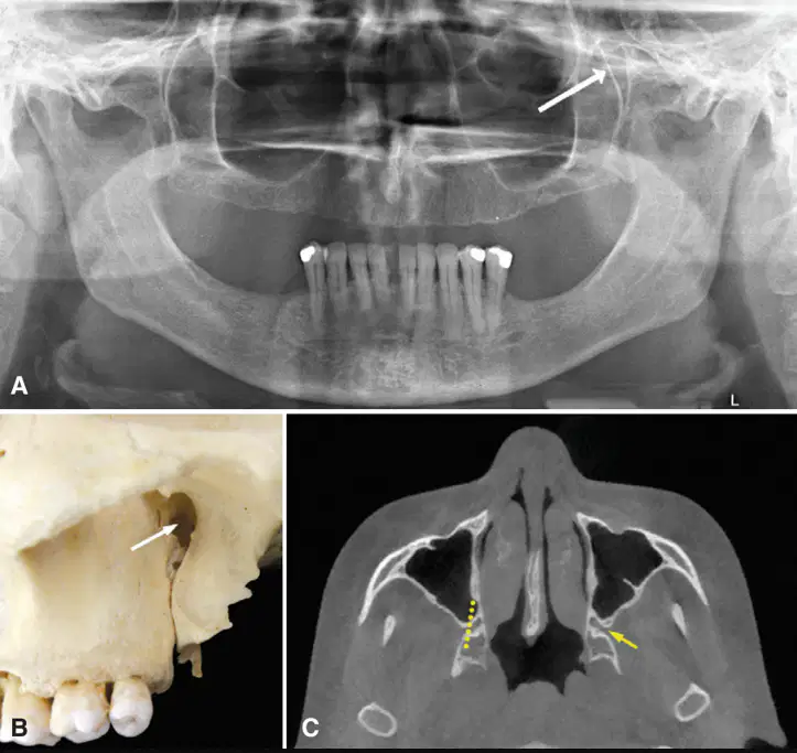
Name the indicated anatomical structure.
Pterygopalatine fossa
Pterygomaxillary fissure
Zygomatic process of the maxilla
Lateral pterygoid plate
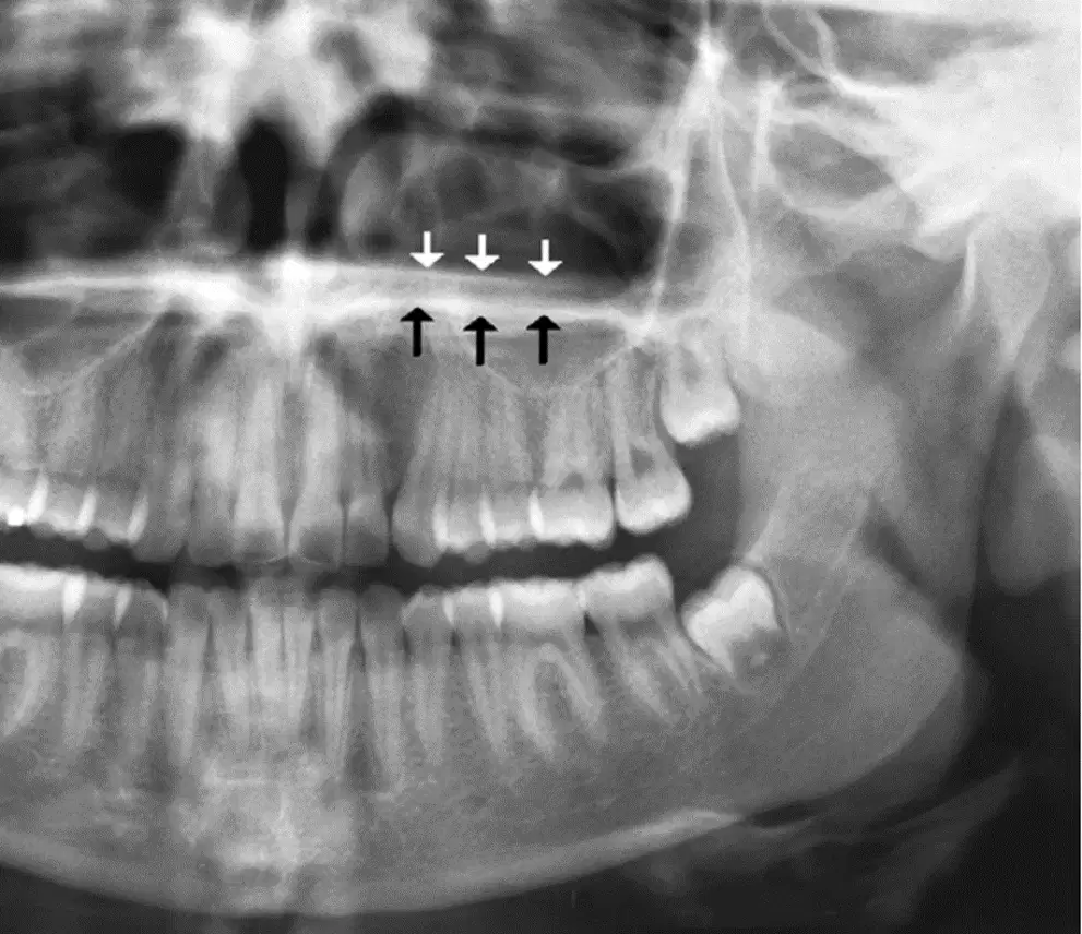
Name the anatomical structure indicated by the white arrows.
Ghost image of the right hard palate
Real image of the left hard palate
Ghost image of the left hard palate

Name the anatomical structure indicated (select all that apply).
Zygomatic process of the maxilla
Zygomatic process of the maxilla
Zygomatic process of the maxilla
Zygomatic process of the maxilla
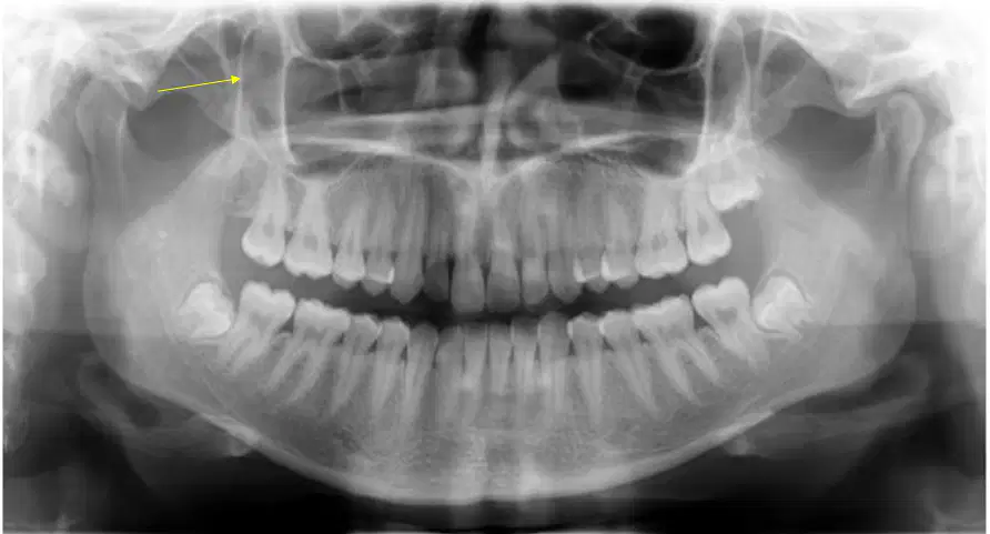
Name the anatomical structure indicated.
Zygomatic process of the maxilla
Posterior border of the maxillary sinus
Rim of the orbit
Coronoid process
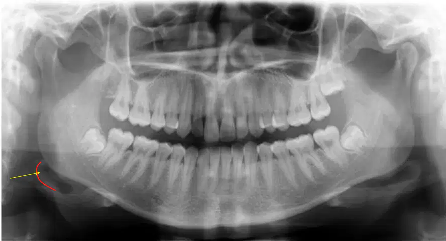
Name the anatomical structure indicated.
Hyoid bone
Not an anatomical structure
Epiglottis
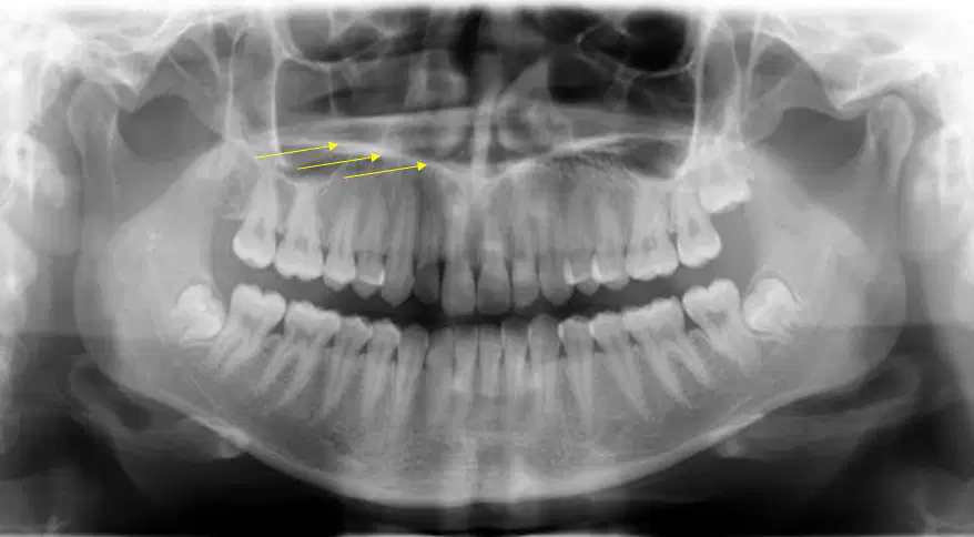
Name the anatomical structure indicated.
Real image of the right hard palate
Ghost image of the right hard palate
Ghost image of the left hard palate
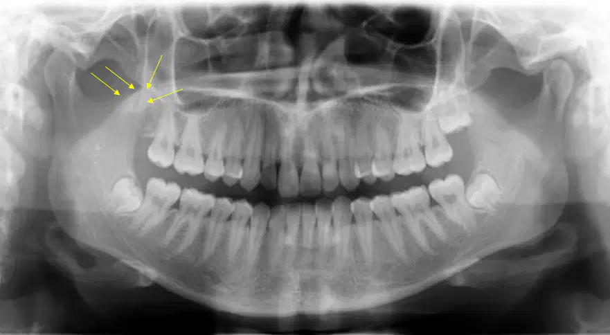
Name the anatomical structure indicated.
Posterior border of the maxillary sinus
Zygomatic process of the maxilla
Coronoid process
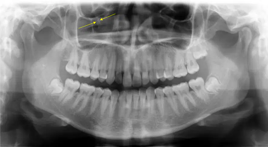
Name the anatomical structure indicated.
Infraorbital rim
Infraorbital canal
Maxillary sinus septum
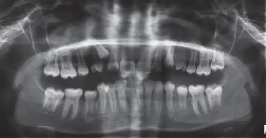
Identify the error seen on the panoramic radiograph
Chin up
Chin down
Too far forward
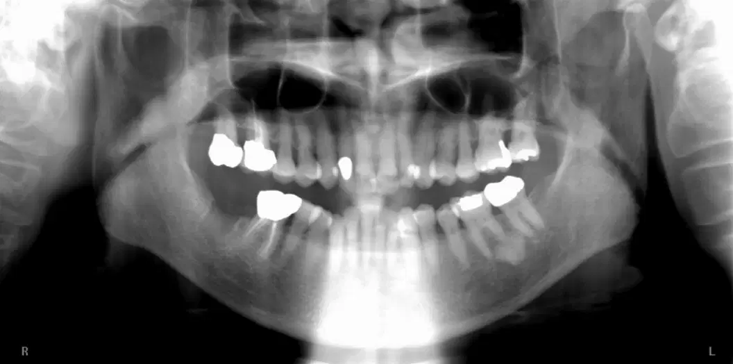
Identify the error seen on the panoramic radiograph
Too far forward
Too far back
Slumped (this means the patient's neck is bent too far forward and it creates a pronounced ghost image of the spine)

