
 Worksheets
Structures of the respiratory system exam review
Total questions: 91
Worksheet time: 46mins
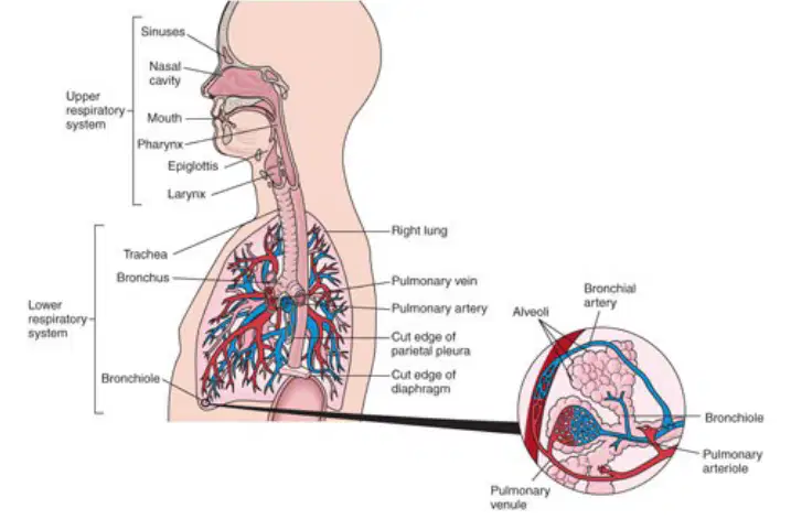
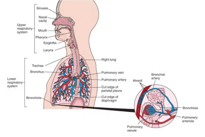
Which of the following is a part of the Upper Respiratory System?
Trachea
Lungs
Nose
Bronchi
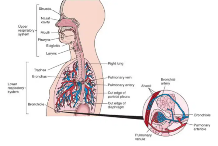
Which of the following is NOT a part of the Upper Respiratory System?
A) Sinuses
B) Pharynx
C) Larynx
D) Lungs
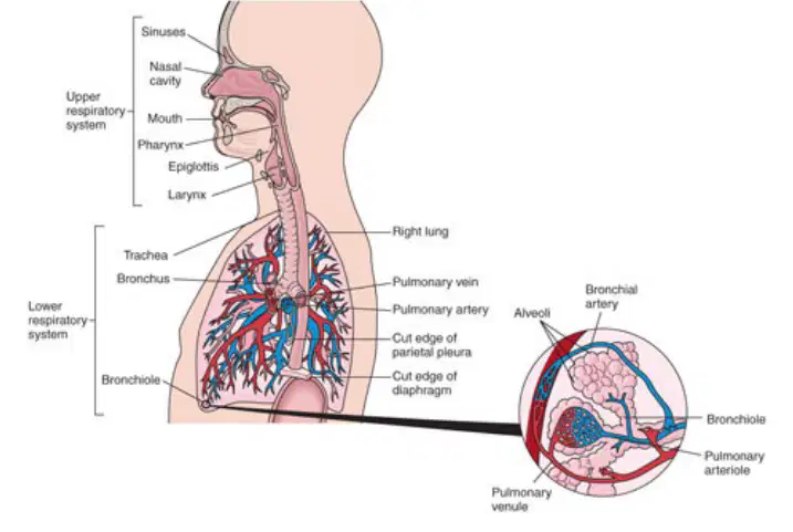
The Trachea is part of the ________ Respiratory System.
Lower
Upper
Central
Peripheral
Which of the following structures is found in the Lower Respiratory System?
Epiglottis
Lungs
Nose
Sinuses
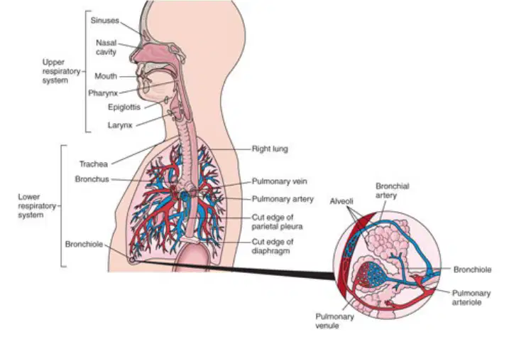
The Larynx is part of the ________ Respiratory System.
Upper
Lower
Central
Peripheral
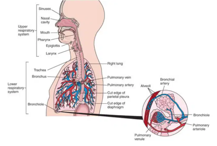
Which of the following is NOT part of the Lower Respiratory System?
A) Trachea
B) Lungs
C) Pharynx
D) None of the above
Which structure is responsible for filtering, warming, and humidifying air in the Upper Respiratory System?
Nose
Lungs
Trachea
Bronchi
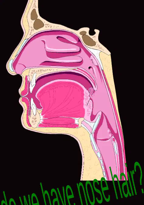
What is the function of the nasal cavity?
The nasal cavity is the space behind the nose, lined with mucous membranes, and warms the air.
It produces digestive enzymes to break down food.
It pumps blood throughout the body.
It filters waste from the blood to form urine.
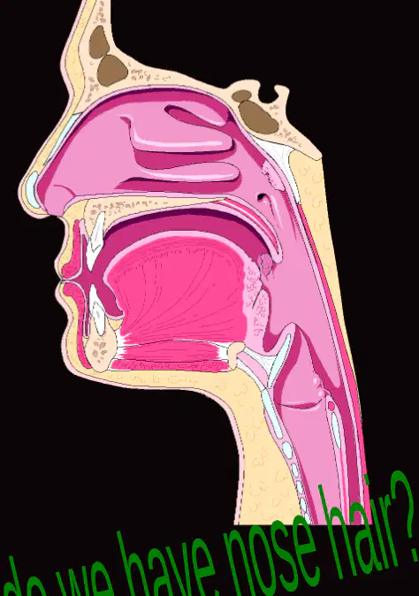
What is the olfactory region responsible for?
The olfactory region is responsible for smell.
The olfactory region is responsible for vision.
The olfactory region is responsible for hearing.
The olfactory region is responsible for taste.

What is the nasal septum?
The nasal septum is cartilage that divides the nose into right and left sides.
The nasal septum is a bone that forms the roof of the mouth.
The nasal septum is a muscle that controls breathing.
The nasal septum is a gland that produces mucus.
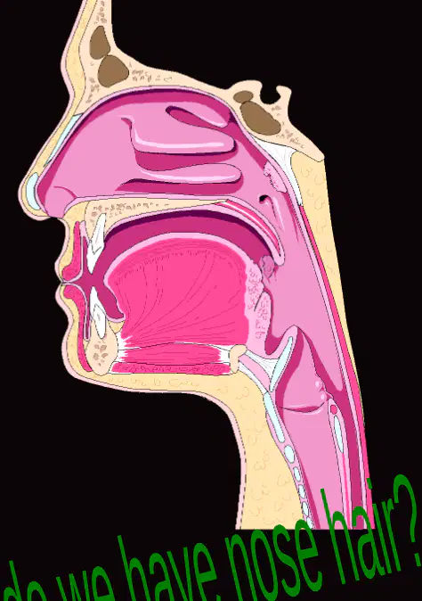
What are turbinates and what is their function?
Turbinates are scroll-like bones in the respiratory region that increase surface area for filtering dust and dirt particles by the mucous membrane.
Turbinates are glands in the digestive system that secrete enzymes for food breakdown.
Turbinates are muscles in the limbs that help in movement and posture.
Turbinates are blood vessels in the heart that regulate blood flow.
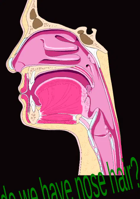
What is the function of cilia in the nose?
Cilia are nose hairs that trap dust and dirt.
Cilia help in producing mucus in the nose.
Cilia are responsible for detecting smells.
Cilia help in warming the air entering the nose.
Air enters the respiratory system passing into 2 oval openings of the nose called ________ (anterior nares).
nostrils
alveoli
bronchioles
pharynx
What filters out dust and dirt in the nose?
cilia
alveoli
bronchi
diaphragm
After passing through the nostrils, air moves into the ________ where additional dust and dirt are filtered out, and the air is warmed and moistened.
nasal cavity
trachea
bronchi
alveoli
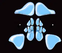
Which structures are cavities in the skull, filled with air in and around the nasal region?
Sinuses
Mandible
Cranial sutures
Mastoid process
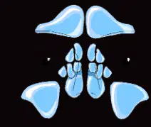
What connects the sinuses to the nasal cavity?
Short ducts
Blood vessels
Nerve fibers
Cartilage plates

What is the function of the mucous membrane lining the sinuses?
To warm and moisten the air
To filter blood
To digest food
To transmit nerve impulses
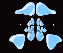
What do the sinuses provide to the voice?
Resonance
Lubrication
Pitch control
Volume increase

The unpleasant voice sound of a nasal cold results from what?
Blocked sinuses
Swollen vocal cords
Dry throat
Excess saliva
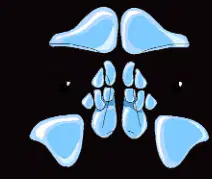
Which of the following is NOT a type of sinus?
Frontal
Maxillary
Sphenoid
Temporal

Fill in the blank: The four main types of sinuses are ________, Maxillary, Sphenoid, and Ethmoid.
Frontal
Temporal
Parietal
Occipital
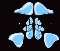
Which sinus is located above the eyes?
Frontal
Maxillary
Sphenoid
Ethmoid
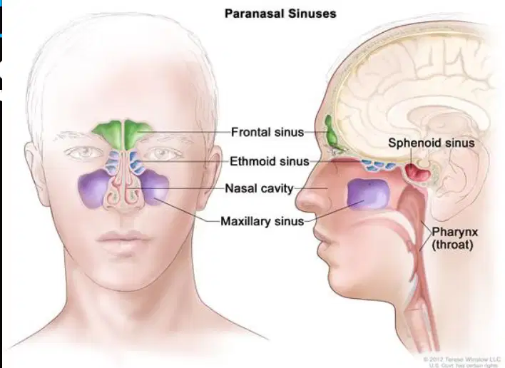
Identify the sinus labeled at the forehead region in the diagram of Paranasal Sinuses.
Frontal sinus
Maxillary sinus
Ethmoid sinus
Sphenoid sinus

Identify the sinus located between the eyes in the diagram of Paranasal Sinuses.
Ethmoid sinus
Maxillary sinus
Frontal sinus
Sphenoid sinus
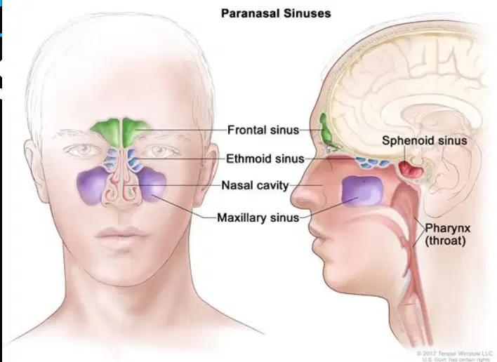
Identify the cavity labeled in the nose area in the diagram of Paranasal Sinuses.
Nasal cavity
Maxillary sinus
Frontal sinus
Sphenoidal sinus
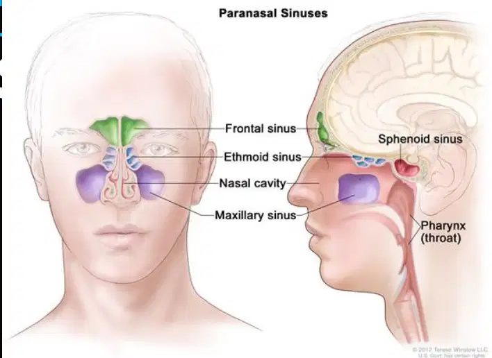
Identify the sinus located in the cheek area in the diagram of Paranasal Sinuses.
Maxillary sinus
Frontal sinus
Ethmoid sinus
Sphenoid sinus
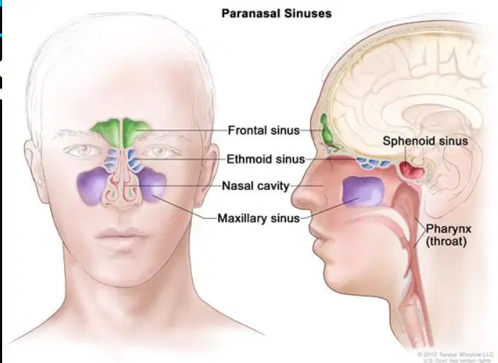
Identify the sinus located deep within the skull, behind the nasal cavity, in the diagram of Paranasal Sinuses.
Sphenoid sinus
Frontal sinus
Maxillary sinus
Ethmoid sinus
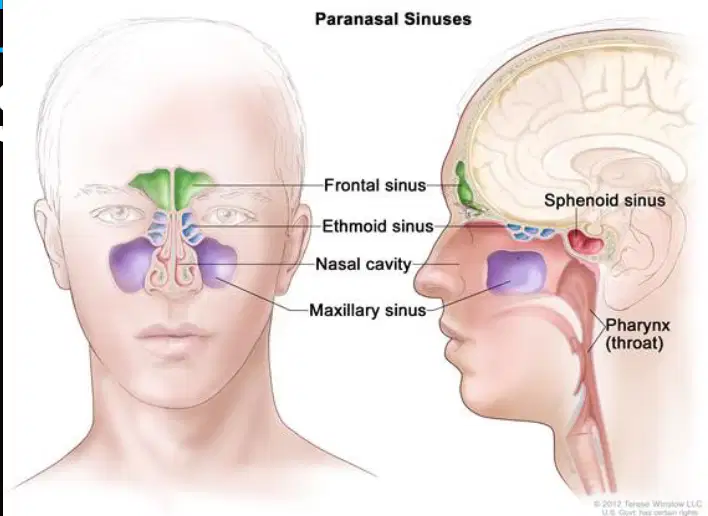
Identify the part labeled as the throat in the diagram of Paranasal Sinuses.
Pharynx (throat)
Maxillary sinus
Frontal sinus
Nasal cavity
What is the pharynx commonly known as?
The nose
The throat
The windpipe
The mouth
How long is the pharynx?
About 2 inches
About 3 inches
About 5 inches
About 7 inches
After air leaves the nasal cavity, it enters the ________.
pharynx (throat)
trachea
larynx
bronchi
The pharynx serves as a common passageway for air to go to the lungs and food to go to the ________.
stomach
kidney
heart
liver
Which section of the pharynx is responsible for air only?
Nasopharynx
Oropharynx
Laryngopharynx
Trachea
The nasopharynx is the uppermost section, just passed the ________.
nasal cavity
oral cavity
larynx
trachea
True or False: The right and left Eustachian tubes open directly into the nasopharynx, connecting the middle ear and nasopharynx.
True
False
Which part of the pharynx allows passage of air, food, and liquid?
Nasopharynx
Oropharynx
Laryngopharynx
Trachea
The oropharynx extends from the soft palate to just above the ________ bone.
hyoid
mandible
zygomatic
temporal
The laryngopharynx is the ______ portion of the pharynx.
lower
upper
middle
anterior
In the laryngopharynx, air travels to the ______.
lungs
stomach
esophagus
liver
In the laryngopharynx, food travels to the ______.
stomach
lungs
esophagus
trachea
The epiglottis is located in the laryngopharynx and closes when you swallow to keep food from going into the ______.
lungs
stomach
esophagus
trachea
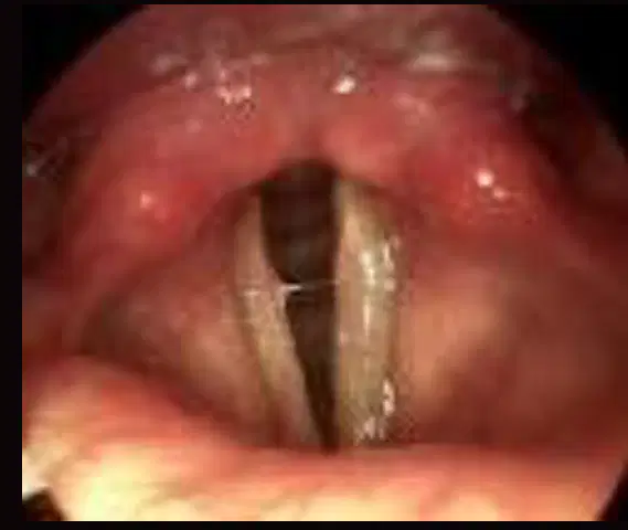
Where does the epiglottis lie? (Fill in the blank) The epiglottis lies between the root of the ______ and the larynx.
tongue
palate
pharynx
esophagus
What is the epiglottis known as? (Fill in the blank) The epiglottis is known as the “________.”
Gatekeeper
Windpipe
Voice box
Food pipe
What is the main function of the epiglottis?
Produces sound
Routes food/air the proper way
Absorbs nutrients
Filters blood
The epiglottis closes to cover the opening of the larynx when you swallow to prevent food from going into the trachea to the lungs.
True
False
What happens if food gets past the epiglottis?
cough
hiccup
sneeze
yawn
What is the common name for the larynx?
Windpipe
Voice Box
Food Pipe
Nasal Cavity
The larynx is a triangular chamber located below the _______.
pharynx
trachea
esophagus
epiglottis
How many fibrocartilaginous plates compose the larynx?
5
7
9
11
During puberty, the vocal cords become larger in males, making the _______ more prominent.
Adam’s Apple
collarbone
jawbone
cheekbone
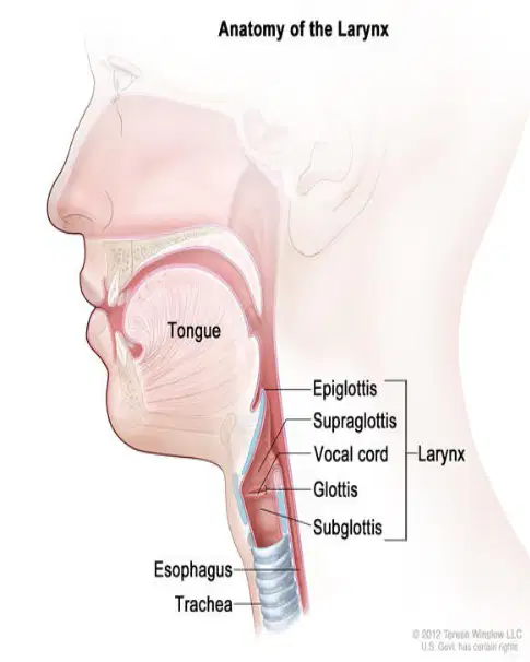
Within the larynx are vocal cords, the space between them is called the ________.
glottis
epiglottis
trachea
pharynx
What causes the vocal cords to vibrate and produce sound?
Air expelled from the lungs causes vibration and produces sound.
Blood flowing through the vocal cords causes vibration and produces sound.
Muscle contractions in the neck cause vibration and produce sound.
Food passing through the throat causes vibration and produces sound.
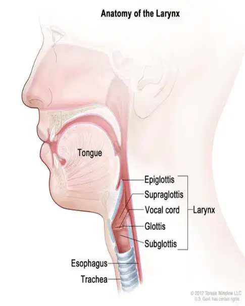
Which of the following is NOT a part of the larynx?
Epiglottis
Glottis
Subglottis
Esophagus
What is the common name for the trachea?
Windpipe
Esophagus
Bronchus
Larynx
Approximately how long is the trachea?
2 inches
4 ½ inches
6 inches
10 inches
The trachea extends from the larynx, passes in front of the esophagus, and continues down to form the R and L ________.
bronchi
alveoli
arteries
ventricles
The walls of the trachea are composed of alternate bands of membrane and how many C-shaped rings of hyaline cartilage?
5-10
10-15
15-20
20-25
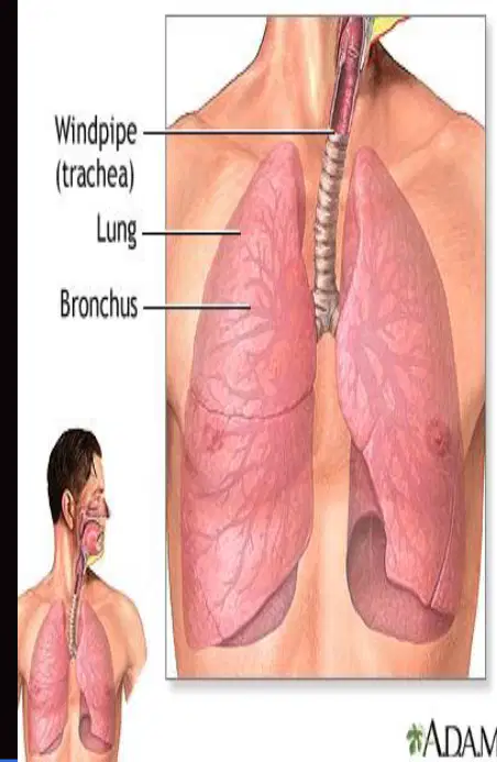
Refer to the diagram. Which structure is labeled as the windpipe?
Lung
Bronchus
Trachea
Esophagus
The trachea is lined with ________ membrane and ciliated epithelium, which trap inhaled dust particles and sweep them to be expelled.
mucous
serous
cartilaginous
fibrous
Coughing and expectoration help to get rid of ________-laden mucous.
dust
water
oil
sugar
The trachea can be obstructed by which of the following?
Large pieces of food
Tumorous growths
Swelling of inflamed lymph nodes in neck
All of the above
Which university is mentioned in relation to the Lower Respiratory Tract video clip?
West Coast University
East Valley University
Northern State College
Southern Tech Institute
At the lower end of the trachea, the bronchus divides into ______ and left branches known as the right and left bronchi.
right
upper
anterior
posterior
What is the bronchi lined with?
Smooth muscle and elastic tissue
Ciliated mucous membrane and hyaline cartilage
Squamous epithelium and connective tissue
Adipose tissue and blood vessels
Which bronchus is somewhat shorter, wider, and more vertical in position?
Left bronchus
Right bronchus
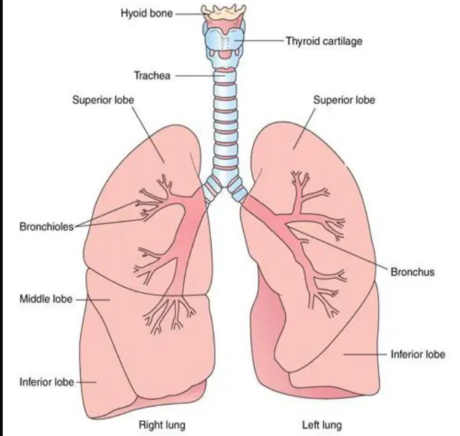
Label the following parts in the diagram:
Trachea, Bronchus, Bronchioles, Superior lobe, Middle lobe, Inferior lobe, Right lung, Left lung (as shown in the diagram)
Femur, Tibia, Fibula, Patella, Humerus, Radius, Ulna, Scapula (as shown in the diagram)
Aorta, Vena cava, Pulmonary artery, Pulmonary vein, Left atrium, Right atrium, Left ventricle, Right ventricle (as shown in the diagram)
Cerebrum, Cerebellum, Medulla oblongata, Spinal cord, Frontal lobe, Parietal lobe, Occipital lobe, Temporal lobe (as shown in the diagram)
Which of the following is a characteristic of bronchial tubes?
Thinner walls of smooth muscle
Cartilaginous plates (instead of C-shaped rings)
Lined with ciliated epithelium
Alveolar sacs
Bronchioles have ______ walls of smooth muscle and are lined with ______ epithelium.
thinner; ciliated
thicker; squamous
thicker; cuboidal
thinner; stratified
At the end of the bronchioles are alveolar ducts and clusters of alveoli called ______.
alveolar sacs
bronchi
capillaries
pleura
Which part of the lower respiratory system is Y shaped?
Bronchioles
Alveoli
Bronchial tubes
Trachea
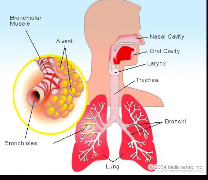
Which structure is directly connected to the alveoli?
Trachea
Bronchi
Bronchioles
Nasal cavity
How many alveoli are present in adult lungs?
500,000,000
50,000,000
5,000,000
1,000,000,000
What type of tissue composes the alveoli?
A single layer of epithelial tissue
Dense connective tissue
Stratified squamous epithelium
Smooth muscle tissue

What is the function of the surfactant covering the inner surface of alveoli?
It helps to stabilize the alveoli, preventing their collapse.
It increases the thickness of the alveolar wall.
It blocks the exchange of gases in the alveoli.
It causes the alveoli to fill with fluid.
Each alveolus is surrounded by what?
Capillaries
Veins
Bronchioles
Arteries

Where does the exchange of O2 and CO2 take place in the lungs?
Between the alveoli and capillaries
In the trachea
Inside the bronchioles
Within the diaphragm
True or False: The alveoli are composed of multiple layers of tissue.
True
False
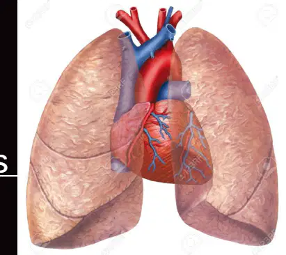
The lungs are ________-shaped organs located in the thoracic cavity.
cone
cube
sphere
cylinder
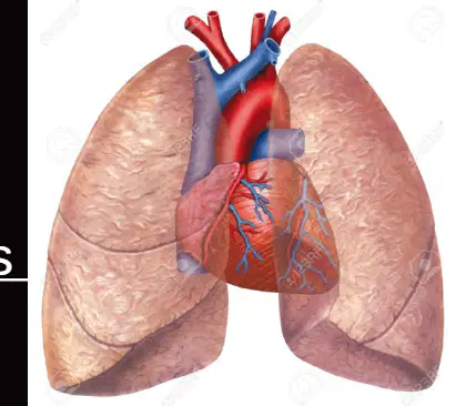
The lungs fill up the 2 lateral chambers of thoracic cavity and are separated by the ________ and heart.
mediastinum
pleura
diaphragm
bronchi
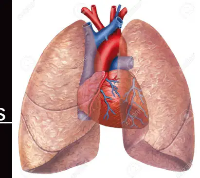
What is the upper, arch part of the lung under the collar bone called?
Base
Apex
Diaphragm
Alveoli
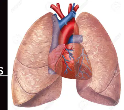
The bottom, broader lower part of the lung is called the ________.
base
apex
lobe
hilum
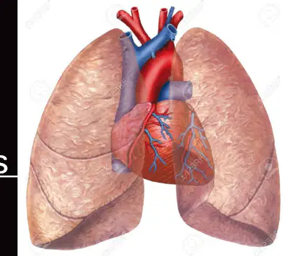
The lungs fit snugly over the ________.
diaphragm
liver
stomach
heart
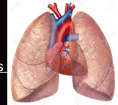
Lung tissue is ________ and spongy due to alveoli and large amounts of air.
porous
rigid
dense
solid
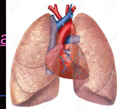
Which lung is larger and shorter than the other?
Right lung
Left lung

Which lung is displaced by the liver?
Right lung
Left lung
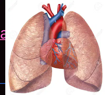
How many lobes does the right lung have?
2
3
4
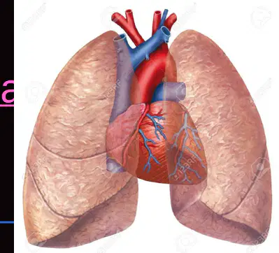
The left lung is displaced by the ________.
heart
liver
stomach
spleen
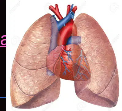
The left lung has 2 lobes, superior and inferior.
True
False
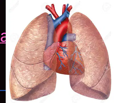
Which lung is smaller, narrower, and longer?
Right lung
Left lung
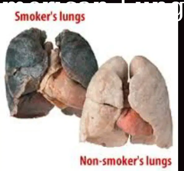
Look at the image comparing smoker's lungs and non-smoker's lungs. Which set of lungs appears darker and more damaged?
Smoker's lungs
Non-smoker's lungs

