
 Worksheets
PRC-HEMA_3MT2
Total questions: 116
Worksheet time: 58mins
Name
Class
Date
1.
Which of the following primary immunodeficiency disorders or inborn errors of immunity is characterized by patients experiencing a marked decrease in circulating T cells, poorly functioning B cells, and hypogammaglobulinemia?
a)
Severe Combined Immune deficiency (SCID)
b)
Wiskott-Aldrich syndrome
c)
DiGeorge syndrome
d)
Bruton Tyrosine Kinase Deficiency
e)
Chediak-Higashi syndrome
2.
Which of the following primary immunodeficiency disorders or inborn errors of immunity is an X-liked recessive disorder, characterized by thrombocytopenia, immunodeficiency, and eczema?
a)
Severe Combined Immune deficiency (SCID)
b)
Wiskott-Aldrich syndrome
c)
DiGeorge syndrome
d)
Bruton Tyrosine Kinase Deficiency
e)
Chediak-Higashi syndrome
3.
Which of the following primary immunodeficiency disorders or inborn errors of immunity shows a decreased T cells, and dysfunctional B, T, and NK cells? Also, here, there is a presence of small and abnormal platelets.
a)
Severe Combined Immune deficiency (SCID)
b)
Wiskott-Aldrich syndrome
c)
DiGeorge syndrome
d)
Bruton Tyrosine Kinase Deficiency
e)
Chediak-Higashi syndrome
4.
Which of the following primary immunodeficiency disorders or inborn errors of immunity is characterized by Thymic hypoplasia, cardiac anomalies, craniofacial deformities, immune dysfunction, developmental delays, and various other systemic issues like hypocalcemia and autoimmune disorders?
a)
Severe Combined Immune deficiency (SCID)
b)
Wiskott-Aldrich syndrome
c)
DiGeorge syndrome
d)
Bruton Tyrosine Kinase Deficiency
e)
Chediak-Higashi syndrome
5.
Which of the following primary immunodeficiency disorders or inborn errors of immunity is caused by a mutation in BTK (a protein that promotes B-cell development)?
a)
Severe Combined Immune deficiency (SCID)
b)
Wiskott-Aldrich syndrome
c)
DiGeorge syndrome
d)
Bruton Tyrosine Kinase Deficiency
e)
Chediak-Higashi syndrome
6.
Which of the following primary immunodeficiency disorders or inborn errors of immunity shows a marked reduction in antibody, absent tonsils, and inability to produce plasma cells; ultimately, a decrease in all immunoglobulin isotypes?
a)
Severe Combined Immune deficiency (SCID)
b)
Wiskott-Aldrich syndrome
c)
DiGeorge syndrome
d)
Bruton Tyrosine Kinase Deficiency
e)
Chediak-Higashi syndrome
7.
Which of the following primary immunodeficiency disorders or inborn errors of immunity is caused by a mutation in the lyst gene on chromosome 1q42.3, leading to giant fused cytoplasmic granules in various cells?
a)
Severe Combined Immune deficiency (SCID)
b)
Wiskott-Aldrich syndrome
c)
DiGeorge syndrome
d)
Bruton Tyrosine Kinase Deficiency
e)
Chediak-Higashi syndrome
8.
Which of the following primary immunodeficiency disorders or inborn errors of immunity presents in infancy with partial albinism, recurrent bacterial infections, mild bleeding, easy bruising, and progressive neurological impairment?
a)
Severe Combined Immune deficiency (SCID)
b)
Wiskott-Aldrich syndrome
c)
DiGeorge syndrome
d)
Bruton Tyrosine Kinase Deficiency
e)
Chediak-Higashi syndrome
9.
Which of the following PSEUDO primary immunodeficiency disorders or inborn errors of immunity resembles the fused lysosomal granules in CHS and most often seen in acute myeloid leukemia (AML)?
a)
Severe Combined Immune deficiency (SCID)
b)
Wiskott-Aldrich syndrome
c)
DiGeorge syndrome
d)
Bruton Tyrosine Kinase Deficiency
e)
Chediak-Higashi syndrome
10.
- ANC of <0.5 x 10⁹ .
a)
Severe congenital neutropenia (CN)
b)
Cyclic Neutropenia
c)
Schwachman-Diamond Syndrome
d)
Leukocyte Adhesion Disorder
e)
Chronic Granulomatous Disease (CGD)
11.
- 50-60% of cases are due to a mutation in the neutrophil elastase gene (ELANE).
a)
Severe congenital neutropenia (CN)
b)
Cyclic Neutropenia
c)
Schwachman-Diamond Syndrome
d)
Leukocyte Adhesion Disorder
e)
Chronic Granulomatous Disease (CGD)
12.
Which of the following congenital defects of phagocyte is also caused by ELANE mutations (but less severe) leading to episodes of severe neutropenia (less than 0.2 × 10⁹/L) with absolute monocytosis occurring in approximately 21-day cycles lasting 3 to 5 days?
a)
Severe congenital neutropenia (CN)
b)
Cyclic Neutropenia
c)
Schwachman-Diamond Syndrome
d)
Leukocyte Adhesion Disorder
e)
Chronic Granulomatous Disease (CGD)
13.
Which of the following congenital defects of phagocyte is caused by mutations in the SBDS gene, leading to neutropenia, pancreatic insufficiency, malabsorption, and skeletal abnormalities?
a)
Severe congenital neutropenia (CN)
b)
Cyclic Neutropenia
c)
Schwachman-Diamond Syndrome
d)
Leukocyte Adhesion Disorder
e)
Chronic Granulomatous Disease (CGD)
14.
Which of the following congenital defects of phagocyte is a rare autosomal recessive disorders characterized by the inability of neutrophils and monocytes to move from peripheral circulation to sides of infection?
a)
Severe congenital neutropenia (CN)
b)
Cyclic Neutropenia
c)
Schwachman-Diamond Syndrome
d)
Leukocyte Adhesion Disorder
e)
Chronic Granulomatous Disease (CGD)
15.
Which of the following congenital defects of phagocyte is caused by the decreased ability of monocytes and granulocytes to kill phagocytized bacteria and yeast (failure in respiratory burst and failed production of antibacterial superoxide anions and reactive oxygen species)?
a)
Severe congenital neutropenia (CN)
b)
Cyclic Neutropenia
c)
Schwachman-Diamond Syndrome
d)
Leukocyte Adhesion Disorder
e)
Chronic Granulomatous Disease (CGD)
16.
Which of the following congenital defects of phagocyte is the most common inherited disorder of phagocytes?
a)
Myeloperoxidase deficiency
b)
Cyclic Neutropenia
c)
Schwachman-Diamond Syndrome
d)
Leukocyte Adhesion Disorder
e)
Chronic Granulomatous Disease (CGD)
17.
Which of the following congenital defects of phagocyte catalyzes the conversion of H2O2 to HOCl and other reactive oxygen species, aiding in the killing of phagocytized bacteria, yeast, and in forming neutrophil extracellular traps (NETs)?
a)
Myeloperoxidase deficiency
b)
Cyclic Neutropenia
c)
Schwachman-Diamond Syndrome
d)
Leukocyte Adhesion Disorder
e)
Chronic Granulomatous Disease (CGD)
18.
Which of the following congenital defects of phagocyte is characterized by impaired ability of neutrophils to eUectively kill microorganisms?
a)
Myeloperoxidase deficiency
b)
Cyclic Neutropenia
c)
Schwachman-Diamond Syndrome
d)
Leukocyte Adhesion Disorder
e)
Chronic Granulomatous Disease (CGD)
19.
Which lysosomal storage disorder is characterized by a deficiency of sphingomyelinase (accumulation of sphingomyelin)?
a)
Niemann-Pick Disease
b)
Gaucher Disease
20.
Which lysosomal storage disorder is common in Ashkenazi Jews?
a)
Niemann-Pick Disease
b)
Gaucher Disease
21.
Which lysosomal storage disorder involved foam cell?
a)
Niemann-Pick Disease
b)
Gaucher Disease
22.
Which lysosomal storage disorder involves defect or deficiency in β-glucocerebrosidase (Accumulation of glucocerebroside)?
a)
Niemann-Pick Disease
b)
Gaucher Disease
23.
Which lysosomal storage disorder is most common of the lipidoses?
a)
Niemann-Pick Disease
b)
Gaucher Disease
24.
Which lysosomal storage disorder has crumpled tissue paper or onion-like appearance?
a)
Niemann-Pick Disease
b)
Gaucher Disease
25.
Large macrophage with small eccentric nucleus.
a)
Foam Cell
b)
Gaucher cell
26.
crumpled tissue paper or onion-like appearance?
a)
Foam Cell
b)
Gaucher cell
27.
Hurler syndrome
a)
MPS I
b)
MPS II
c)
MPS III
d)
MPS IV
28.
Deficiency in Alpha-L-iduronidase
a)
MPS I
b)
MPS II
c)
MPS III
d)
MPS IV
29.
Hunter syndrome
a)
MPS I
b)
MPS II
c)
MPS III
d)
MPS IV
30.
Deficiency in Iduronate-2-sulfatase
a)
MPS I
b)
MPS II
c)
MPS III
d)
MPS IV
31.
Sanfilippo syndrome
a)
MPS I
b)
MPS II
c)
MPS III
d)
MPS IV
32.
Morquio syndrome
a)
MPS I
b)
MPS II
c)
MPS III
d)
MPS IV
33.
Which subtype of MPS III is characterized by a deficiency in N-sulfoglucosamine sulfohydrolase?
a)
MPS IIIA
b)
MPS IIIB
c)
MPS IIIC
34.
Which subtype of MPS III is characterized by a deficiency in Alpha-Nacetylglucosaminidase?
a)
MPS IIIA
b)
MPS IIIB
c)
MPS IIIC
35.
Which subtype of MPS III is characterized by a deficiency in Heparan-alphaglucosaminide N-acetyltransferase?
a)
MPS IIIA
b)
MPS IIIB
c)
MPS IIIC
36.
Which subtype of MPS IV is characterized by a deficiency in N-acetylgalactosamine-6- sulfatase AS: Keratan sulfate (KS), Chondroidin sulfate (CS) ?
a)
MPS IVA
b)
MPS IVB
37.
Which subtype of MPS IV is characterized by a deficiency in Beta-galactosidase?
a)
MPS IVA
b)
MPS IVB
38.
Maroteaux-Lamy syndrome
a)
MPS VI
b)
MPS VII
c)
MPS IX
39.
Sly disease
a)
MPS VI
b)
MPS VII
c)
MPS IX
40.
Natowicz syndrome
a)
MPS VI
b)
MPS VII
c)
MPS IX
41.
Deficiency in Arylsulfatase B
a)
MPS VI
b)
MPS VII
c)
MPS IX
42.
Deficiency in Beta-glucuronidase
a)
MPS VI
b)
MPS VII
c)
MPS IX
43.
Deficiency in Hyaluronidase-1
a)
MPS VI
b)
MPS VII
c)
MPS IX
44.
The nucleus has > 6 lobes
a)
Hypersegmented neutrophils
b)
Pelger-Huet Anomaly
c)
Alder-Reilly Anomaly
d)
May-Hegglin Anomaly
45.
Associated conditions: Megaloblastic anemia Myelodysplastic syndromes Hereditary neutrophil hypersegmentation, Myelokathexis
a)
Hypersegmented neutrophils
b)
Pelger-Huet Anomaly
c)
Alder-Reilly Anomaly
d)
May-Hegglin Anomaly
46.
Most common WBC genetic disorder.
a)
Hypersegmented neutrophils
b)
Pelger-Huet Anomaly
c)
Alder-Reilly Anomaly
d)
May-Hegglin Anomaly
47.
Results from failure of the neutrophil nucleus to segment.
a)
Hypersegmented neutrophils
b)
Pelger-Huet Anomaly
c)
Alder-Reilly Anomaly
d)
May-Hegglin Anomaly
48.
Result from a mutation in the lamin β-receptor gene.
a)
Hypersegmented neutrophils
b)
Pelger-Huet Anomaly
c)
Alder-Reilly Anomaly
d)
May-Hegglin Anomaly
49.
May be unilobed or bilobed.
a)
Hypersegmented neutrophils
b)
Pelger-Huet Anomaly
c)
Alder-Reilly Anomaly
d)
May-Hegglin Anomaly
50.
Pseudo type of this disease is characterized by hyposegmented and hyogranular cells (contrary to the true version of the disease, which has normal granulation). This is usually seen in AML, chronic myeloproliferative neoplasms, MDS, HIV infection, TB, M. pneumoniae infection.
a)
Hypersegmented neutrophils
b)
Pelger-Huet Anomaly
c)
Alder-Reilly Anomaly
d)
May-Hegglin Anomaly
51.
Characterized by dense azurophilic granulation in all types of leukocytes
a)
Hypersegmented neutrophils
b)
Pelger-Huet Anomaly
c)
Alder-Reilly Anomaly
d)
May-Hegglin Anomaly
52.
Results from abnormal deposition and storage of mucopolysaccharides
a)
Hypersegmented neutrophils
b)
Pelger-Huet Anomaly
c)
Alder-Reilly Anomaly
d)
May-Hegglin Anomaly
53.
An autosomal dominant disorder characterized by the presence of grey-blue spindle-shaped inclusions (Dohle body-like inclusions) in the cytoplasm of granulocytes and monocytes.
a)
Hypersegmented neutrophils
b)
Pelger-Huet Anomaly
c)
Alder-Reilly Anomaly
d)
May-Hegglin Anomaly
54.
Associated conditions: Pregnancy, severe burns, aplastic anemia, scarlet fever, other infectious diseases, and administration of toxic agents
a)
Hypersegmented neutrophils
b)
Pelger-Huet Anomaly
c)
Alder-Reilly Anomaly
d)
May-Hegglin Anomaly
55.
Altered primary granules that are present due to rapid cell maturation.
a)
Toxic granulations
b)
Dohle bodies
c)
Neutrophil Vacuolation
56.
Blue-black cytoplasmic granules in neutrophils.
a)
Toxic granulations
b)
Dohle bodies
c)
Neutrophil Vacuolation
57.
Seen in severe infections and chemical poisoning, such as lead poisoning
a)
Toxic granulations
b)
Dohle bodies
c)
Neutrophil Vacuolation
58.
Round/oval bluestaining cytoplasmic inclusions found in neutrophils (arranged in parallel rows and consists of ribosomal RNA or rRNA)
a)
Toxic granulations
b)
Dohle bodies
c)
Neutrophil Vacuolation
59.
Includes toxic vacuolation, and autophagic vacuolation
a)
Toxic granulations
b)
Dohle bodies
c)
Neutrophil Vacuolation
60.
Indicate phagocytic activity, are often seen with infections, and may coalesce or contain microorganisms, sometimes distorting cell morphology
a)
Toxic Vacuolation
b)
Autophagic Vacuolation
61.
Autophagic vacuoles (2 μm in diameter) form during cell death and are associated with conditions like hypoxia, chemical exposure, and VEXAS syndrome, often seen with delayed blood film preparation.
a)
Toxic Vacuolation
b)
Autophagic Vacuolation
62.
Smaller
a)
Dohle bodies
b)
Dohle-Body-like inclusions in MHA
63.
Round
a)
Dohle bodies
b)
Dohle-Body-like inclusions in MHA
64.
Positive (+) in Periodic Acid Schiff
a)
Dohle bodies
b)
Dohle-Body-like inclusions in MHA
65.
Contains rRNA
a)
Dohle bodies
b)
Dohle-Body-like inclusions in MHA
66.
Larger
a)
Dohle bodies
b)
Dohle-Body-like inclusions in MHA
67.
Spindle-shaped
a)
Dohle bodies
b)
Dohle-Body-like inclusions in MHA
68.
Negative (-) in Periodic Acid Schiff
a)
Dohle bodies
b)
Dohle-Body-like inclusions in MHA
69.
Contains mRNA
a)
Dohle bodies
b)
Dohle-Body-like inclusions in MHA
70.
Human monocytic ehrlichiosis
a)
Ehrlichia chageensis
b)
Anaplasma phagocytophilum
c)
Histoplasma capsulatum
d)
Candida spp
71.
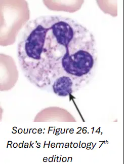
Human granulocytic anaplasmosis
a)
Ehrlichia chageensis
b)
Anaplasma phagocytophilum
c)
Histoplasma capsulatum
d)
Candida spp
72.
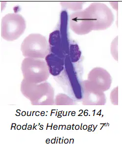
Candidiasis
a)
Ehrlichia chageensis
b)
Anaplasma phagocytophilum
c)
Histoplasma capsulatum
d)
Candida spp
73.
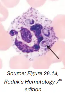
Histoplasmosis
a)
Ehrlichia chageensis
b)
Anaplasma phagocytophilum
c)
Histoplasma capsulatum
d)
Candida spp
74.
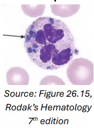
Described as bluishgreen or mostly green amorphous, refractile, shiny bodies of irregular shape, size, and number, contained in cytoplasm of neutrophils.
a)
Green-Crystals of death
b)
Reactive Monocytes
c)
Ruptured, Smudged Eosinophils
d)
Hypogranular Basophils
75.

A sign of impending death in most cases.
a)
Green-Crystals of death
b)
Reactive Monocytes
c)
Ruptured, Smudged Eosinophils
d)
Hypogranular Basophils
76.
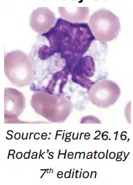
This monocytic morphology is associated with infections (including COVID-19), recovery from myelosuppression, and administration of GMCSF
a)
Green-Crystals of death
b)
Reactive Monocytes
c)
Ruptured, Smudged Eosinophils
d)
Hypogranular Basophils
77.
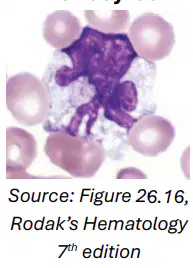
Difficult to differentiate from benign reactive monocytes.
a)
Green-Crystals of death
b)
Reactive Monocytes
c)
Ruptured, Smudged Eosinophils
d)
Hypogranular Basophils
78.
Eosinophils are fragile and often rupture during slide preparation.
a)
Green-Crystals of death
b)
Reactive Monocytes
c)
Ruptured, Smudged Eosinophils
d)
Hypogranular Basophils
79.
Regardless, guidelines state that they are to be counted along with intact eosinophils.
a)
Green-Crystals of death
b)
Reactive Monocytes
c)
Ruptured, Smudged Eosinophils
d)
Hypogranular Basophils
80.
water-soluble and easily washes away during staining. - Also associated with myeloid neoplasms.
a)
Green-Crystals of death
b)
Reactive Monocytes
c)
Ruptured, Smudged Eosinophils
d)
Hypogranular Basophils
81.
Commonly a neutrophil that has ingested the antibody-coated nucleus of another neutrophil.
a)
Lupus Erythematosus (LE) cell
b)
Rieder cell
c)
Flame cell
d)
Grape cell
e)
Russell bodies
82.
Commonly a neutrophil that has ingested the antibody-coated nucleus of another neutrophil.
a)
Lupus Erythematosus (LE) cell
b)
Rieder cell
c)
Flame cell
d)
Grape cell
e)
Russell bodies
83.
- Similar to normal lymphocytes, but the nucleus is notched, lobulated, and cloverleaf-like
a)
Lupus Erythematosus (LE) cell
b)
Rieder cell
c)
Flame cell
d)
Grape cell
e)
Russell bodies
84.
Abnormal plasma cell with intensely eosinophilic or “flaming” cytoplasm.
a)
Lupus Erythematosus (LE) cell
b)
Rieder cell
c)
Flame cell
d)
Grape cell
e)
Russell bodies
85.
Associated condition: - IgA myeloma
a)
Lupus Erythematosus (LE) cell
b)
Rieder cell
c)
Flame cell
d)
Grape cell
e)
Russell bodies
86.
a.k.a. Thesaurocyte
a)
Lupus Erythematosus (LE) cell
b)
Rieder cell
c)
Flame cell
d)
Grape cell
e)
Russell bodies
87.
- Abnormal plasma cells with cytoplasm filled with Russell bodies.
a)
Lupus Erythematosus (LE) cell
b)
Rieder cell
c)
Flame cell
d)
Grape cell
e)
Russell bodies
88.
Associated condition: - Multiple myeloma
a)
Lupus Erythematosus (LE) cell
b)
Rieder cell
c)
Flame cell
d)
Grape cell
e)
Russell bodies
89.
a.k.a. Morula/Mott cell
a)
Lupus Erythematosus (LE) cell
b)
Rieder cell
c)
Flame cell
d)
Grape cell
e)
Russell bodies
90.
Plasma cell cytoplasmic inclusions that appear like vacuoles containing aggregates of immunoglobulins.
a)
Lupus Erythematosus (LE) cell
b)
Rieder cell
c)
Flame cell
d)
Grape cell
e)
Russell bodies
91.
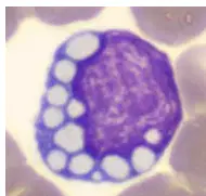
a)
Lupus Erythematosus (LE) cell
b)
Rieder cell
c)
Flame cell
d)
Grape cell
e)
Russell bodies
92.
Small lymphocytes (B-cell lineage) with little cytoplasmic projections.
a)
Hairy Cells
b)
Tart Cells
c)
Reed-Sternberg Cells
d)
Flower Cells
93.
Tartrate Resistant Acid Phosphatase (TRAP) Positive (+)
a)
Hairy Cells
b)
Tart Cells
c)
Reed-Sternberg Cells
d)
Flower Cells
94.
Annexin A is specific to HCL and can be used to differentiate HCL from other B-cell neoplasms.
a)
Hairy Cells
b)
Tart Cells
c)
Reed-Sternberg Cells
d)
Flower Cells
95.
A monocyte that has ingested a whole lymphocyte or a nucleus.
a)
Hairy Cells
b)
Tart Cells
c)
Reed-Sternberg Cells
d)
Flower Cells
96.
May be seen in drug sensitivity
a)
Hairy Cells
b)
Tart Cells
c)
Reed-Sternberg Cells
d)
Flower Cells
97.
A large lymphoid cell which may demonstrate two nuclei (with eosinophilic nucleoli) and an abundant cytoplasm).
a)
Hairy Cells
b)
Tart Cells
c)
Reed-Sternberg Cells
d)
Flower Cells
98.
With “Owl’s eye” appearance.
a)
Hairy Cells
b)
Tart Cells
c)
Reed-Sternberg Cells
d)
Flower Cells
99.
- Its presence is the definitive histologic characteristic of Hodgkin's lymphoma
a)
Hairy Cells
b)
Tart Cells
c)
Reed-Sternberg Cells
d)
Flower Cells
100.
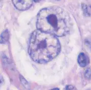
- Its presence is the definitive histologic characteristic of Hodgkin's lymphoma
a)
Hairy Cells
b)
Tart Cells
c)
Reed-Sternberg Cells
d)
Flower Cells
101.
Seen in Adult T cell leukemia or lymphoma
a)
Hairy Cells
b)
Tart Cells
c)
Reed-Sternberg Cells
d)
Flower Cells
102.
s a post thymic neoplastic disorder of T cells that is associated with retroviral infection by the Human T lymphotropic virus type I (HTLV-1).
a)
Hairy Cells
b)
Tart Cells
c)
Reed-Sternberg Cells
d)
Flower Cells
103.
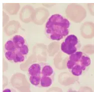
a)
Hairy Cells
b)
Tart Cells
c)
Reed-Sternberg Cells
d)
Flower Cells
104.
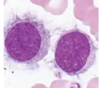
a)
Hairy Cells
b)
Tart Cells
c)
Reed-Sternberg Cells
d)
Flower Cells
105.
Seen in Nodular Lymphocyte Predominant Hodgkin’s Lymphoma (NLPHL)
a)
Popcorn Cells
b)
Sezary Cells
c)
Faggot cells
106.
Instead of the RS cell found in CHL, the malignant cell is a lymphocytic histiocytic (L&H) variant.
a)
Popcorn Cells
b)
Sezary Cells
c)
Faggot cells
107.
The L&H tumor cells are large, with scant cytoplasm and a folded single nucleus, and are labeled
a)
Popcorn Cells
b)
Sezary Cells
c)
Faggot cells
108.
L&H cells
a)
Popcorn Cells
b)
Sezary Cells
c)
Faggot cells
109.
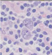
L&H cells
a)
Popcorn Cells
b)
Sezary Cells
c)
Faggot cells
110.
Characteristic cell found in Mycosis fungoides and Sezary syndrome. They are the most familiar form of cutaneous T-cell lymphoma.
a)
Popcorn Cells
b)
Sezary Cells
c)
Faggot cells
111.
The malignant T-cells in these conditions have an abnormal appearance (scant cytoplasm, cerebriform and folded nucleus, variably condensed chromatin, and inconspicuous nucleoli.
a)
Popcorn Cells
b)
Sezary Cells
c)
Faggot cells
112.
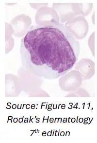
The malignant T-cells in these conditions have an abnormal appearance (scant cytoplasm, cerebriform and folded nucleus, variably condensed chromatin, and inconspicuous nucleoli.
a)
Popcorn Cells
b)
Sezary Cells
c)
Faggot cells
113.
A cell with bundles of Auer Rods
a)
Popcorn Cells
b)
Sezary Cells
c)
Faggot cells
114.
Auer rods - Linear projections of primary granules.
a)
Popcorn Cells
b)
Sezary Cells
c)
Faggot cells
115.
Seen in certain acute myeloid leukemia (AML) types
a)
Popcorn Cells
b)
Sezary Cells
c)
Faggot cells
116.
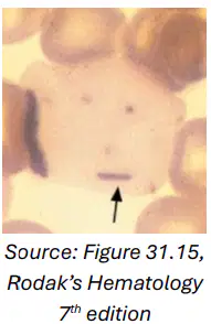
Seen in certain acute myeloid leukemia (AML) types
a)
Popcorn Cells
b)
Sezary Cells
c)
Faggot cells
100 %

