
 Worksheets
Pulmonary
Total questions: 176
Worksheet time: 29hrs 20mins
What precautions should be taken for contact isolation?
Gown and gloves; no treatment outside the room
Mask required; no treatment outside the room
N95 required; no treatment outside the room
Protect immunocompromised patient; gown and gloves
What is required for droplet isolation?
Gown and gloves; no treatment outside the room
Mask required; no treatment outside the room
N95 required; no treatment outside the room
Protect immunocompromised patient; gown and gloves
What is the requirement for airborne isolation?
Protect immunocompromised patient; gown and gloves
Gown and gloves; no treatment outside the room
Mask required; no treatment outside the room
N95 required; no treatment outside the room
What is the purpose of neutropenic isolation?
Protect immunocompromised patients; gown and gloves, limited treatment in room
Gown and gloves; no treatment outside the room
N95 required; no treatment outside the room
Mask required; no treatment outside the room
MRSA requires which type of precautions?
Contact precautions
Droplet precautions
Airborne precautions
Neutropenic precautions
VRE requires which type of precautions?
Droplet precautions
Contact precautions
Airborne precautions
Neutropenic precautions
C. diff requires which type of precautions?
Airborne precautions
Neutropenic precautions
Contact/Enteric precautions
Droplet precautions
Scabies requires which type of precautions?
Neutropenic precautions
Airborne precautions
Droplet precautions
Contact precautions
Norovirus requires which type of precautions?
Contact/Enteric precautions
Droplet precautions
Airborne precautions
Neutropenic precautions
Rotavirus requires which type of precautions?
Droplet precautions
Airborne precautions
Contact/Enteric precautions
Neutropenic precautions
Influenza requires which type of precautions?
Contact precautions
Neutropenic precautions
Droplet precautions
Airborne precautions
COVID-19 most commonly requires which type of precautions?
Droplet precautions
Airborne precautions
Neutropenic precautions
Contact precautions
Mumps requires which type of precautions?
Droplet precautions
Airborne precautions
Contact precautions
Neutropenic precautions
Rubella requires which type of precautions?
Airborne precautions
Droplet precautions
Neutropenic precautions
Contact precautions
Meningitis requires which type of precautions?
Contact precautions
Neutropenic precautions
Airborne precautions
Droplet precautions
Tuberculosis requires which type of precautions?
Droplet precautions
Airborne precautions
Contact precautions
Neutropenic precautions
Measles requires which type of precautions?
Neutropenic precautions
Contact precautions
Airborne precautions
Droplet precautions
Chickenpox (Varicella) requires which type of precautions?
Airborne precautions
Droplet precautions
Contact precautions
Neutropenic precautions
SARS/MERS require which type of precautions?
Contact precautions
Droplet precautions
Neutropenic precautions
Airborne precautions
A chemotherapy patient would most likely be placed under which precautions?
Droplet precautions
Neutropenic precautions
Contact precautions
Airborne precautions
A bone marrow transplant patient would most likely be placed under which precautions?
Contact precautions
Droplet precautions
Neutropenic precautions
Airborne precautions
A patient with severe burns may require which precautions?
Airborne precautions
Contact precautions
Neutropenic precautions
Droplet precautions
A patient with aplastic anemia may require which precautions?
Contact precautions
Neutropenic precautions
Droplet precautions
Airborne precautions
Leukemia patients often require which precautions?
Droplet precautions
Contact precautions
Airborne precautions
Neutropenic precautions
What is external respiration?
Gas exchange at the alveolar-capillary membrane and pulmonary capillaries
Gas exchange at tissue capillaries
The movement of air through conducting airways
Temporary cessation of breathing during sleep
What occurs during internal respiration?
Movement of air through the conducting airways
Gas exchange at the tissue capillary level
Relaxed, passive expiration
Accessory muscle recruitment during breathing
What is ventilation?
Gas exchange at alveolar-capillary level
Gas exchange at tissue capillary level
Movement of air through conducting airways
Passive expiration
What is sleep apnea?
A passive process of expiration
A condition where breathing stops or becomes shallow during sleep
Gas exchange at alveolar level
Forceful exhalation during coughing
What nerve supplies the diaphragm?
Vagus nerve
Phrenic nerve (C3–C5)
Accessory nerve
Intercostal nerves
What are the primary muscles of inspiration?
Sternocleidomastoid, scalenes, pecs, upper trapezius, levator scapulae, rhomboids
Diaphragm and external intercostals
Abdominals, internal intercostals, quadratus lumborum
Internal intercostals and quadratus lumborum
Which muscles are primary for expiration?
Sternocleidomastoid, scalenes, pecs, upper trapezius, levator scapulae, rhomboids
Diaphragm and external intercostals
Abdominals, internal intercostals, quadratus lumborum
Internal intercostals and quadratus lumborum
Which muscles are accessory muscles of inspiration?
Sternocleidomastoid, scalenes, pecs, upper trapezius, levator scapulae, rhomboids
Diaphragm and external intercostals
Rectus abdominis and transverse abdominis
Abdominals, internal intercostals, quadratus lumborum
What is quiet inspiration?
Breathing at rest with minimal effort
Gas exchange at tissue level
Forceful inhalation requiring accessory muscles
Passive expiration
What occurs during forced inspiration?
Relaxed passive expiration
Engages accessory muscles during heavy oxygen debt
Movement of air through conducting airways
Gas exchange at the alveoli
What is the primary process of expiration at rest?
Forceful exhalation
Relaxed, passive process
Movement of air through airways
Use of accessory muscles
What is quiet expiration?
Active process with muscle use
Passive process of exhalation
Gas exchange at alveoli
Use of sternocleidomastoid
What is forced expiration?
Passive process at rest
Gas exchange at tissues
Active exhalation during coughing or exertion
Air exchange at alveoli
How many lobes does the right lung have?
Three lobes: upper, middle, and lower
Two lobes: upper and lower, with the lingula
One lobe
Four lobes
How many lobes does the left lung have?
Three lobes: upper, middle, and lower
Two lobes: upper and lower, with the lingula
One lobe
Four lobes
What covers each lung?
Visceral pleura
Parietal pleura
Pleural fluid
Intercostal fascia
What lines the thoracic wall?
Visceral pleura
Parietal pleura
Pleural fluid
Intercostal fascia
How are the lungs inflated?
By positive pressure
By negative pressure
By muscle contraction alone
By pleural fluid volume
What is the function of pleural fluid?
Provides oxygen for gas exchange
Lubricates pleurae during ventilation
Maintains negative pressure
Protects lungs from infection
What are the components of the upper respiratory tract?
Nasal cavity, pharynx, larynx
Trachea, bronchi, alveoli
Bronchioles, alveoli, lungs
Pharynx, trachea, bronchi
What are the functions of the nasal cavity?
To control airflow and prevent aspiration
To warm air and filter/remove particles
To lubricate pleura during breathing
To exchange oxygen and carbon dioxide
What is the function of the larynx?
Gas exchange at alveoli
To warm air before lungs
To control airflow and prevent aspiration
To lubricate the pleurae
Tumor is classified as which type of lung disorder?
Restrictive disorder
Obstructive disorder
Interstitial pulmonary fibrosis is classified as which type of lung disorder?
Restrictive disorder
Obstructive disorder
Pulmonary edema is classified as which type of lung disorder?
Restrictive disorder
Obstructive disorder
Pulmonary embolism is classified as which type of lung disorder?
Restrictive disorder
Obstructive disorder
Atelectasis is classified as which type of lung disorder?
Restrictive disorder
Obstructive disorder
Inadequate pulmonary development is classified as which type of lung disorder?
Restrictive disorder
Obstructive disorder
Postural deformities are classified as which type of lung disorder?
Restrictive disorder
Obstructive disorder
Scleroderma is classified as which type of lung disorder?
Restrictive disorder
Obstructive disorder
Ankylosing spondylitis is classified as which type of lung disorder?
Restrictive disorder
Obstructive disorder
Neurologic disorders can cause which type of lung disorder?
Restrictive disorder
Obstructive disorder
Chest wall trauma is classified as which type of lung disorder?
Restrictive disorder
Obstructive disorder
What are the classic symptoms of restrictive pulmonary disorders?
Wheezing and chest tightness
Dyspnea with activity and a persistent non-productive cough
Productive cough and prolonged expiration
Hemoptysis and pleural pain
What are some other signs of restrictive pulmonary disorders?
Rapid shallow breathing, limited chest expansion, inspiratory crackles, clubbing, cyanosis
Wheezing, prolonged expiration, pursed-lip breathing
Cough with sputum, barrel chest, orthopnea
Chest pain, hemoptysis, productive cough
What do pulmonary function tests (PFTs) show in restrictive pulmonary disorders?
Increased vital capacity and increased residual volume
Increased vital capacity and decreased total lung capacity
Decreased inspiratory reserve volume and decreased vital capacity
Decreased total lung capacity and increased functional residual capacity
What is a common arterial blood gas (ABG) finding in restrictive pulmonary disorders?
Hypercapnia
Hypoxemia
Normal oxygen saturation
Respiratory alkalosis
Chronic Obstructive Pulmonary Disease (COPD) is best described as:
A restrictive lung disease with limited chest expansion
A condition causing lung hyperinflation and airflow limitation that is not fully reversible
A neuromuscular disorder leading to reduced tidal volume
A surgical removal of diseased lung tissue
What is the function of the alveoli?
Tiny air sacs in the lungs where oxygen and carbon dioxide are exchanged
Structures that prevent food from entering the trachea
Muscles that drive breathing
Tubes that transport air to the lungs
What is tidal volume (resting tidal volume)?
Maximum amount of air inhaled after quiet inspiration
Amount of air exchanged during quiet inspiration and expiration (~500 mL)
Amount of air remaining after maximum expiration
Total volume of air after maximum inspiration
What is inspiratory reserve volume (IRV)?
Amount of air left after max exhalation
Maximum amount of air a person can breathe in after quiet inspiration
Volume exchanged during quiet breathing
Total lung capacity after maximum inspiration
What is expiratory reserve volume (ERV)?
Max amount of air exhaled after quiet expiration
Max amount of air inhaled after quiet inspiration
Amount of air left after max exhalation
Total amount of air in lungs after max inspiration
What is residual volume (RV)?
Amount of air exchanged during quiet breathing
Amount of air left in lungs after maximum expiration
Maximum inhalation after quiet breathing
Total amount of air in lungs after max inspiration
What is total lung capacity (TLC)?
Amount left after quiet expiration (RV+ERV)
Total amount of air after maximum inspiration (TV+IRV+ERV+RV)
Maximum inspiration after quiet expiration (TV+IRV)
Maximum inspiration + expiration (IRV+TV+ERV)
What is inspiratory capacity (IC)?
TV + IRV, maximum inhalation after quiet expiration
RV + ERV, maximum inhalation after forced expiration
RV + ERV, maximum inhalation after quiet expiration
TV + IRV, maximum inhalation after forced expiration
What is functional residual capacity (FRC)?
TV + IRV, amount left in lungs after resting expiration
RV + ERV, amount left in lungs after forced expiration
RV + ERV, amount left in lungs after resting expiration
IRV + TV + ERV, max inspiration followed by max expiration
What is vital capacity (VC)?
TV + IRV, amount left in lungs after resting expiration
RV + ERV, amount left in lungs after forced expiration
RV + ERV, amount left in lungs after resting expiration
TV + IRV, amount left in lungs after forced expiration
What effect do obstructive lung diseases have on lung volumes?
Increased TLC, TV, RV, FRC; decreased VC, IRV, IC, ERV
Decreased TLC, TV, RV, FRC; increased VC
Increased VC and IRV
Decreased inspiratory reserve volume and decreased vital capacity
What effect does aging have on lung volumes?
Max O₂ uptake decreases, VC decreases, RV increases
TLC increases, VC increases
IRV increases, FRC decreases
Lung volumes remain constant
What does FEV1 measure?
Volume of air forcefully exhaled in 1 second, reflects airway status
Maximal volume inhaled after quiet expiration
Greatest flow during forced expiration; drop shows airway narrowing
Residual air left after maximal expiration
What does peak expiratory flow rate (PEF) indicate?
Volume of air forcefully exhaled in 1 second, reflects airway status
Maximal volume inhaled after quiet expiration
Greatest flow during forced expiration; drop shows airway narrowing
Residual air left after maximal expiration
What defines restrictive pulmonary disorders?
Any condition reducing lung volume and compliance, making deep breathing difficult
Air trapping with increased lung volumes
Acute infections of the pleura
Airway dilation due to chronic infections
A group of lung diseases that cause airflow blockage and breathing problems.
Chronic obstructive pulmonary disease (COPD)
Chronic bronchitis
Emphysema
Asthma
A condition of productive cough lasting more than 3 months per year for 2 years.
Pneumonia
Chronic bronchitis
Sleep apnea
Pulmonary abscess
Abnormal enlargement of the distal respiratory unit (alveoli) with destructive changes of the alveolar walls without obvious fibrosis.
Pleurisy
Bronchiectasis
Emphysema
Pulmonary embolism
Inflammation of the pleura caused by viral or bacterial infection, injury, or tumor. Presents with sharp chest pain that increases with inspiration and coughing.
Pneumothorax
Hemothorax
Asthma
Pleurisy (pleuritis)
Accumulation of air or gas in the pleural space caused by a defect in the visceral pleura or chest wall.
Pneumothorax
Atelectasis
Bronchiolitis
Pulmonary tuberculosis
Accumulation of air and blood in the pleural space, often after chest trauma.
Pulmonary edema
Hemothorax
Cystic fibrosis
Lung cancer
Accumulation of fluid in the pleural space.
Pulmonary effusion (hydrothorax)
Influenza
Pulmonary abscess
Bronchitis
A chronic respiratory condition that affects the airways, causing inflammation and making it difficult to breathe.
Tuberculosis
Pneumonia
Pulmonary edema
Asthma
Collapse of normally expanded and aerated lung tissue, often due to obstruction of a bronchus. Can be caused by immobility, over-sedation, or paralysis.
Atelectasis
Bronchiolitis
COPD
Influenza
A sleep-disordered breathing condition with intermittent disruption of gas exchange and sleep interruption.
Pulmonary embolism
Sleep apnea
Pleural effusion
Bronchiectasis
A progressive obstructive lung disease with irreversible destruction and dilation of airways due to chronic bacterial infections.
Bronchiectasis
Pulmonary abscess
Cystic fibrosis
Emphysema
Severe inflammation of the bronchioles (lower airways).
Bronchitis
Bronchiolitis
Pleurisy
Pneumonia
Inflammation of the bronchial lining, usually short-term (1–3 weeks) and self-limiting.
Pulmonary abscess
Chronic bronchitis
Acute bronchitis
Pleurisy
A disease with exocrine gland dysfunction causing abnormally viscous secretions that narrow or obstruct airways, leading to hyperinflation, infection, and tissue destruction.
Cystic fibrosis
Pulmonary embolism
COPD
Tuberculosis
A highly contagious viral respiratory infection with many strains, spread by respiratory droplets.
Influenza (flu)
Pulmonary edema
Sleep apnea
Pleurisy
A malignancy of the respiratory tract epithelium, often asymptomatic until metastasis. Most often caused by cigarette smoking.
Tuberculosis
Pulmonary embolism
Pulmonary abscess
Lung cancer
What are the most common sites of metastases?
Lungs, liver, bone, brain
Heart, kidneys, pancreas, stomach
Skin, spleen, bladder, spine
Esophagus, stomach, intestines, thyroid
An infectious systemic disease caused by Mycobacterium tuberculosis, primarily affecting the lungs.
Pulmonary tuberculosis
Pneumonia
Pulmonary edema
Asthma
Inflammation of the lungs.
Pneumothorax
Pneumonia
Pulmonary embolism
Bronchiectasis
An infection a patient contracts while receiving care in a healthcare setting (e.g., pneumonia, UTI).
Nosocomial infection
Influenza
Pulmonary abscess
Emphysema
Localized collection of infectious material within the lung that may rupture and cause empyema or septicemia.
Pulmonary abscess
Pulmonary edema
Hemothorax
Bronchiolitis
Tissues and air spaces of the lungs fill with fluid that leaks from the vascular system, often due to cardiovascular disease, CHF, hypertension, PE, or renal failure.
Pulmonary edema
Pneumothorax
Pleural effusion
Lung cancer
Sudden blockage of a pulmonary artery by an embolism (clot), potentially life-threatening and may cause cardiac arrest.
Pulmonary embolism (PE)
Pneumonia
Tuberculosis
Atelectasis
Severe and highly contagious viral lung infection with high fever caused by coronavirus. Spread by respiratory droplets.
Pneumonia
Severe acute respiratory syndrome (SARS)
Influenza
Atelectasis
Surgical removal of a whole lung.
Thoracotomy
Tracheotomy
Pneumonectomy
Lobectomy
Surgical removal of a lobe of the lung.
Lobectomy
Pneumonectomy
Thoracotomy
Tracheostomy
Surgical incision into the chest wall.
Lobectomy
Pneumonectomy
Thoracotomy
Tracheostomy
Placement of a tracheal tube directly into the trachea to form an airway.
Lobectomy
Pneumonectomy
Thoracotomy
Tracheostomy
Drug used to suppress coughing, helpful for dry cough but not justified for productive cough.
Antitussive drug
Antihistamine
Bronchodilator
Mucolytic/Expectorants
Drug used to dry up excess mucus produced in the upper respiratory tract.
Bronchodilator
Decongestant
Antitussive
Glucocorticoid
Drug used to treat the respiratory allergic response to seasonal allergies and asthma.
Antihistamines
Mucolytic/Expectorants
Decongestants
Steroids
Drug used to decrease the viscosity of respiratory secretions (e.g., in Chronic Bronchitis or Cystic Fibrosis).
Pulmonary embolism
Antihistamines
Antitussives
Mucolytics/Expectorants
Drug used to control inflammation-mediated bronchospasm; can be given orally, inhaled, or IV (asthma).
Bronchodilator
Glucocorticoids (steroids)
Decongestant
Antihistamine
Drug that dilates the bronchi in the lungs, decreasing resistance and inducing smooth muscle relaxation (asthma, emphysema).
Antihistamines
Glucocorticoids
Bronchodilators
Antitussives
Normal hemoglobin (Hgb) values for men.
7–10 mg/dL
10–12 mg/dL
13–18 mg/dL
20–24 mg/dL
Normal hemoglobin (Hgb) values for women.
8–11 mg/dL
12–16 mg/dL
17–20 mg/dL
22–25 mg/dL
Normal value for PaCO2 (partial pressure of carbon dioxide).
35–45 mmHg
75–100 mmHg
60–70 mmHg
45–55 mmHg
Normal value for PaO2 (arterial oxygen pressure).
45–55 mmHg
60–70 mmHg
75–100 mmHg
100–120 mmHg
Normal blood pH range.
7.10–7.20
7.25–7.30
7.36–7.44
7.50–7.60
Normal white blood cell (WBC) count.
1,000–3,000 cells/mm³
4,500–11,000 cells/mm³
15,000–20,000 cells/mm³
25,000–30,000 cells/mm³
Normal fasting blood glucose value.
40–60 mg/dL
70–100 mg/dL
120–150 mg/dL
160–200 mg/dL
Normal adult respiration rate (RR).
6–10 bpm
12–24 bpm
25–30 bpm
35–40 bpm
High PaCO2 is called what?
Hypocapnia
Hypercapnia
Hypoxemia
Hypoxia
Low PaCO2 is called what?
Hypoxemia
Alkalemia
Hypocapnia
Leukopenia
PaO2 less than 40 mmHg is called what?
Hypoxemia
Hypoxia
Anemia
Hypocapnia
High blood pH is called what?
Acidemia
Alkalemia
Hyperglycemia
Hypoxemia
Low blood pH is called what?
Hypoxemia
Acidemia
Hypoglycemia
Leukopenia
High hemoglobin is called what?
Leukocytosis
Anemia
Polycythemia
Hypoxemia
Low hemoglobin is called what?
Anemia
Hypoxemia
Leukopenia
Hypocapnia
High white blood cell count is called what?
Hyperglycemia
Leukocytosis
Leukopenia
Anemia
Low white blood cell count is called what?
Leukopenia
Polycythemia
Acidemia
Hypoxemia
High blood glucose is called what?
Hypoglycemia
Hyperglycemia
Anemia
Hypoxia
Low blood glucose is called what?
Hypoglycemia
Leukopenia
Hypoxemia
Acidemia
Which of the following is considered a normal breathing sound heard over most of the lung fields?
Crackles
Vesicular
Wheezes
Rhonchi
Which breathing sound is normally heard over the trachea?
Vesicular
Bronchial
Crackles
Wheezes
Which breathing sound is normally heard between the scapulae or near the sternum?
Bronchovesicular
Vesicular
Stridor
Crackles
Which normal breathing sound is soft and low-pitched, heard during inspiration and fading quickly during expiration, and is heard over most of the peripheral lung fields?
Bronchial
Vesicular
Crackles
Wheezes
Which normal breathing sound is high-pitched, loud, and hollow, with a longer expiratory phase than inspiratory, and is normally heard over the trachea?
Vesicular
Bronchial
Bronchovesicular
Crackles
Which normal breathing sound has equal inspiratory and expiratory phases with a medium pitch and intensity, and is best heard near the sternum or between the scapulae?
Crackles
Wheezes
Bronchovesicular
Vesicular
Which adventitious breathing sound is described as discontinuous, brief, popping sounds that may be fine or coarse, and are commonly associated with pneumonia, heart failure, or pulmonary fibrosis?
Wheezes
Crackles
Vesicular
Bronchial
Which adventitious breathing sound is described as continuous, high-pitched, musical or squeaky sounds, more prominent during expiration, and commonly associated with asthma or chronic obstructive pulmonary disease (COPD)?
Crackles
Wheezes
Bronchovesicular
Vesicular
Which adventitious breathing sound is a low-pitched, snoring or gurgling noise often caused by secretions in larger airways, and may clear with coughing?
Rhonchi
Crackles
Wheezes
Vesicular
Which adventitious breathing sound is a harsh, high-pitched, crowing sound heard primarily during inspiration, and usually indicates upper airway obstruction such as croup or a foreign body?
Crackles
Stridor
Bronchial
Wheezes
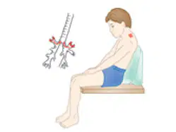
The positioning of the patient in the photograph is MOST likely to be recommended for postural drainage of which of the following areas of the lungs?
Upper Lobes Apical segments
Upper Lobes Posterior segments
Upper Lobes Anterior segments
Upper Lobes Lateral segments
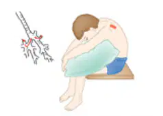
The positioning of the patient in the photograph is MOST likely to be recommended for postural drainage of which of the following areas of the lungs?
Upper Lobes Apical segments
Upper Lobes Posterior segments
Upper Lobes Anterior segments
Upper Lobes Lateral segments
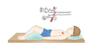
The positioning of the patient in the photograph is MOST likely to be recommended for postural drainage of which of the following areas of the lungs?
Upper Lobes Apical segments
Upper Lobes Posterior segments
Upper Lobes Anterior segments
Upper Lobes Lateral segments
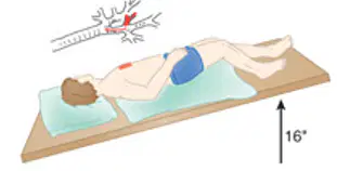
The positioning of the patient in the photograph is MOST likely to be recommended for postural drainage of which of the following areas of the lungs?
Right Upper Lobe
Right Middle Lobe
Right Lower Lobe
Upper Lobes Lateral segments
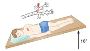
The positioning of the patient in the photograph is MOST likely to be recommended for postural drainage of which of the following areas of the lungs?
Left Upper Lobe
Left Middle Lobe
Left Lower Lobe Anterior Basal Segments
Left Lower Lobe Apical Basal Segments
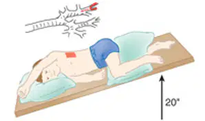
The positioning of the patient in the photograph is MOST likely to be recommended for postural drainage of which of the following areas of the lungs?
Left Upper Lobe
Left Middle Lobe
Lower Lobe Anterior Basal Segments
Lower Lobe Lateral Basal Segments
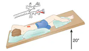
The positioning of the patient in the photograph is MOST likely to be recommended for postural drainage of which of the following areas of the lungs?
Left Upper Lobe
Left Middle Lobe
Lower Lobe Anterior Basal Segments
Lower Lobe Lateral Basal Segments
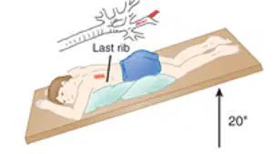
The positioning of the patient in the photograph is MOST likely to be recommended for postural drainage of which of the following areas of the lungs?
Lower Lobe Superior Segments
Lower Lobe Posterior Basal Segments
Lower Lobe Anterior Basal Segments
Lower Lobe Lateral Basal Segments
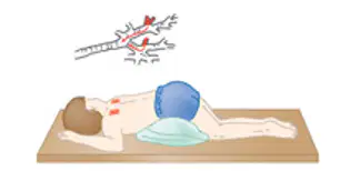
The positioning of the patient in the photograph is MOST likely to be recommended for postural drainage of which of the following areas of the lungs?
Lower Lobe Superior Segments
Lower Lobe Posterior Basal Segments
Lower Lobe Anterior Basal Segments
Lower Lobe Lateral Basal Segments
Which of the following is NOT the most common post-op complications?
Atelectasis
Pneumonia
PE/DVT
Asthma
On the Borg Scale, which rating corresponds to "no exertion"?
6
7
8
9
On the Borg Scale, which rating corresponds to "extremely light"?
7
9
11
13
On the Borg Scale, which rating corresponds to "very light"?
9
11
13
15
On the Borg Scale, which rating corresponds to "light"?
11
13
15
17
On the Borg Scale, which rating corresponds to "somewhat hard"?
11
13
15
17
On the Borg Scale, which rating corresponds to "hard"?
13
15
17
19
On the Borg Scale, which rating corresponds to "very hard"?
15
17
19
20
On the Borg Scale, which rating corresponds to "extremely hard"?
17
18
19
20
On the Borg Scale, which rating corresponds to "maximal exertion"?
15
17
19
20
During exercise training in patients with mild to moderate pulmonary disease, what percentage of maximum heart rate is typically recommended for intensity?
50%-60% of maximum heart rate
70%-85% of maximum heart rate
30%-40% of maximum heart rate
90%-100% of maximum heart rate
What is the recommended rating of perceived exertion (RPE) for moderate intensity exercise in pulmonary rehabilitation, corresponding to 40%-60% of VO₂ max?
RPE 6-9
RPE 12-13
RPE 15-17
RPE 19-20
What is the recommended rating of perceived exertion (RPE) for vigorous intensity exercise in pulmonary rehabilitation, corresponding to 60%-90% of VO₂ max?
RPE 12-13
RPE 14-17
RPE 6-9
RPE 19-20
What is the typical prescribed duration of exercise sessions in pulmonary rehabilitation programs?
10-15 minutes
20-30 minutes
45-60 minutes
90 minutes
How often should patients in pulmonary rehabilitation perform moderate intensity exercise to achieve benefits?
1-2 times per week
3-5 times per week
6-7 times per week
Only once weekly
A patient presents with dyspnea with activity, persistent non-productive cough, rapid shallow breathing, limited chest expansion, inspiratory crackles, clubbing/cyanosis, and ABGs showing hypoxemia. What is the most likely condition?
Emphysema
Asthma
Restrictive pulmonary disorder
Sleep apnea
A patient with a history of smoking presents with dyspnea, chronic cough, chronic sputum production, recurrent lower respiratory tract infections, and FEV1 < 70%. What condition is most likely?
Asthma
COPD
Cystic fibrosis
Acute bronchitis
A patient presents with dyspnea, recurrent infections, decreased chest expansion, wheezing, cyanosis, hypoxemia, and decreased exercise tolerance. What is the most likely condition?
Chronic bronchitis
COVID-19
Restrictive pulmonary disorder
Bronchiectasis
A thin-built patient presents with dyspnea, tachypnea with prolonged expiration, accessory muscle use, and a barrel chest. What condition is most likely?
Acute bronchitis
Emphysema
Sleep apnea
Influenza
A patient presents with wheezing, dyspnea, chest tightness, and cough. Which condition does this presentation most likely indicate?
Asthma
COPD
Acute respiratory distress syndrome
Pulmonary embolism
A patient reports interruptions of deep restorative sleep, sluggishness, headaches, daytime fatigue, cognitive impairment, weight gain, and sexual impotence. What condition is most likely?
Influenza
Sleep apnea
COVID-19
Acute bronchitis
A patient presents with a persistent cough producing large amounts of purulent sputum, dyspnea, and fatigue. What is the most likely condition?
Cystic fibrosis
Bronchiectasis
COPD
Asthma
A patient reports a recent upper respiratory infection, fever, tight feeling behind the sternum, and a dry cough that later became productive. What is the most likely condition?
Influenza
Acute bronchitis
COVID-19
Restrictive pulmonary disorder
A patient presents with frequent pulmonary infections and chronic cough due to pulmonary dysfunction, failure to thrive due to GI dysfunction, and diabetes mellitus due to pancreatic dysfunction. Which condition does this describe?
Emphysema
Sleep apnea
Cystic fibrosis
Bronchiectasis
A patient presents with sudden onset fever, chills, headache, back and muscle pain, cough, runny nose, sore throat, sneezing, hoarseness, nausea, vomiting, and diarrhea. Which condition is most likely?
Influenza (Flu)
COVID-19
Acute bronchitis
ARDS
A patient presents with dyspnea, pleuritic chest pain, apprehension, cough, and tachypnea. Risk factors include blood stasis, local trauma, hypercoagulable states, immobility, recent abdominal or pelvic surgery, THR/TKR, late pregnancy, lower limb fracture, or previous PE. What condition is most likely?
Pulmonary embolism
COPD
Restrictive pulmonary disorder
ARDS
A patient develops acute respiratory failure following severe trauma, sepsis, or major surgery. What is the most likely condition?
ARDS (Acute respiratory distress syndrome)
COPD
COVID-19
Sleep apnea
A patient presents with dry cough, fever, fatigue, and shortness of breath. What condition is most likely?
Influenza
COVID-19
Acute bronchitis
Restrictive pulmonary disorder

