
 Worksheets
EPS0 N3UR0 C0MP1LE
Total questions: 124
Worksheet time: 1hrs 6mins
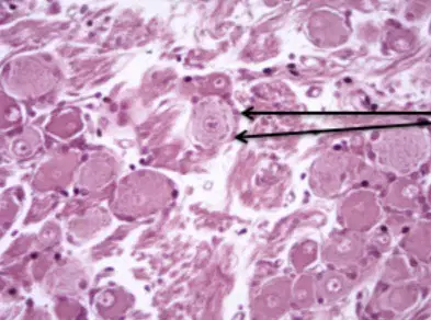
What is the function of this cell ?
Conduct nerve impulses
Produce myelin sheath
Supporting ganglion cells
Which of the following medium is used for isolating Mycobacterium tuberculosis?
Manitol salt agar
Sabouraud dextrose
Thioglycolate broth
Chocolate agar
Lowenstein-Jensen
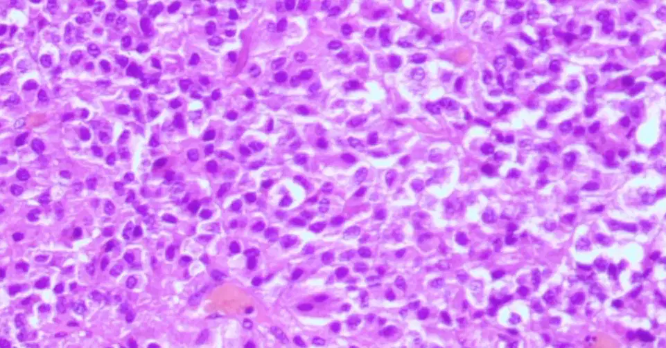
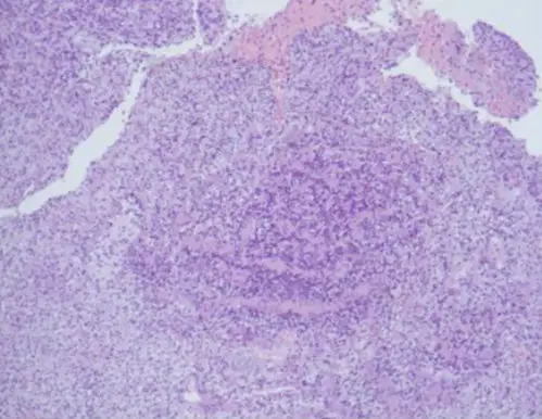
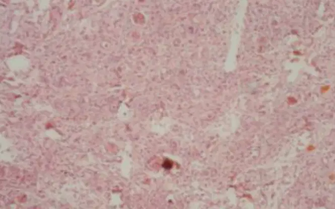
A 27-year-old woman had persistent headache and amenorrhea. She was also diagnosed with acromegaly based on various symptoms and clinical findings. Brain MRI showed a giant tumor around pituitary fossa, pressuring on optic chiasm from the middle. Based on the result of biopsy presented below, what is her diagnosis?
Craniopharyngioma
Pituitary adenoma
Ependymoma
Medulloblastoma
Hemangioblastoma
On the bed side LCS examination then
Nonne's examination is to determine whether there is a qualitative increase in protein levels
Nonne's examination is to determine whether there is an increase in Globulin levels qualitatively
Pandi examination to determine whether there is a quantitative increase in protein levels
Nonne's examination is to determine whether there is a qualitative increase in Globulin levels
Pandi examination to determine whether there is a qualitative increase in Globulin levels

What is the function of this cell ?
Conduct impulses from receptor to spinal cord
Conduct impulses from central nervous system to peripheral nervous system
Conduct impulses from spinal cord to muscle
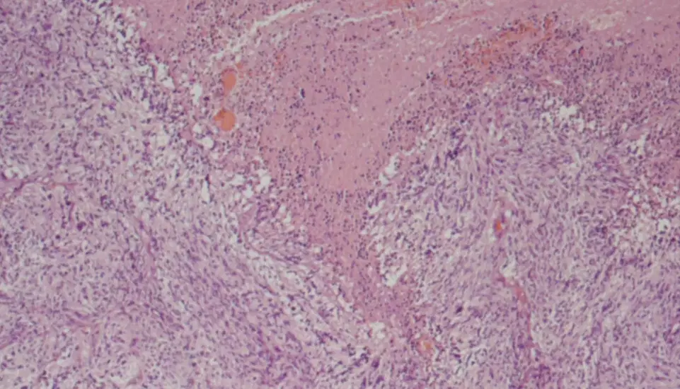
A 60-year-old man presented with altered level of consciousness and right sided hemiplegia which was progressive over four days. MRI showed left occipitoparietal space occupying lesion with multiple linear projections. Based on the result of biopsy presented below, what is his diagnosis?
Gliosarcoma
Astrocytoma
Glioblastoma multiforme
Ependymoma
Cerebral metastasis
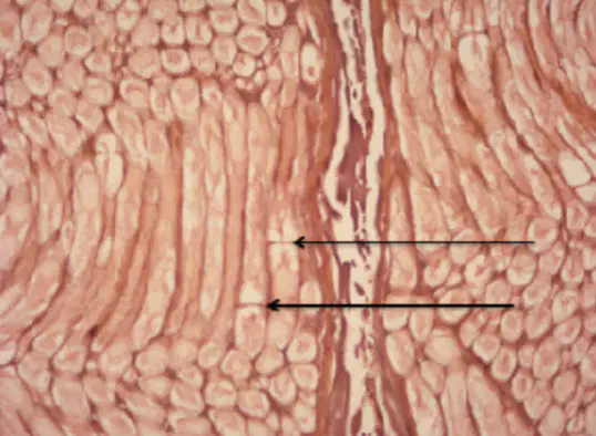
What is the name of this structure?
Ranvier Node
Neurolemma
Axolemma
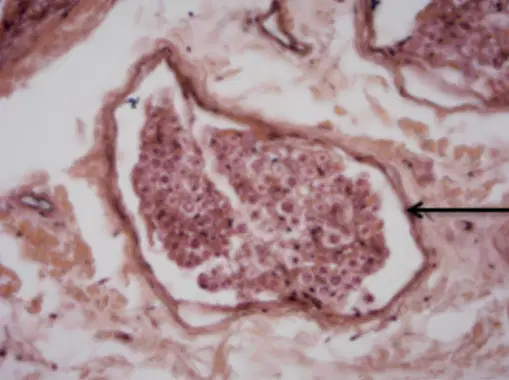
What is the name of this tissue?
Endoneurium
Epineurium
Perineurium
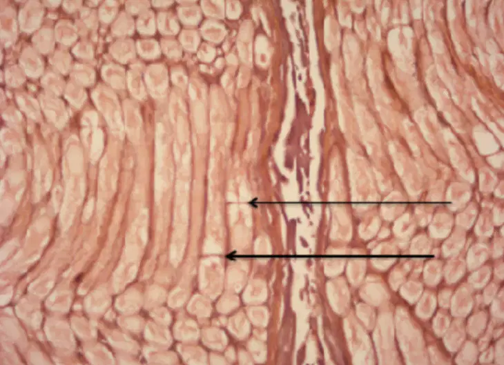
Where is the structure occurs?
Myelin sheath and axolemma are discontinous
Myelin sheath is discontinous
Axolemma is absent
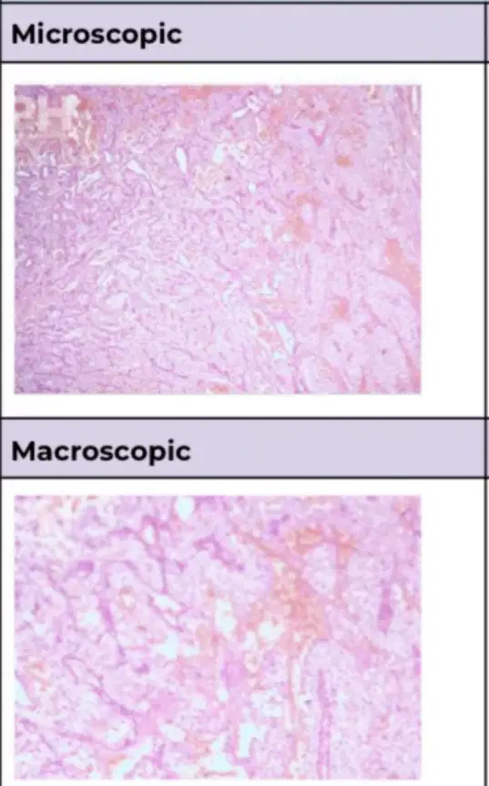
A 54-year-old man presented with persistent headache associated with vomiting, balance disorder with ataxia, recurrent tremor, as well as nystagmus for the last 2 months. MRI showed a lesion of the left cerebellar hemisphere. Based on the result of biopsy presented below, what is his diagnosis?
Pilocytic astrocytoma
Hemangioblastoma
Glioblastoma
Medulloblastoma
Arteriovenous malformation
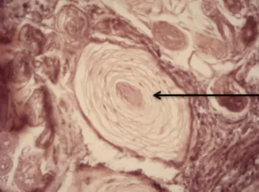
What is the function of this structure?
Gross pressure / vibration receptor
Light touch receptor
Pain receptor
Wrist drop is a disorder caused by …………… palsy
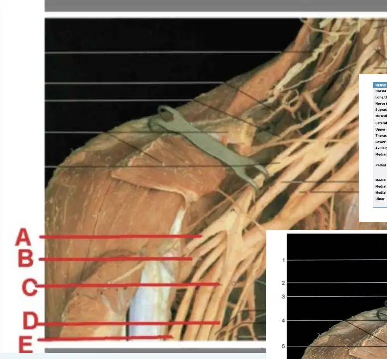
Choose one from the five options in the image below and write the name of the nerve!
The brachial plexus is visible from the anterolateral view
A,C: from posterior division
B,D,E : from anterior division
A. N. Axillary
B. N. Median
C. N. Radialis
D. N. Musculocutaneous
E. N. Ulnaris
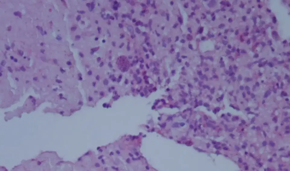
A 30-year-old man presented with fever, dizziness and vomiting, with bout of seizures in the last 7 days. He has history of HIV infection and was on antiretroviral drugs. CT scan of the brain showed an irregular ring enhancing lesion in the right basal ganglia with surrounding marked white matter edema and mass effect. Based on the result of biopsy presented below, what is his diagnosis?
Progressive multifocal encephalopathy
Primary lymphoma
Metastatic brain lesion
Toxoplasmosis
Tuberculoma
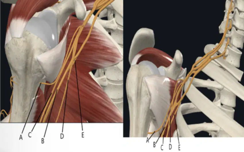
From the image below, which nerve will continue as the lateral cutaneous nerve of the forearm (n. cutaneus antebrachii lateralis)? Choose one from the five options in the image below and write the name of the nerve!
The brachial plexus is visible from the anterolateral view
A,C: from posterior division
B,D,E : from anterior division
A. N. Axillary
B. N. Median
C. N. Radialis
D. N. Musculocutaneous
E. N. Ulnaris
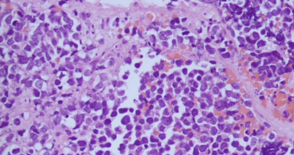
A 12-year-old boy presented with progressive loss of vision in both eyes since 2 months with headache and nausea. Brain MRI revealed large ill-defined lobulated solid cystic extra axial space occupying lesion in posterior aspect of third ventricle involving pineal gland. Based on the result of biopsy presented below, what is his diagnosis?
Diffuse midline glioma
Pineocytoma
Germinoma
Pineoblastoma
Medulloblastoma
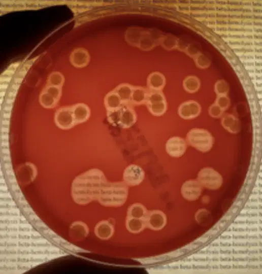
Specify the type of hemolysis produced by bacteria as illustrated in figure below:
γ-hemolysis
β-hemolysis
Incomplete hemolysis
µ-hemolysis
α-hemolysis
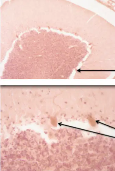
What type of cell is it?
Multipolar neuron
Bipolar neuron
Unipolar neuron
The results of the CSF examination that support the diagnosis of Tuberculous Meningitis are
Cells dominated by PMN and opalescent, prot – (neg)
Cells dominated by PMN and purulent, prot – (neg)
Cells are predominantly lymphocyte and purulent, prot + 2
Cells dominated by PMN and purulent, prot + 3
Cells are predominantly lymphocyte and opalescent, prot + 2
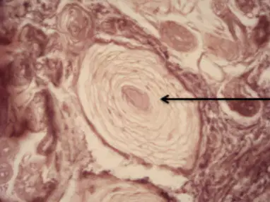
What is the name os this structure?
Meissner corpuscle
Merkel cospuscle
Pacini corpuscle
A bacterium with less than a complete twist or comma shaped is known as?
Vibrioid
Spirilla
Helical
Flagellated
Spirochetes
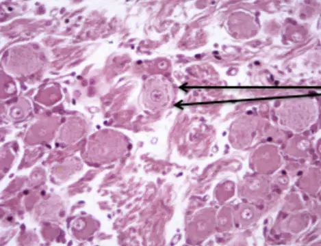
What is the name of this cell?
Satellite cell
Purkinje cell
Ganglion cell

This nerve (?) leaves the base of the cranium through a structure; state the full name of that structure
(a)
Normal Cebro Spinalis liquor is
Gross appearance : clear and white Culture : positive Gram stain : positive
Gross appearance : clear and colorless Culture : steril Gram stain : negative
Gross appearance : clear and white Culture : steril
Gross appearance : clear and white Culture : positive Gram stain : negative
Gross appearance : clear and colorless Culture : steril Gram stain : positive
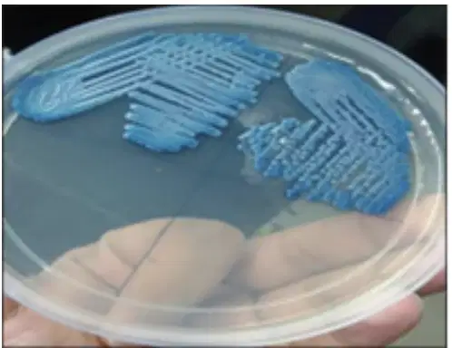
A baby was diagnosed with ventriculitis. Cerebro Spinal Fluid was taken for microbiology
examination, and the culture result on was performed on figure (A). The Gram stain was showed on figure (B). What is the best media for this organism!
LJ media
TSIA
ChromAgar
MSA
Blood agar
Which of the following is transport medium most frequently used?
Amies
ChromAgar
MacConkey
Nutrient agar
Columbia blood agar
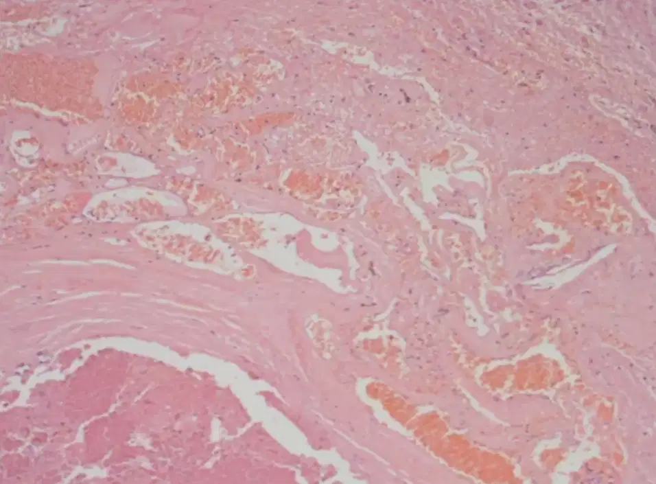
A 20-year-old woman presented with two episodes of generalized seizure. There were no
neurological deficits on admission, and CT scan revealed a 5 cm sized mixed density lesion with multifocal calcification in the left frontal and basal ganglia region. Based on the result of biopsy presented below, what is her diagnosis?
Venous angiomas
Dural arteriovenous fistulas
Capillary telangiectasias
Arteriovenous malformations
Cavernoma
Circulus arteriosus cerebri is composed of ……. arteries. Name all the arteries!
(a)

What is the name of this cell ?
Satellite cell
Ganglion cell
Purkinje cell

A 32-year-old man presented with complaints of vertigo and tinnitus for the last month. Magnetic resonance imaging revealed a large mass lesion in the right frontal region with an extra-axial localization. Based on the result of biopsy presented below, what is his diagnosis?
Intracranial Hodgkin lymphoma
Meningioma
Gliosarcoma
Plasmacytoma
Intracranial hemangiopericytoma
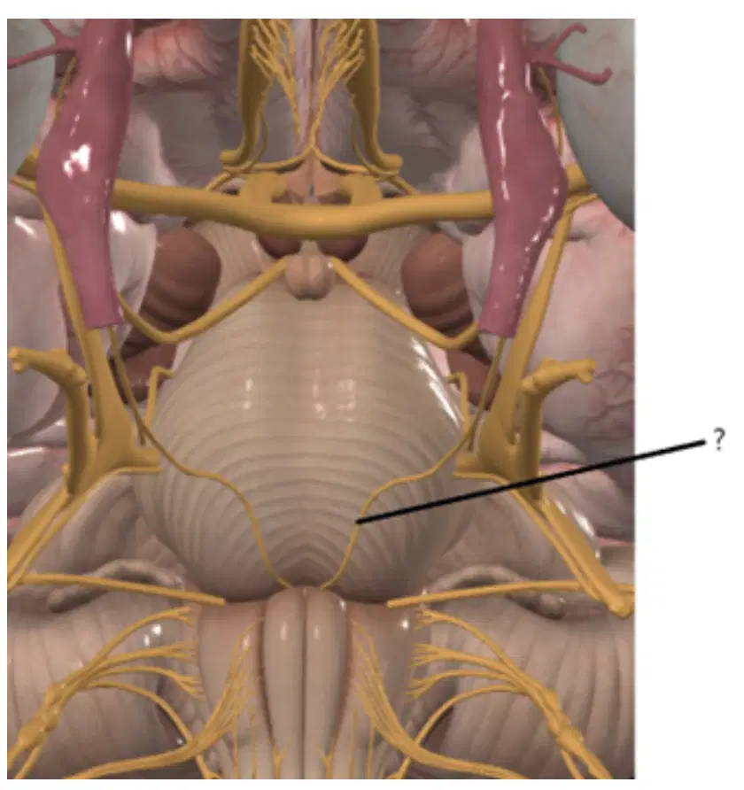
What is the name of this marked (?) nerve ?
(a)

Culture on nutrient slant agar (18 h at 25°C) shown production of a green fluorescent pigment (siderophore)
Klebsiella pneumoniae
Serratia marcescens
Escherichia coli
Candida albicans
Pseudomonas aeruginosa
LCS in GBS sufferers (guillain barre syndrome)
There is a decrease in protein levels accompanied by an increase in cell number
There is an increase in protein levels without an increase in cell number
There is a decrease in protein levels accompanied by an increase in cell number
There is a decrease in protein levels accompanied by a decrease in cell number
protein accompanied by an increase in cell number
The dural infoldings divide the cranial cavity into compartments. How many folds of the
duramater are there?
(a)

A 9-year-old girl was admitted to the hospital with a 15-day-long hemiparesis and disorientation. CT scan showed a deep-seated expansive lesion on the right brain. Based on the result of biopsy presented below, what is his diagnosis?
Cerebral abscess
Astrocytoma
Glioblastoma
Toxoplasmosis
Oligodendroglioma

A 34 years-old woman presented with frontal headache with progression of 7 months and
diplopia that appeared in the last month. Clinical evaluation revealed left abducens nerve palsy.
Coronal CT scan showed a large sphenoid sinus heterogeneous mass with destruction of the
ethmoid sinus and sella turcica. Based on the result of biopsy presented below, what is her
diagnosis?
Craniopharyngioma
Hemangioblastoma
Pituitary adenoma
Meningioma
Oligodendroglioma
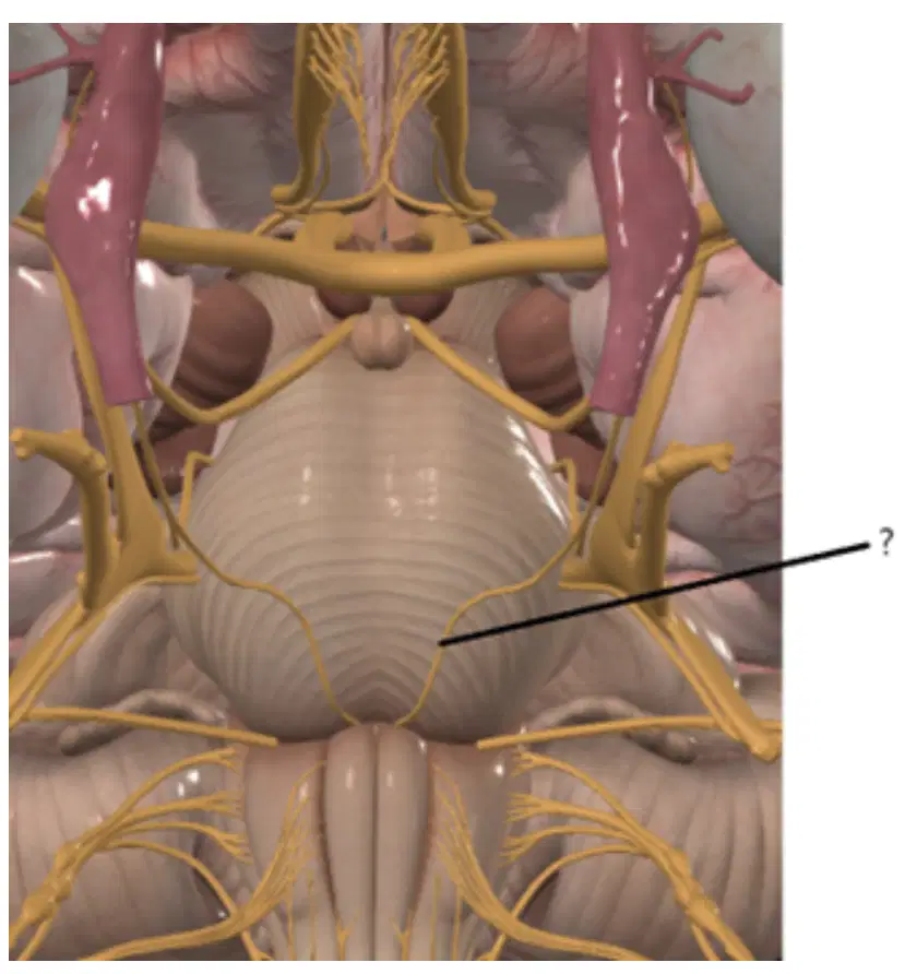
This nerve(?) leaves basis cranii through a hole. Name this hole !
(a)
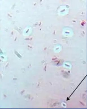
According to the capsule staining, which of the following organism has the white glycocalyx surrounding purple cell (arrow).
Staphylococcus epidermidis
Mycobacterium tuberculosis
Candida albicans
Klebsiella pneumoniae
Pseudomonas aeruginosa
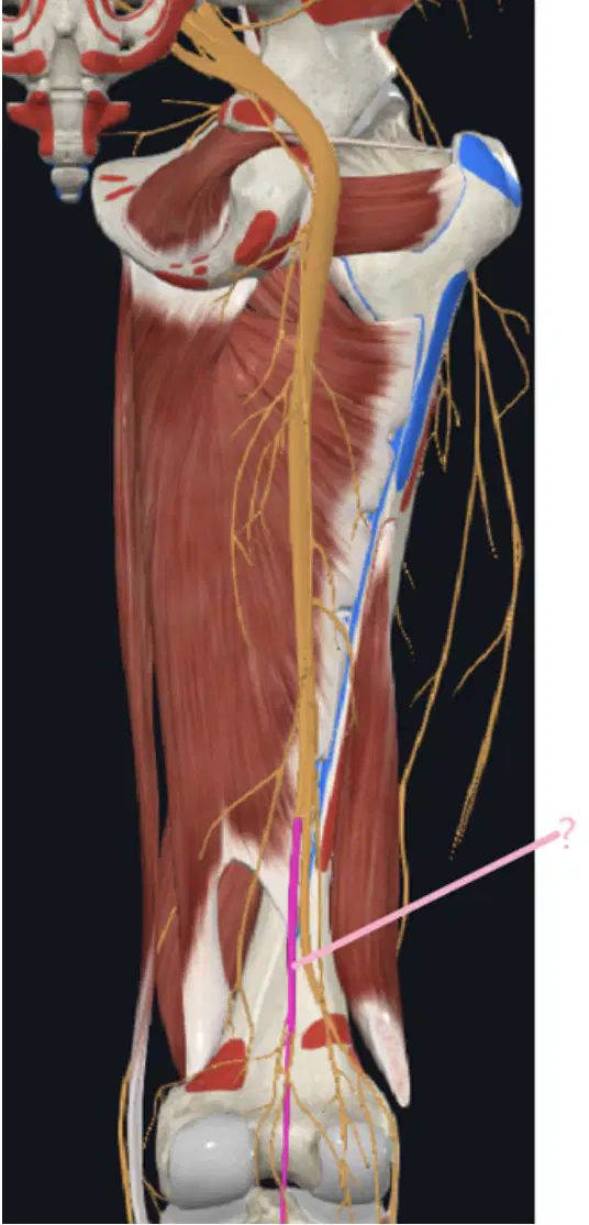
What is the name of the nerve colored in pink(?) ?
(a)

A 32-year-old man experienced an episode of concurrent bilateral tinnitus and left hemi-body
paraesthesia involving the face, arm, and leg. An MRI revealed a non-enhancing, T1-hyperintense
lesion within in Heschl’s Gyrus of the right cerebral hemisphere, extending into the insular cortex.
Based on the result of biopsy presented below, what is his diagnosis?
Multiple sclerosis
Toxoplasmosis
Diffuse astrocytoma
Glioblastoma
Encephalitis
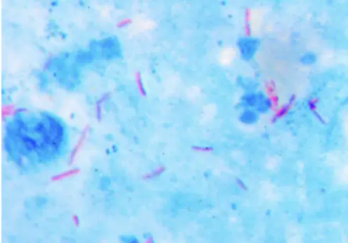
The result of bacteria staining showed pink bacilli against blue background of the cells. State the name of this staining.
ZN
Albert
Gram
Gins Bury
India Ink
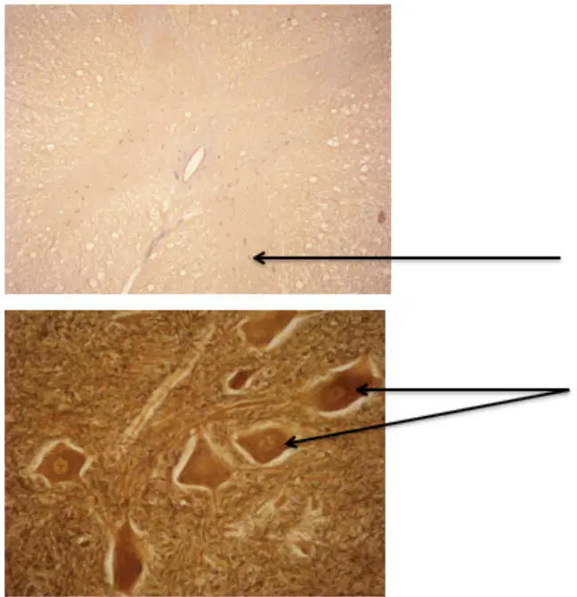
What is the name of this cell ?
Sensory neuron cells
Connecting neuron cells
Motor neuron cells

What is the function of this cell ?
Sensory neuron cells
Connecting neuron cells
Motor neuron cells
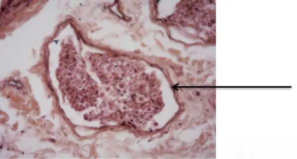
Where is the location of this tissue ?
Central nervous system
Autonomic nervous system
Peripheral nervous system
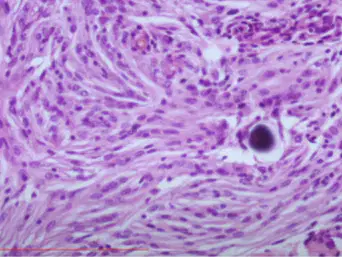
A 45-year-old woman has had unilateral headaches on the right for the past 5 months. Physical examination yields no remarkable findings. The mass is surgically removed. What is the most likely diagnosis?
Astrocytoma
Ependymoma
Metastasis
Meningioma
Tuberculoma
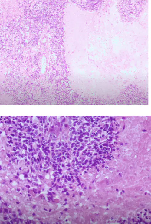
45-year man addressed complaint for his unilateral headaches. The head CT scan showed a 2-cm mass, well circumscribed, in his temporal cerebral hemispheres. After been removed by neurosurgeon, the mass
was sent to a pathologic laboratory.
From the microscopic appearance here, which of the following will best describe your diagnosis?
Oligodendroglioma
Meningioma
Astrocytoma
Glioblastoma
Ependymoma
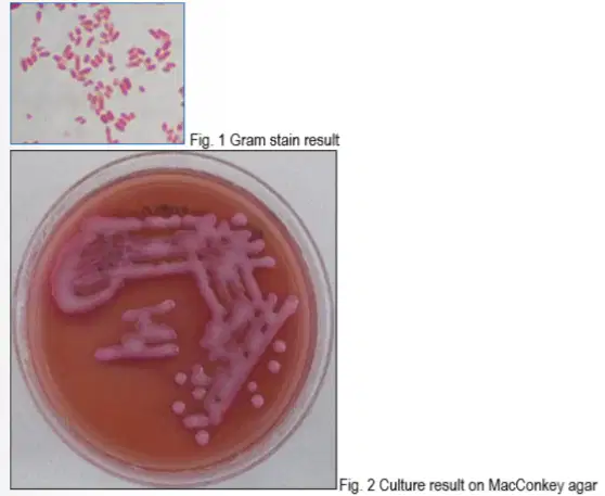
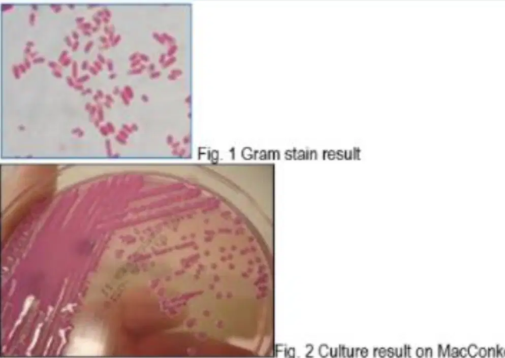
A young girl was diagnosed with ventriculitis after a VP shunt surgery. LCS specimen was taken for
microbiology examination. The result are performed on fig. 1, and 2
QUESTION:
According to the microbiology examination, what is the causative microorganism?
Cryptococcus neoformans
Klebsiella pneumoniae
Salmonella typhi
Pseudomonas aeruginosa
Escherichia coli
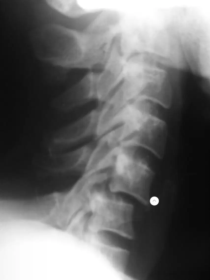
Patients with a history of head trauma and weakness in the limbs. In the photo to the side, you can find an abnormality in the form of?
Scoliosis
Cervical Fracture
Cervical Subluxation
Grade II spondylolisthesis
None of the above
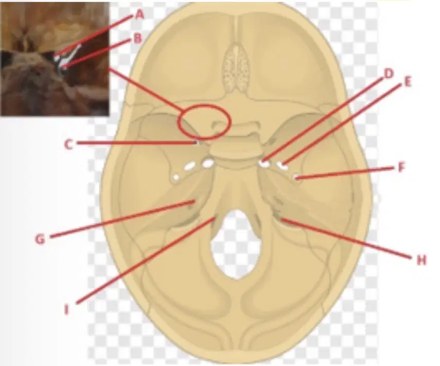
The nerve, which is medial to olive of the medulla oblongata, exit from the basis cranii through:
Location F
Location C
Location B
Location |
Location E
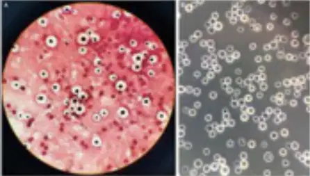
A young boy was diagnosed with ventriculitis after a VP shunt surgery. LCS specimen was taken for direct
stain. The result is performed on fig. 1.
Please state the name of this direct stain.
Periodic-Acid Schiff
20% Potassium Hydroxide
India Ink
Gram
Grocott's Methenamine Silver
The anatomical structure that forms the basis of the ventriculus quartus is
Brachium conjunctivum
Pedunculus cerebrii
Vellum medullare superius
Fossa rhomboidea
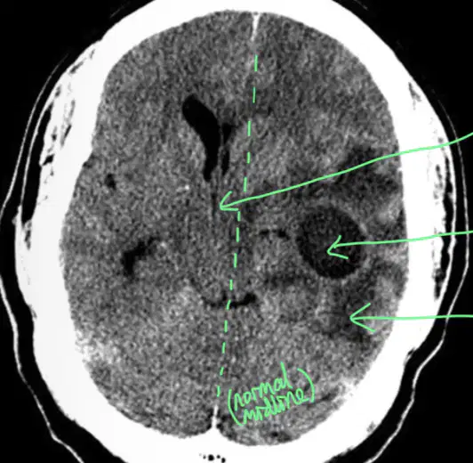
A 40-year-old woman with complaints of chronic headaches. A CT scan of the head with contrast was carried out, as shown in the image below. What picture do you see?
Vasogenic edema with abscess and midline shift to the right
Cytotoxic edema with midline shift to the right
Cerebral Infarction
Intracranial Hemorrhage
Not one of the above
Hasil pemeriksaan LCS yang mendukung diagnosis meningitis tuberculosa adalah:
Sel di dominasi oleh PMN dan purulent, prot — (neg)
Sel di dominasi oleh PMN dan purulent, prot +3
Sel di dominasi oleh PMN dan opalecent, prot — (neg)
Sel di dominasi lymphocyte dan purulent, prot + 2
Sel di dominasi lymphocyte dan opalecent, prot + 2
LCS pada penderita gbs (guillain barre syndrome):
Terdapat peningkatan kadar protein tanpa peningkatan jumlah sel
Terdapat peningkatan kadar protein disertai peningkatan jumlah sel
Terdapat penurunan kadar protein disertai penurunan jumlah sel
Terdapat penurunan kadar protein disertai peningkatan jumlah sel
Terdapat penurunan kadar protein disertai peningkatan jumlah sel
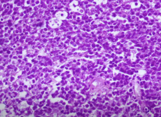
Having been experiencing headaches for three weeks now, a 58-year age boy then has sudden onset of
vomiting and comatous. The head CT scan showed the presence of a 3-cm mass in the region left frontal.
Cytology to CSF that produced after a lumbal puncture apparently showed no malignant cells. An operation
on his mass was then performed.
The best diagnostic is
Malignant lymphomas
Adenoma hipofisis
Meningioma
Astrocytoma
Glioblastoma multiforme
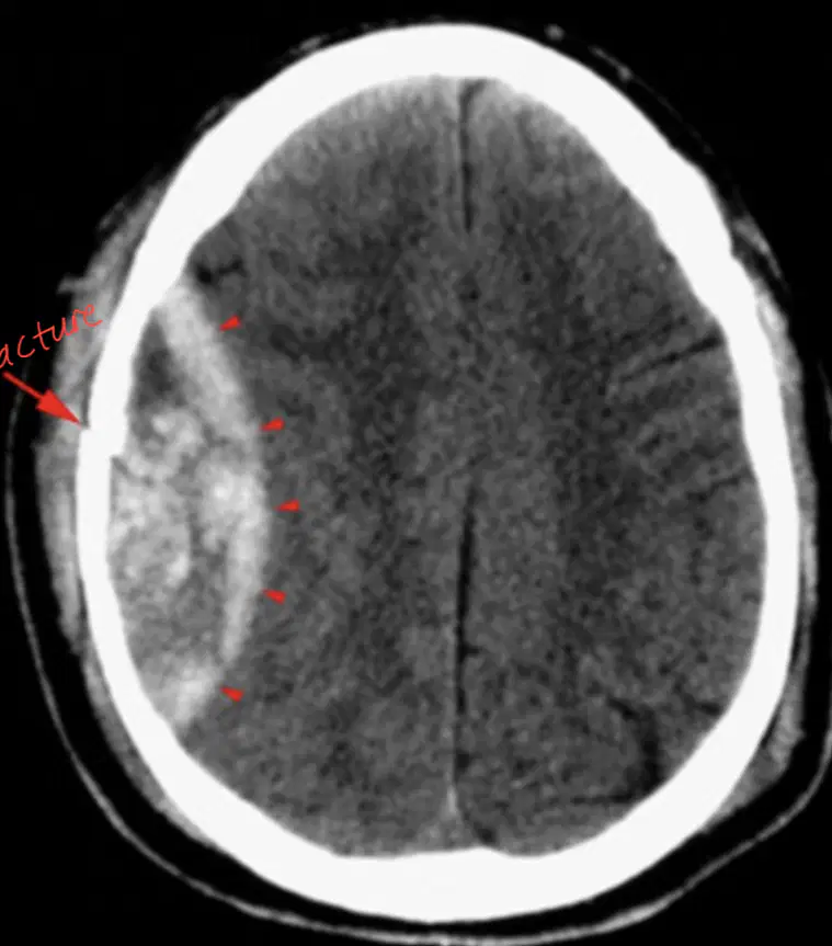
A boy presented with a history of Capitis trauma.
A CT scan was carried out. What do you see?
Old subdural hematoma
Acute epidural hematoma
Subarachnoid hemorrhage
Intracerebral Hematoma
Not one of the above
Nervus, that arises proximal to olive and pyramides and caudal to fibrae pontis trasnversae:
N. trochlearis
N. mandibularis
N. facialis
N.abducens
N. trigeminus
Carpal Tunnel syndrome is a neurological disorder that occurs when nerve “X” becomes pressed or squezzed.
This nerve “X” originates from:
Fasciculus medialis
Fasciculus lateralis and fasciculus medialis
Fasciculus lateralis
Fasciculus posterior
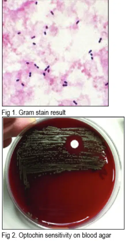
A boy was diagnosed with meningitis. LCS specimen was taken for microbiology examination. The results are
performed on fig. 1 and 2.
QUESTION:
According to the microbiology examination, what is the causative microorganism?
Streptococcus mitis
Escherichia coli
Candida albicans
Streptococcus pneumoniae
Methicillin Resistant Staphylococcus aureus
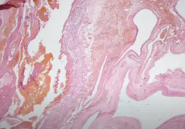
A-27 year old female complained of headaches and decreased mental function for several months and later she has experienced onset of generalized seizures and this been persisted for several weeks. A Head CT scan showed a 2 cm mass in the right cerebral hemisphere. The Sterotactic biopsy of this lesion shown in this microscopic.
Based upon your microscopic examination, which of following that most likely did happened?
Hemangioma Cavernosum
Arteriovenous malformation
Ruptured aneurysm
Gliosis
Abscess Cerebri
Liquour CebroSpinalis yang normal adalah:
Gross appearance: clear and colorless
Culture: steril
Gram stain: positive
Gross appearance: clear and white
Culture: positive
Gram stain: positive
Gross appearance: clear and colorless
Culture: steril
Gram stain: negative
Gross appearance: clear and white
Culture: positive
Gram stain: negative
Gross appearance: clear and white
Culture: steril
Gram stain: negative
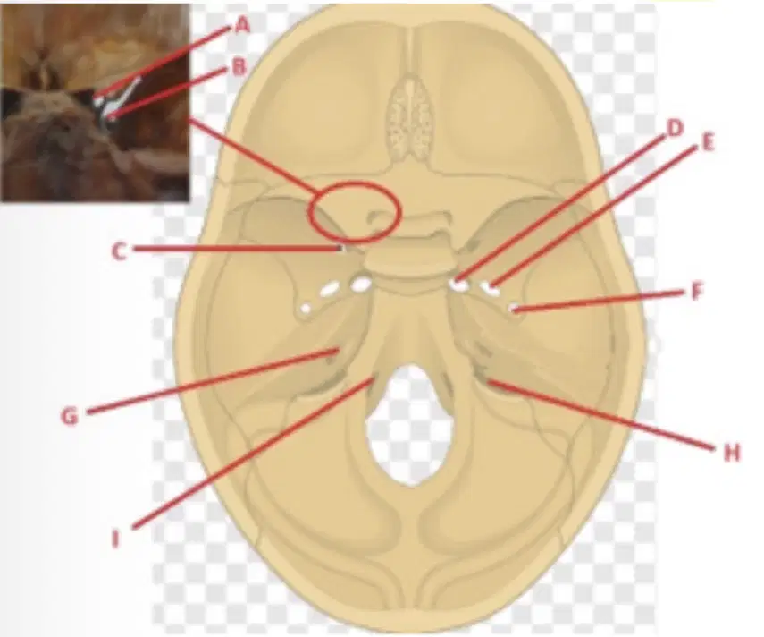
Nerve that arises on each caudal from colliculus caudalis midbrain, exit from the basis cranii through:
Location D
Location B
Location E
Location H
Location A
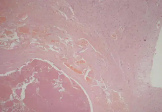
A 65-year old male was headaches, dizziness, dysarthria, double vision, balance problems and tremor. While
being examined in ER, the MRI examination showed a cluster of abnormal blood vessels in brain. From the
microscopic examination here, which of the following that most likely did happened?
Arteriovenous malformation
Intra-cerebral hematoma
Subdural hematoma
Ruptured saccular aneurysm
Cavernous Hemangioma
A. vertebralis runs through foramina transversaria of the ......... CO veces
Vertebrae thoracal | to vertebrae cervical IV
Vertebrae cervical VI to vertebrae cervical |
Vertebrae cervical VII to vertebrae cervical II
Vertebrae cervical VI to vertebrae cervical III
Vertebrae cervical | to vertebrae cervical VII
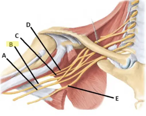
Nervus that runs posteriorly and runs in a sulcus in the posterior part of the humerus bone is:
NerveA
NerveB
NerveD
NerveE
Nerve C
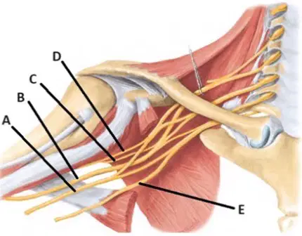
N. cutaneus antebrachii lateralis is a continuation of............... :
Nerve C
Nerve E
Nerve D
Nerve A
Nerve B
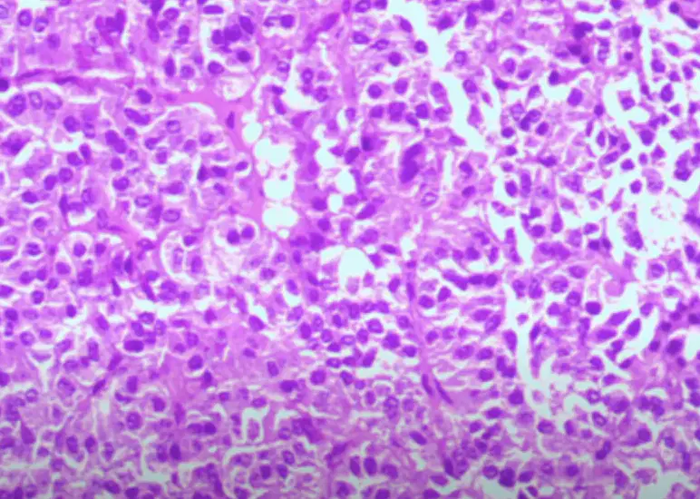
A 48-year-old, previously healthy woman presented to her gynecologist with a complaint of headaches,
amenorrhea, and galactorrhea for approximately 1 year. Physical examination was unremarkable, and she
had no neurological deficits. Pertinent laboratory values included an elevated prolactin level of 52.5 ng/mL
(normal value for a postmenopausal female reported as 1.8-20.3 ng/mL). Whole-brain magnetic resonance
imaging showed a 1.5-cm mass within the pituitary fossa that diffusely enhanced following administration of
gadolinium contrast material. Which of the following neoplasm lesions is he most likely to have?
Pituitary adenoma
Gliosis
Metastatic carcinoma
Astrocytoma
Ependymoma
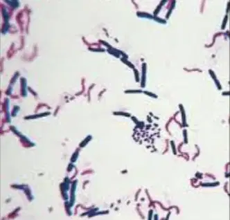
Specify your interpretation of bacteria in the figure 1 according the result of Gram-stain!
Gram-negative fusiform rods, budding spores, and Gram-positive bacillus
Gram-positive cocci in cluster, Gram-negative bacilli, and acid-fast bacilli
Gram-positive spore-forming, Gram-negative bacilli, and Gram-positive cocci in chain
Gram-positive cocci in pairs, budding spores, and Gram-negative bacilli
Gram-positive rod, Gram-positive cocci in pairs, and Gram-negative comma
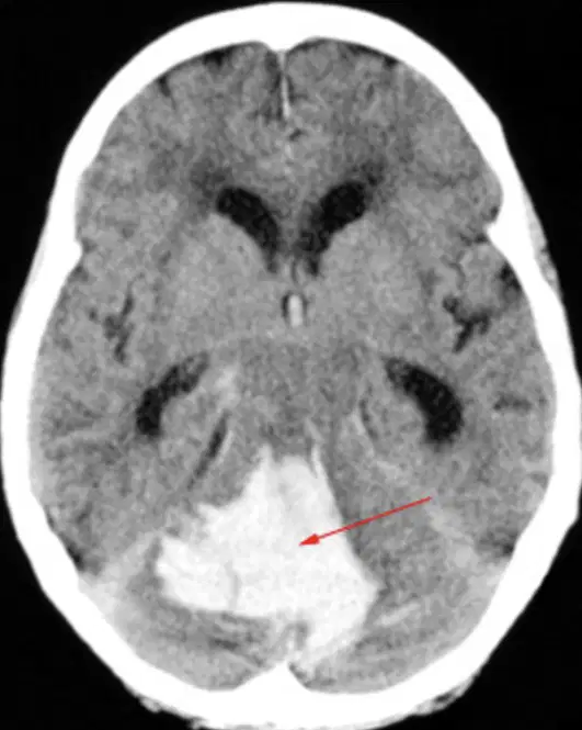
A man came with decreased unconsciousness and a history of previous complaints of severe headaches. On a CT scan, results were obtained, as shown in the image below. What do you see?
Acute infarction in the cerebellum accompanied by non-communicating hydrocephalus
Cerebellar tumor accompanied by non-communicating hydrocephalus
Bleeding in the cerebellum accompanied by non-communicating hydrocephalus
Cerebellar Meningioma
None of the above
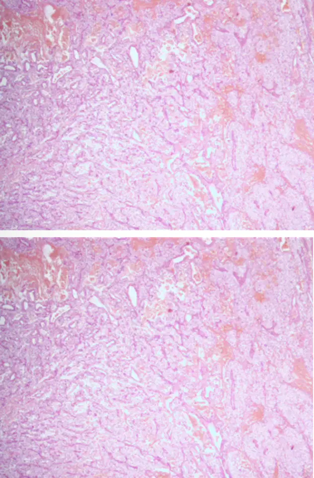
A 40-year old female has a headache, loss of coordination, imbalance, nausea and vomiting symptoms. The MRI showed a cystic tumor in cerebellum. The best diagnostic of her disease based on microscopic here is
Ependymoma
Glioblastoma
Haemangioblastoma
Neurofibroma
Schwannoma
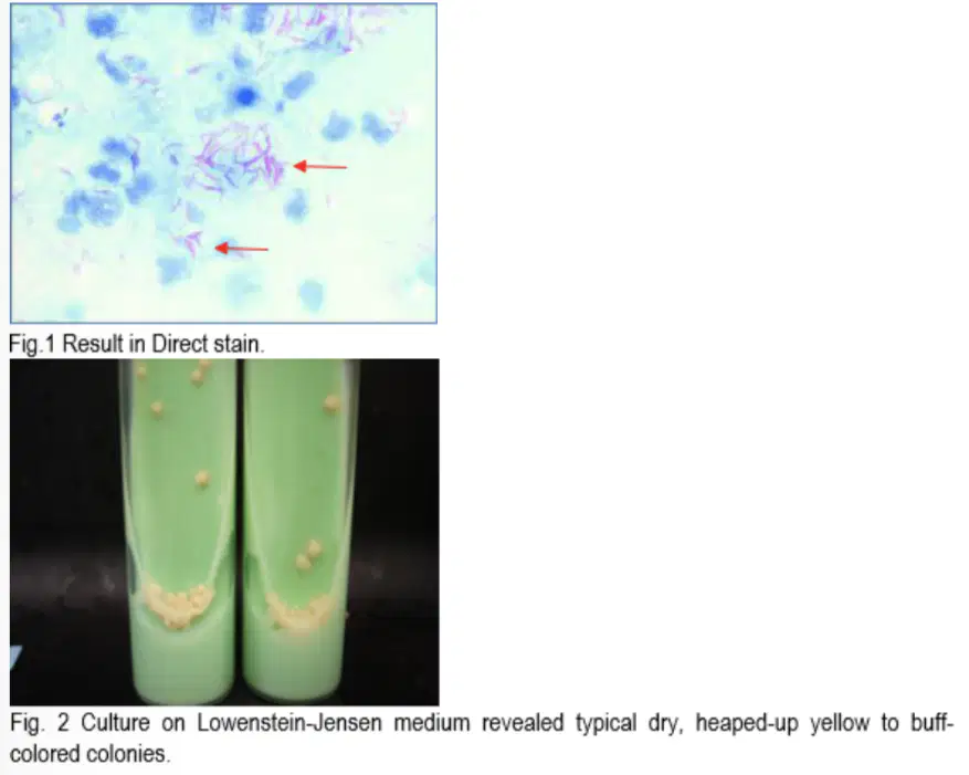
A young girl was diagnosed with ventriculitis after a VP shunt surgery. LCS specimen was taken for
microbiology examination. The result are performed on fig. 1 and 2
QUESTION:
According to the microbiology examination, what is the causative microorganism?
Cryptococcus neoformans
Candida albicans
Aspergillus flavus
Streptococcus pyogenes
Mycobacterium tuberculosis
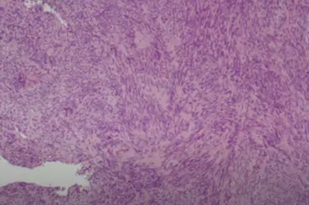
A 49-year-old woman with a long history of a subcutaneous mass on the dorsal side of her 4th finger of the right hand visited a dermatologist because of slight enlargement of the mass. Physical examination revealed a small, round, firm subcutaneous mass on the dorsal side of her proximal interphalangeal joint of the right 4" finger. The mass was immobile and nontender and its overlying skin was intact. An excisional biopsy was done for the patient and the specimen was sent for pathologic evaluation. What is the most likely diagnosis?
Schwannoma
Elastofibtoma
Fibroma
Fibrosarcoma
Neurofibroma
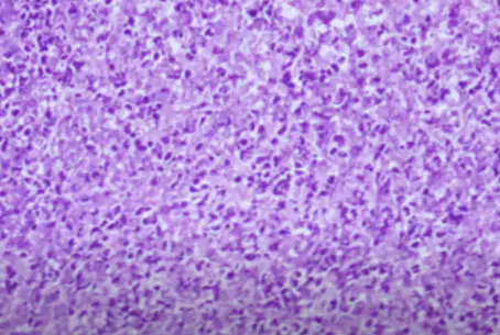
A healthy 20-year-age male has a mild fever for a few days and then followed by a sudden onset of severe headache. The Head CT Scan reveals a cystic mass, irregular borders in left temporal lobe. An operation was then perfomed and the microscopic is as shown here.
Your best diagnostic is
Meningioma
Oligodendroglioma
Brain abscess
Astrocytoma
Ependymoma
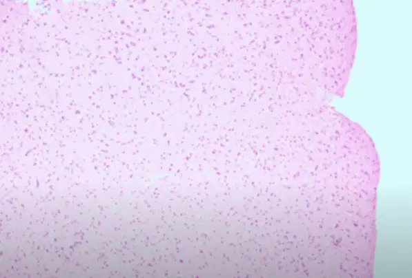
A 65-year age of healthy woman was driving car when she got sudden onset of severe headache. She pulled on into a gas station, stop the car but while trying to exit then slumped over the wheel. When taken to ER, the MRI show a mass in right parietal region. The microscopic appearance shown here.
The best diagnostic is:
Schwannoma
Diffuse astrocytoma
Subdural hematoma
Oligodendroglioma
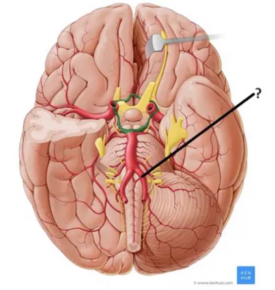
The source of this artery (black line):
A.subclavia
A. axillaris
A.carotis communis
A. thyroidea inferior
Pada pemeriksaan LCS bed side maka:
Pemeriksaan Nonne untuk mengetahui adanya peningkatan kadar Globulin secara kwanlitative
Pemeriksaan Pandi untuk mengetahui adanya peningkatan kadar Globulin secara kwalitative
Pemeriksaan Pandi untuk mengetahui adanya peningkatan kadar protein secara kwanlitative
Pemeriksaan Nonne untuk mengetahui adanya peningkatan kadar Globulin secara kwalitative
Pemeriksaan Nonne untuk mengetahui adanya peningkatan kadar protein secara kwalitative
The most common cause of foot drop is …. injury.
N. saphenus
N. peroneus communis
N. Tibialis
N. Femoralis
N. Suralis
A basilaris is located at:
3
1
2
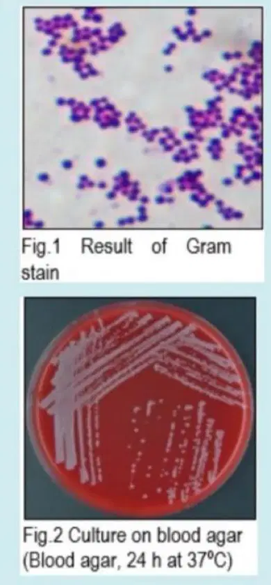
A young boy was diagnosed with ventriculitis after a VP shunt surgery. LCS specimen was taken for microbiology examination. The result are performed on fig. 1, 2, and 3 QUESTION:
According to the microbiology examination, what is the causative microorganism?
Staphylococcus epidermidis
Streptococcus pneumoniae
Staphylococcus aureus
Escherichia coli
Candida albicans

This marked anatomical structure (red line) is ?
Select one:
N.oculomotorius
N.trochlearis
N.abducens
N.trigeminus
N.facialis
PADA PEMERIKSAAN LCS BED SIDE MAKA:
Select one:
Pemeriksaan Nonne untuk mengetahui adanya peningkatan kadar Globulin secara kwalitative
Pemeriksaan Pandi untuk mengetahui adanya peningkatan kadar protein secara kwanlitative
Pemeriksaan Pandi untuk mengetahui adanya peningkatan kadar Globulin secara kwalitative
Pemeriksaan Nonne untuk mengetahui adanya peningkatan kadar Globulin secara kwanlitative
Pemeriksaan Nonne untuk mengetahui adanya peningkatan kadar protein secara kwalitative
Which examination that you choose in the probable neck trauma case?
Cervical MRI
Cervical myelography
Open mouth AP position cervical X ray
3D cervical MS CT scan
Lateral position cervical X ray
Wrist drop is a disorder of a nerve that innervating the ...... muscles of the wrist and digits
Flexor
Extensor
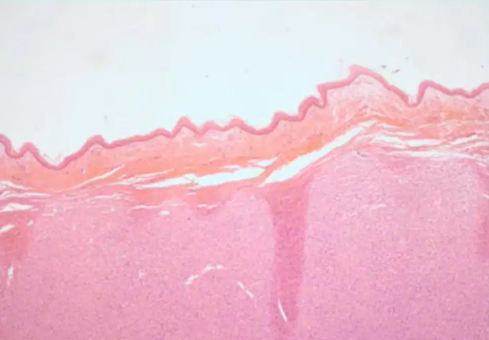
A 20-year old female has 7 scattered of 1-3 cm nodule with irregular borders hyperpigmented skin lesions on her extremities and torso. The best diagnostic of her disease based on microscopic here is,
(a)
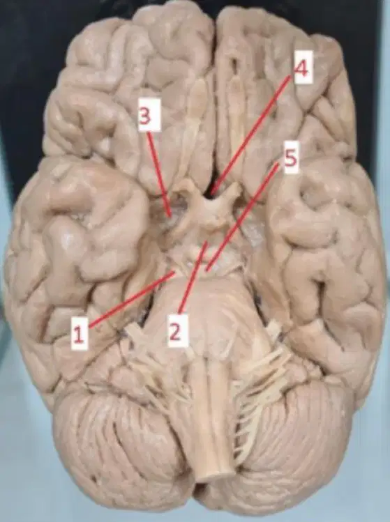
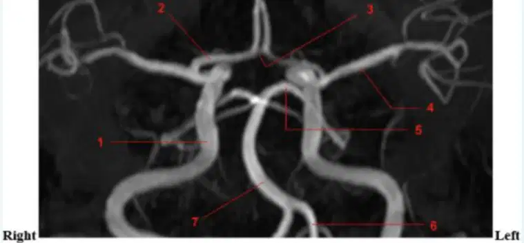
The internal carotid artery terminates and branches into the anterior cerebral artery and the posterior cerebral artery at:
1
2
3
4
5

Wrist drop is a disorder causes by ...... nerve palsy:
A
B
C
D
E
A Male, came to emergency department with blood from the left ear. Which is the best examination for this patient?
Mastoid T1 MRI
UHR mastoid MS CT Scan
Mastoid T2 MRI
Maxillo-facial MRI
Photo mastoid (Schuller)
Cranial nerve that supplies the sternocleidomastoid and trapezius muscles, leave the basis cranii on which part of basis cranii?
Fossa cranii media
Fossa cranii anterior
Fossa cranii posterior
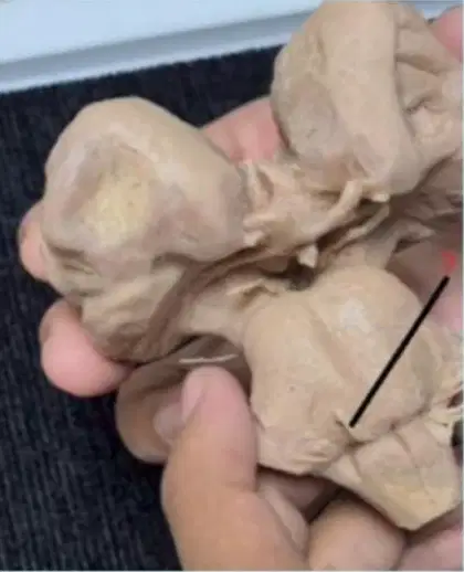
This marked anatomical structure (red line) is:
Select one:
N.trochlearis
N.oculomotorius
N.facialis
N.trigeminus
N.abducens
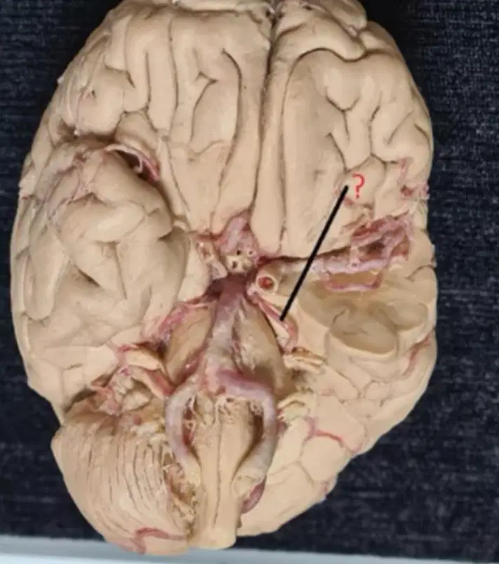
This marked artery is: Select one:
A.cerebri communicans anterior
A.cerebri anterior
A.cerebri posterior
A.cerebri communicans posterior
A.basilaris

Which is the most possible affected arteries in saccular aneurysm case?
A
B
C
D
E
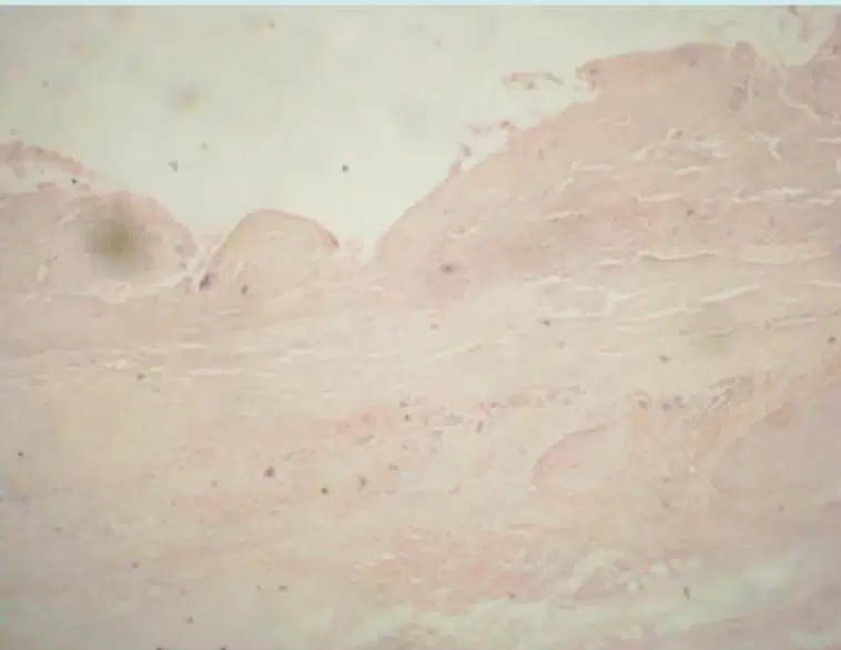
A 65-year age of healthy woman was driving car when she got sudden onset of severe headache. She pulled on into a gas station, stop the car but while trying to exit then slumped over the wheel. When taken to ER, she was remain comatose but died hours later. At autopsy, the microscopic appearance of the right middle cerebral artery is as shown here. The best diagnostic Is:
A-V malformation
Subdural hematoma
Epidural hematoma
Aneurysm
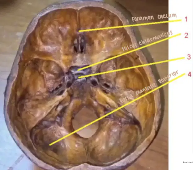
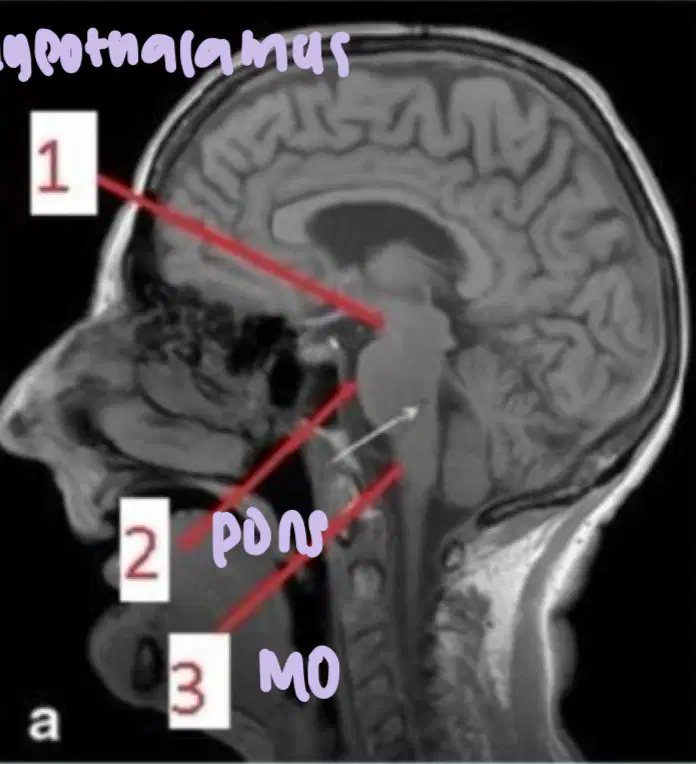
Fossa hypophysialis is located at number:
1
2
3
4
Termination of internal carotid artery is located caudally to:
Substansia perforate posterior
Corpus mammillare
Substansia perforata anterior
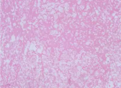
A healthy 20-year-age male has a mild fever for a few days and then
followed by a sudden onset of severe headache. The Head CT Scan reveals a cystic mass, irregular borders in left temporal lobe. An operation was then perfomed and the microscopic is as shown here.
Your best diagnostic is
Oligodendroglioma
Astrocytoma
Brain abscess
Ependymoma
Meningioma
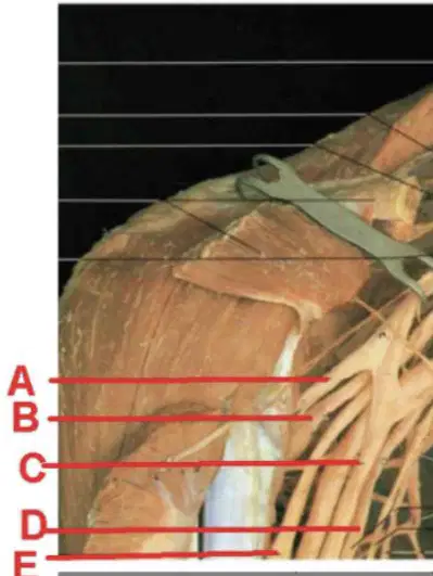
Carpal Tunnel Syndrome occurs when …………… ( Choose the correct one
from the picture) is compresed as it travels through the wrist
A
B
C
D
E
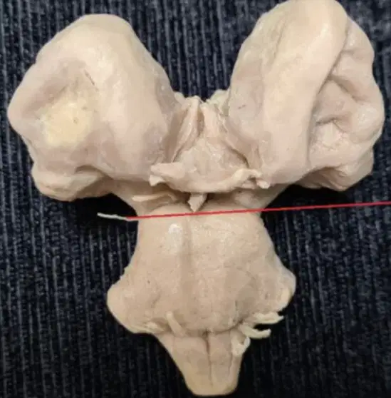
This nerve is (red line):
.N.abducens
.N.trigeminus
N. Oculomotorius
N. Trochlearis
N. Fascialis
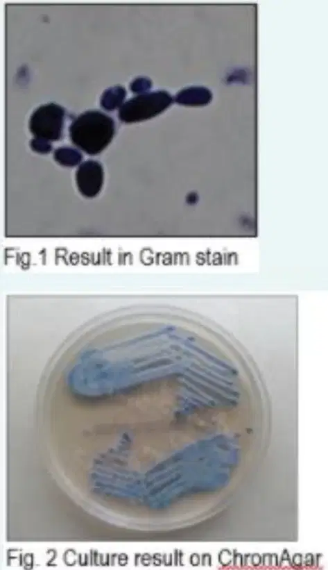
A young girl was diagnosed with ventriculitis after a VP shunt surgery. LCS specimen was taken for microbiology examination. The result are
performed on fig. 1 and 2
According to the microbiology examination, what is the causative
microorganism?
Crytococcus neoformans
Aspergillus flavus
Candida tropicalis
Mycobacterium tuberculosis
Streptococcus pyogenes
Male, 60 years old, came to the emergency department with sudden onset of left sided weakness. There were no movement in his left leg and only minimal movement in his left arm. The patient was diagnosed with ischemic stroke.
Which is the most possible occluded blood vessel for this patient? (See the
image below)
The affected blood vessel was marked with number:
1
2
3
4
5
What is the name of this cell?
Oligodendroglia cell
Microglia
Schwann cell
What is the name of this cell?
Motor neuron cells
Connecting neuron cells
Sensory neuron cells
What is the function of this cell?
Conduct impulses from spinal cord to muscle
Conduct impulses from peripheral nervous system to central nervous system
Conduct impulses from receptor to spinal cord
49-year-old woman with a long history of a subcutaneous mass on the dorsal side of her 4th finger of the right hand visited a dermatologist because of slight enlargement of the mass. Physical examination revealed a small, round, firm subcutaneous mass on the dorsal side of her proximal interphalangeal joint of the right 4th finger. The mass was immobile and nontender and its overlying skin was intact. An excisional biopsy was done for the patient and the specimen was sent for pathologic evaluation. What is the most likely diagnosis?
Neurofibroma
Elastofibtoma
Schwannoma
Fibrosarcoma
Fibroma
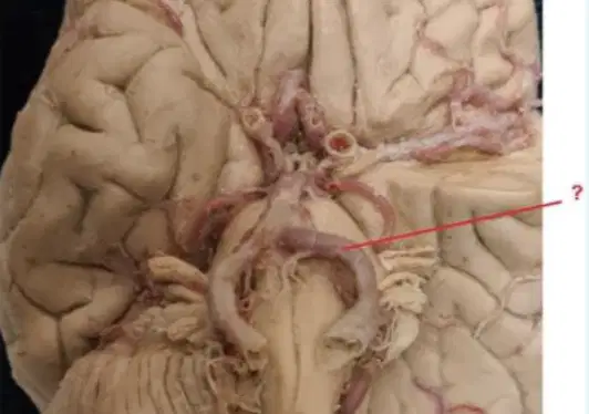
This anatomical structure is (red line):
A. communicans posterior
A. carotis interna
A. vertebralis
A. basilaris
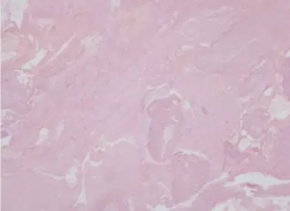
A-27 year old female complained of headaches and decreased mental function for several months and later she has experienced onset of generalized seizures and this been persisted for several weeks. A Head CTscan showed a 2 cm mass in the right cerebral hemisphere. The Sterotactic biopsy of this lesion shown in this microscopic
Based upon your microscopic examination, which of following that most
likely did happened?
Abscess Cerebri
Hemangioma Cavernosum
Ruptured aneurysm
Arteriovenous malformation
Gliosis
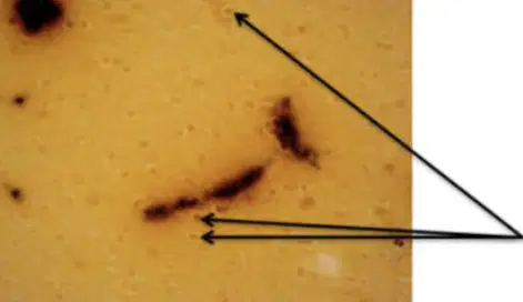
What is the function of this cell?
Supporting cell
Forming myeling sheath
Immune system in central nervous system
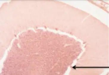
What is the name of this cell?
Purkinje cell
Satellite cell
Ganglion cell
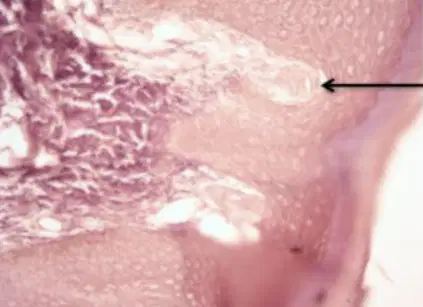
What is the function of this structure?
Gross pressure/vibration receptor
Light touch receptor
Pain receptor
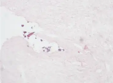
A 65-year old male was fallen from a stair but not losing consciousness.
While being examined in ER, about 4-hoursafter the incident he was then
unconscious state. The examination observed a scalp contusion on the left
temporal. When taken to surgery, the exploration found a blood clot.
From the microscopic examination here, which of the following that most
likely did happened?
Subdura hematoma
Arteriovvenous malformation
Intra-cerebral hematoma
Epidural hematoma
Ruptured saccular aneurysm
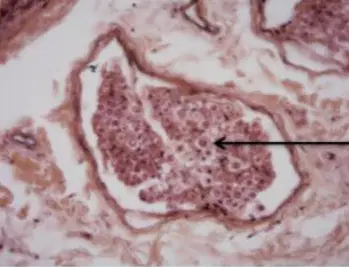
Where is the location of this tissue?
Peripheral nervous system
Autonomic nervous system
Central nervous system
A young girl was diagnosed with ventriculitis after an VP shunt surgery.
LCS specimen was taken for microbiology examination. The result are
performed on fig. 1, and 2QUESTION:
According to the microbiology examination, what is the causative
microorganism?
.Cryptococcus neoformans
.Pseudomonas aeruginosa
.Escherichia coli
Salmonella typhi
Klebsiella pneumoniae
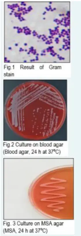
A young boy was diagnosed with ventriculitis after a VP shunt surgery. LCS
specimen was taken for microbiology examination. The result are
performed on fig. 1, 2, and 3
QUESTION:
According to the microbiology examination, what is the causative
microorganism?
.Candida albicans
Streptococcus pneumoniae
.Escherichia coli
Staphylococcus epidermidis
.Methicillin Resistant Staphylocococcus aureus
A 45-year-old woman has had unilateral headaches on the right for the
past 5 months. Physical examination yields no remarkable findings. The
mass is surgically removed. What is the most likely diagnosis
Meningioma
Astrocytoma
Metastasis
Ependymoma
Tuberculoma

A 45-year man addressed complaint for his unilateral headaches. The
head CT scan showed a 2-cm mass, well circumscribed, in his temporal
cerebral hemispheres. After been removed by neurosurgeon, the mass
was sent to a pathologic laboratory.
From the microscopic appearance here, which of the following will best
describe your diagnosis?
Astrocytoma
Meningioma
Oligodendroglioma
Glioblastoma
Ependymoma
Wrist drop is a disorder caused by ……………… (Choose the correct one from
the picture) palsy.
A
B
C
D
E
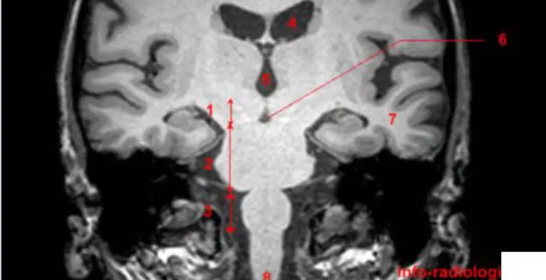
Female, 58 years old, with prior history of diabetes mellitus, suddenly had
complaint of double vision. The physician reported there were infarct areain left abducens nerve nuclei that caused inability of the left eye to move
laterally. Where the infarct lesion is possibly located?
1
2
3
7
8
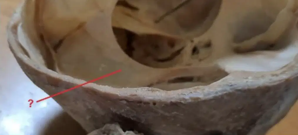
This anatomical structure is:
Falx Cerebelli
Tentorium Cerebelli
Falx Cerebri
Diaphragma sellae

What is the name of this tissue?
Perineurium
Epineurium
Endoneurium

Male, 62 years old, presented with sudden onset of speech disturbance
and right sided weakness. He suddenly felt that he could not understand
the conversation, but his wife noticed he still be able to form complete
words without context. Which structure of the brain that might
correspond as the causes of the speech disturbance?
A
B
C
D
E
A 20-year old female has 7 scattered of 1-3 cm nodule with irregular borders hyperpigmented skin lesions on her extremities and torso. The best diagnostic of her disease based on microscopic here is,
Depth Fibromatosis
Neurofibroma
Schwannoma
Dermatofirbromatosis
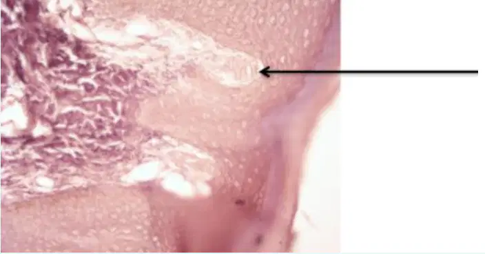
What is the name of this structure?
Meissner corpuscle
Pacini corpuscle
Merkel cospuscle
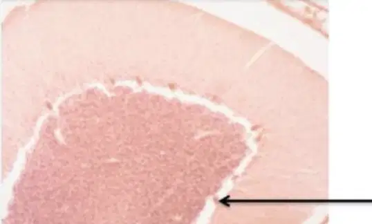
Which layer is this cell are located?
Molecular layer
Ganglionar layer
Granular layer
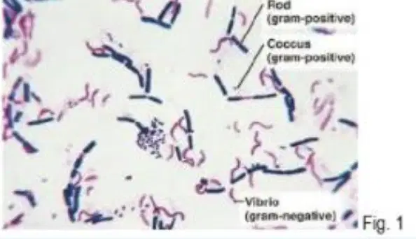
Specify your interpretation of bacteria in the figure 1 according the result of
Gram-stain!
Gram positive rod, Gram positive cocci in pairs, and Gram negative
comma
Gram positive cocci in cluster, Gram negative bacilli, and acid fast
bacilli
Gram positive spore-forming, Gram negative bacilli, and Gram positive
cocci in chain
Gram negative fusiform rods, budding spores, and Gram positive
bacillus
Gram positive cocci in pairs, budding spores, and Gram negative
bacilli
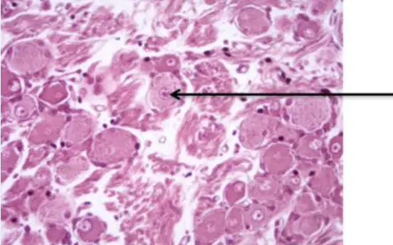
What is the name of this cell?
Satellite cell
Ganglion cell
Purkinje cell
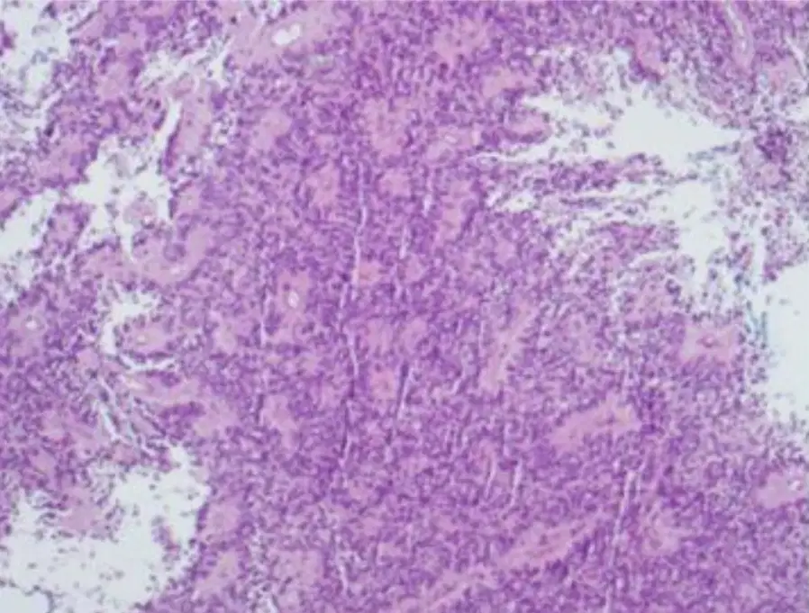
Having been experiencing headaches for three weeks now, a 8-year age
boy then has sudden onset of vomiting and comatous. The head CT scan
showed the presence of a 3-cm mass in the region left ventricle
lateralis. Cytology to CSF that produced after a lumbal puncture
apparently showed no malignant cells. An operation on his mass was then
performed.
The best diagnostic is
Ependymoma
Glioblastoma multiforme
Adenoma hipofisis
Astrocytoma
Meningioma

