
 Worksheets
Functions of Digestion
Total questions: 128
Worksheet time: 1hrs 5mins
Fill in the blank: ________ of the food is the process of taking food into the body.
Ingestion
Digestion
Absorption
Assimilation
Fill in the blank: ________ the food is the process of moving food through the digestive system.
Transport
Digesting
Absorbing
Chewing
Fill in the blank: ________ of the food into smaller usable components is an important function of digestion.
Digestion
Absorption
Excretion
Respiration
Fill in the blank: ________ of the necessary nutrients into the bloodstream is a function of digestion.
Absorption
Excretion
Filtration
Evaporation
Fill in the blank: ________ of the waste products from the body is the final function of digestion.
Elimination
Absorption
Secretion
Ingestion
Which of the following are the two separate categories of organs in the digestive system?
Digestive organs and respiratory organs
Digestive organs and accessory digestive organs
Accessory digestive organs and circulatory organs
Respiratory organs and alimentary canal
Digestive organs collectively make up the _________?
gastrointestinal (GI) tract
respiratory system
nervous system
urinary tract
The gastrointestinal (GI) tract is also called the ________ or ________.
digestive tract, alimentary canal
respiratory tract, bronchial tube
urinary tract, nephron canal
circulatory tract, vascular tube
Which of the following is NOT a GI tract organ?
A) Oral cavity
B) Pharynx
C) Esophagus
D) Kidney
Fill in the blank: The GI tract organ that comes after the stomach is the _________.
small intestine
large intestine
esophagus
rectum
Fill in the blank: The GI tract is a continuous tube about ________ feet (9-10 meters) long.
30
15
50
100
The GI tract runs from the mouth to the anus.
True
False
What type of muscle is found in the wall of the GI tract?
Skeletal muscle
Cardiac muscle
Smooth muscle
Striated muscle
Fill in the blank: Smooth muscle in the wall of the GI tract is responsible for ________.
motility
digestion of proteins
hormone secretion
absorption of nutrients
Smooth muscle in the GI tract wall pushes materials from one end to the other.
True
False
Which of the following is NOT a characteristic of accessory digestive organs?
A) Do not form the GI tube
B) Can develop as outgrowths
C) Are connected to the GI tract (some by ducts)
D) Form the GI tube
Accessory digestive organs assist the GI tract in the digestion of _______.
food
water
air
minerals
Which of the following is an accessory digestive organ?
Heart
Teeth
Kidney
Lungs
Accessory digestive organs include which of the following? (Select all that apply)
Tongue
Salivary glands
Liver
Pancreas
Fill in the blank: The _______ is an accessory digestive organ that stores bile produced by the liver.
Gallbladder
Pancreas
Stomach
Small intestine
True or False: Accessory digestive organs form the GI tube.
True
False

What is the name of the gland labeled as the largest salivary gland in the accessory digestive organs?
Parotid salivary gland
Sublingual salivary gland
Submandibular salivary gland
Pineal gland
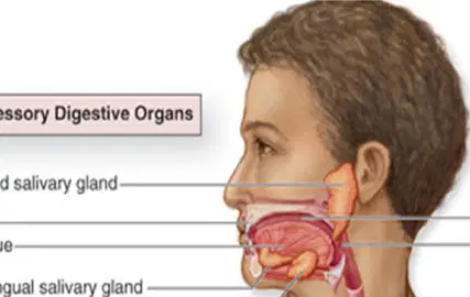
Which organ in the gastrointestinal tract is responsible for the initial breakdown of food?
Oral cavity
Esophagus
Stomach
Large intestine
Name the tube that connects the pharynx to the stomach in the digestive system.
Esophagus
Trachea
Bronchus
Duodenum

Which organ stores bile produced by the liver?
Gallbladder
Stomach
Pancreas
Small intestine
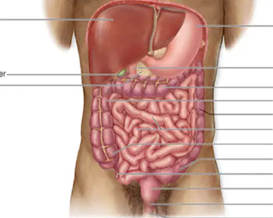
What is the function of the pancreas in the digestive system?
The pancreas produces digestive enzymes and hormones such as insulin.
The pancreas stores bile produced by the liver.
The pancreas absorbs nutrients from digested food.
The pancreas churns food to mix it with gastric juices.
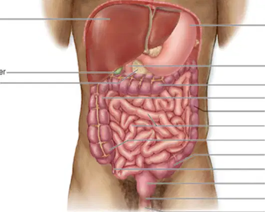
Which part of the digestive system is responsible for most nutrient absorption?
Small intestine
Stomach
Large intestine
Esophagus
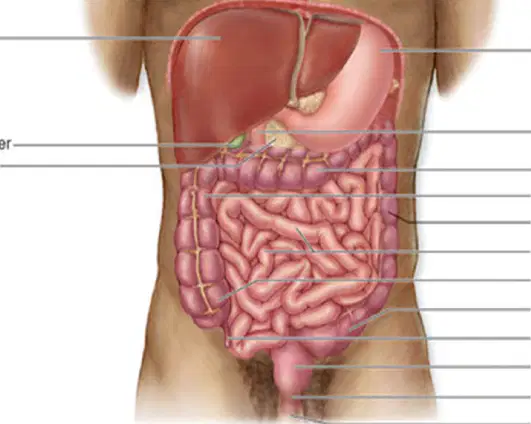
What is the final section of the gastrointestinal tract called, where waste is expelled from the body?
Anus
Esophagus
Stomach
Duodenum
Match the following digestive organs to their functions:
Stomach
Absorbs nutrients
Liver
Stores and expels feces
Small intestine
Produces bile
Rectum
Mixes and digests food
What is the main function of the salivary glands in the oral cavity?
Absorb nutrients
Produce and secrete saliva
Move food to the stomach
Filter air
What is the name of the fluid produced by the salivary glands that assists in the initial activities of digestion?
Saliva
Bile
Mucus
Gastric juice
What is the process of mechanical digestion in the oral cavity called?
Peristalsis
Mastication
Absorption
Secretion
The tongue moves food to the pharynx during digestion.
True
False
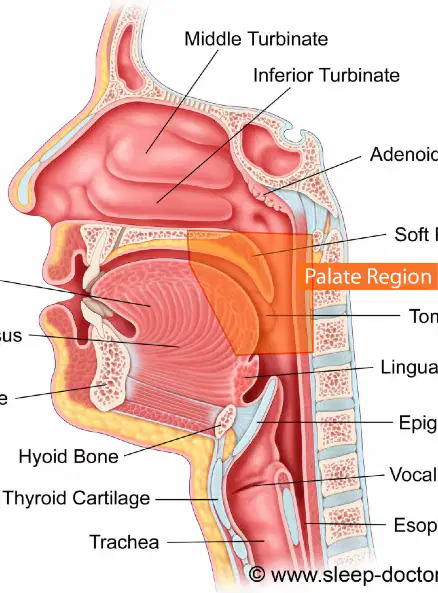
Label the following parts in the diagram of the oral cavity:
Tongue, Soft Palate, Epiglottis, Esophagus (as labeled in the diagram)
Teeth, Hard Palate, Larynx, Trachea
Tongue, Uvula, Pharynx, Stomach
Gums, Tonsils, Nasal Cavity, Duodenum
What is the hard palate?
A soft tissue at the back of the mouth
A thin horizontal bony plate of the skull, located in the roof of the mouth
The muscle that controls the tongue
The structure responsible for the gag reflex
The hard palate aids in _______ and speech.
mastication
digestion
respiration
circulation
Which of the following is NOT a characteristic of the soft palate?
Contains bone
Uvula
Gag reflex
Soft tissue on the back of the roof of the mouth
The soft palate is responsible for the gag reflex.
True
False
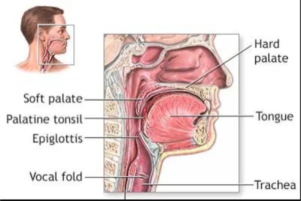
Refer to the diagram. Which structure is labeled as the 'hard palate'?
The hard palate is the bony structure located at the front part of the roof of the mouth, as labeled in the diagram.
The hard palate is the muscular structure at the back of the roof of the mouth, as labeled in the diagram.
The hard palate is the flap that covers the windpipe during swallowing, as labeled in the diagram.
The hard palate is the structure that forms the lower jaw, as labeled in the diagram.
Which systems is the pharynx common to?
A) Respiratory and digestive systems
B) Circulatory and nervous systems
C) Muscular and skeletal systems
D) Endocrine and excretory systems
What is the pharynx commonly called?
Nose
Throat
Esophagus
Larynx
Name the region of the pharynx that is posterior to the oral cavity.
Oropharynx
Nasopharynx
Laryngopharynx
Hypopharynx
Which region of the pharynx is posterior to the larynx and connects to the esophagus?
Laryngopharynx
Nasopharynx
Oropharynx
Hypopharynx
What is the function of the epiglottis?
A flap that prevents food from entering the larynx
It produces digestive enzymes
It absorbs nutrients from food
It pumps blood throughout the body
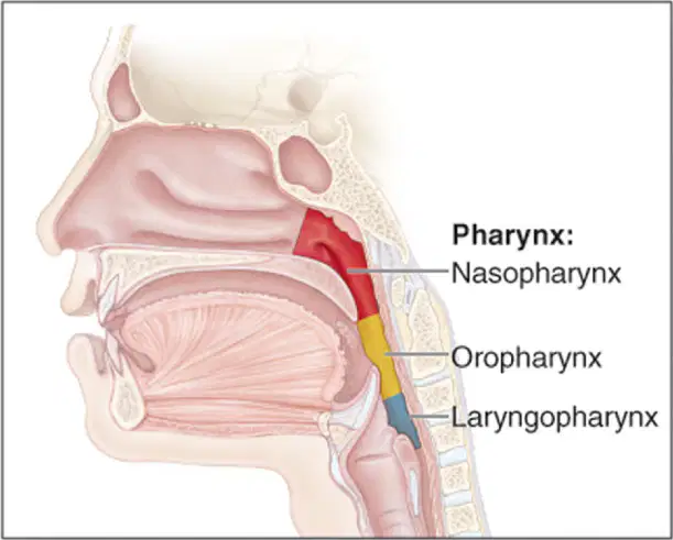
Label the regions of the pharynx in the diagram.
Nasopharynx (upper), Oropharynx (middle), Laryngopharynx (lower)
Oropharynx (upper), Nasopharynx (middle), Laryngopharynx (lower)
Laryngopharynx (upper), Oropharynx (middle), Nasopharynx (lower)
Nasopharynx (lower), Laryngopharynx (middle), Oropharynx (upper)
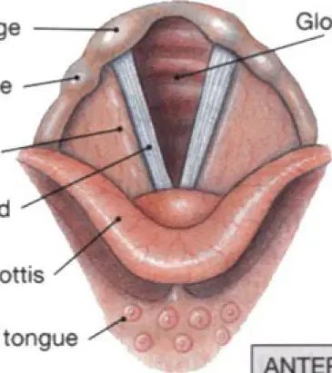
Identify the part labeled as 'Corniculate cartilage' in the diagram of the larynx.
Corniculate cartilage
Thyroid cartilage
Cricoid cartilage
Arytenoid cartilage
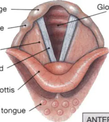
Identify the part labeled as 'Cuneiform cartilage' in the diagram of the larynx.
Cuneiform cartilage
Thyroid cartilage
Cricoid cartilage
Arytenoid cartilage
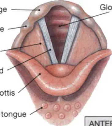
Identify the part labeled as 'False vocal cord' in the diagram of the larynx.
False vocal cord
True vocal cord
Epiglottis
Thyroid cartilage
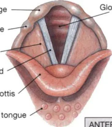
Identify the part labeled as 'Vocal cord' in the diagram of the larynx.
Vocal cord
Epiglottis
Trachea
Thyroid cartilage
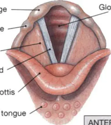
Identify the part labeled as 'Epiglottis' in the diagram of the larynx.
Epiglottis
Trachea
Vocal cords
Thyroid cartilage
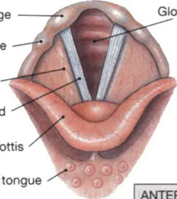
Identify the part labeled as 'Root of tongue' in the diagram of the larynx.
Root of tongue
Epiglottis
Vocal cords
Cricoid cartilage
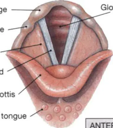
Identify the part labeled as 'Glottis' in the diagram of the larynx.
Glottis
Epiglottis
Vocal cords
Trachea
What is the function of the esophagus?
It produces digestive enzymes
It is a tubular passageway that connects the pharynx to the stomach
It absorbs nutrients
It stores bile
Fill in the blank: The esophagus connects the _______ to the stomach.
pharynx
trachea
larynx
bronchi
What is peristalsis?
The wave like motion that moves the bolus down the esophagus.
The process of breaking down food using enzymes.
The absorption of nutrients in the small intestine.
The production of saliva in the mouth.
The bolus is moved down the esophagus by peristalsis.
True
False
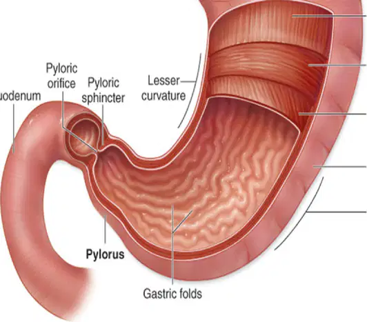
Which of the following is NOT one of the four parts of the stomach?
Cardia
Fundus
Duodenum
Pylorus
Fill in the blank: The stomach is a ______-shaped organ.
J
S
O
L
Which structure prevents the backflow of food from the stomach into the esophagus?
Pyloric sphincter
Cardiac (lower esophageal) sphincter
Rugae
Fundus
What are the folds in the stomach called?
Villi
Rugae
Chyme
Sphincters
Fill in the blank: The stomach produces ______, a semi-liquid mixture of partially digested food.
chyme
bile
saliva
mucus
Which of the following is a function of the stomach?
Chemical digestion
Oxygen transport
Hormone production
Blood filtration
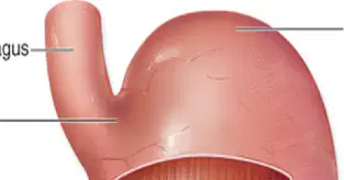
Name the four parts of the stomach as shown in the diagram.
Cardia, Fundus, Body, Pylorus
Duodenum, Jejunum, Ileum, Cecum
Esophagus, Trachea, Bronchi, Alveoli
Cortex, Medulla, Pelvis, Ureter
What is the main function of the small intestine?
Starts chemical digestion
Finishes chemical digestion
Stores waste
Produces bile
The small intestine is responsible for most of the _______ of nutrients.
absorption
digestion
excretion
production
How many hours does food spend in the small intestine?
12 hours
2 hours
24 hours
6 hours
What are villi in the small intestine?
Fingerlike projections to increase the surface area for absorption
Digestive enzymes
Blood vessels
Muscles
Which of the following is NOT a part of the small intestine?
Duodenum
Jejunum
Ileum
Colon
Name the three parts of the small intestine.
Duodenum, Jejunum, Ileum
Cecum, Colon, Rectum
Fundus, Body, Pylorus
Ascending, Transverse, Descending
Identify the part labeled as 'Duodenum' in the diagram of the human digestive system.
Duodenum
Esophagus
Rectum
Pancreas
What is the name of the flexure between the duodenum and jejunum as shown in the diagram?
Duodenojejunal flexure
Hepatic flexure
Splenic flexure
Ileocecal flexure
Which part of the small intestine is labeled as 'Jejunum' in the diagram?
Jejunum
Duodenum
Ileum
Cecum
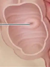
Name the valve that separates the small intestine from the large intestine.
Ileocecal valve
Pyloric valve
Mitral valve
Tricuspid valve
What is the last part of the small intestine labeled in the diagram?
Ileum
Duodenum
Jejunum
Cecum
Fill in the blank: The large intestine absorbs most of the ______ and electrolytes from the remaining digested material.
water
protein
fiber
glucose
Fill in the blank: Watery material that first enters the large intestine soon solidifies and becomes ______.
feces
urine
plasma
chyme
Fill in the blank: The large intestine stores fecal material until the body is ready to ______.
defecate
digest
inhale
urinate
Fill in the blank: The large intestine absorbs a very small percentage of ______ still remaining in the digested material.
nutrients
enzymes
hormones
proteins
How many parts does the large intestine consist of?
3
4
5
6
Which of the following is NOT a part of the large intestine?
Cecum
Appendix
Stomach
Rectum
Fill in the blank: The large intestine consists of the Cecum, Appendix, Colon, Rectum, and _______.
Anal canal
Esophagus
Duodenum
Gallbladder
The colon is divided into how many parts?
2
3
4
5
Which of the following is a part of the colon?
Ascending colon
Descending colon
Sigmoid colon
All of the above
Fill in the blank: The four parts of the colon are Ascending colon, Transverse colon, Descending colon, and _______.
Sigmoid colon
Cecum
Rectum
Ileum
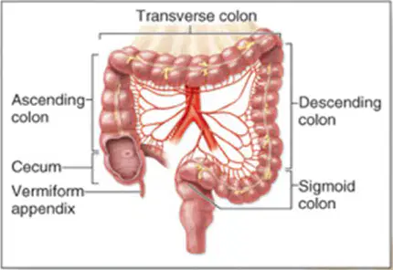
What is the name of the structure labeled as the 'Transverse colon' in the diagram of the large intestine?
Transverse colon
Ascending colon
Sigmoid colon
Cecum
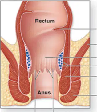
What is the name of the structure labeled as the 'Rectum' in the diagram of the anal canal?
Rectum
Anus
Sigmoid colon
Ileum
Refer to the diagram of the human abdomen. What anatomical part is labeled as 1?
Ascending Colon
Descending Colon
Transverse Colon
Sigmoid Colon
Refer to the diagram of the human abdomen. What anatomical part is labeled as 2?
Transverse colon
Gallbladder
Pancreas
Descending colon
Refer to the diagram of the human abdomen. What anatomical part is labeled as 3?
Descending colon
Liver
Stomach
Pancreas
What anatomical part is labeled as 4?
Stomach
Liver
Pancreas
Small intestine
Refer to the diagram of the human abdomen. What anatomical part is labeled as 5?
Liver
Stomach
Pancreas
Gallbladder
Refer to the diagram of the human abdomen. What anatomical part is labeled as 6?
Gall bladder
Pancreas
Appendix
Spleen
How many lobes does the liver have?
2
3
4
5
Which lobe of the liver is visible from the anterior view?
Right lobe
Left lobe
Caudate lobe
Quadrate lobe
True or False: The left lobe of the liver is larger than the right lobe.
True
False
Name the two subdivisions of the liver that are only seen from the posterior inferior view.
Caudate lobe and Quadrate lobe
Right lobe and Left lobe
Falciform ligament and Ligamentum teres
Hepatic artery and Portal vein
Label the following parts on the diagram:
Liver, Gallbladder, Stomach, Spleen, Duodenum, Common bile duct, Portal vein, Hepatic artery, Splenic vein, Inferior vena cava, Hepatic vein, Bile ducts.
Liver, Pancreas, Kidney, Appendix, Colon, Esophagus, Jugular vein, Femoral artery, Renal vein, Superior vena cava, Pulmonary vein, Lymph nodes.
Heart, Gallbladder, Stomach, Spleen, Jejunum, Pancreatic duct, Carotid artery, Hepatic artery, Splenic artery, Inferior vena cava, Hepatic vein, Bile ducts.
Liver, Gallbladder, Stomach, Pancreas, Duodenum, Common bile duct, Portal vein, Hepatic artery, Splenic vein, Inferior vena cava, Hepatic vein, Ureter.
Fill in the blank: The liver produces ______, a greenish fluid that breaks down fats into small droplets to assist in their chemical digestion.
bile
insulin
amylase
pepsin
Fill in the blank: The liver helps to ______ drugs, metabolites, and poisons.
detoxify
digest
absorb
excrete
Fill in the blank: The liver ______ excess nutrients and vitamins and releases them when they are needed.
stores
destroys
ignores
filters


Which numbers point to the right and left lungs?
5 and 10
10 and 11
5 and 15
15 and 16

What is illustrated with #2?
Bronchi
Bronchioles
Trachea
Alveoli

Identify structure #12
epiglottis
glottis
pharynx
larynx
Identify structure #16
Intercostal muscles
Pleura membrane
Ribs
Left lung
Right lung

Name the structure labeled 8
lungs
trachea
diaphragm
alveoli
Identify structure #13
epiglottis
esophagus
glottis
larynx

What side of your lung is the arrow pointing to?
(a)
Identify structure #11
epiglottis
glottis
pharynx
larynx
Identify structure #17
bronchi
trachea
diaphragm
bronchiole
Identify structure #4
epiglottis
glottis
pharynx
larynx
Identify structure #14
epiglottis
esophagus
vocal cords
larynx
Identify structure #8
Diaphragm
Intercostal muscles
Pleura membrane
Ribs
Identify structure #8
Intercostal muscles
Pleura membrane
Ribs
Left lung
Right lung
Identify structure #7
Diaphragm
Intercostal muscles
Pleura membrane
Ribs

Another name for your voice box.
pharynx
larynx
trachea
lung

The space or indent in the left lung to make room for the heart.
(a)

What is C pointing to in the diagram?
bronchioles
bronchi
alveoli
trachea

Number 4 is a lung. Which lung is it?
Left
Right

What is number one?
Bronchi
Alveoli
Trachea
Bronchioles

The lungs are covered with a thin membrane called the
bronchioles
synovial
serous
pleura


What is the scientific term for the tube that is commonly known as your wind pipe?
esophogus
bronchi
trachea
alveoli
Identify structure #6
Diaphragm
Intercostal muscles
Pleura membrane
Ribs

