
 Worksheets
Unit 4 Transport Test — Part 1 Multiple Choice
Total questions: 43
Worksheet time: 22mins
Which of the following statements about mature red blood cells is not true?
Life span of around 120 days
No nuclei
Contain hemoglobin
Divide to make new cells when old ones need to be replaced
If your blood type is O- that means
you have no agglutinogens on your red blood cells.
you have the rhesus antigens.
you only have anti-A antibodies in your blood plasma.
all of the above.
Which ion allows hemoglobin to readily bind with oxygen?
Fe2+
Na+
K+
Ca2+
The ______ side of the heart is the ______ circuit and pumps oxygen-poor blood to the ________.
right; systemic; lungs
right; pulmonary; lungs
left; systemic; body
left; pulmonary; body
The ______ side of the heart is the ______ circuit and pumps oxygen-rich blood to the ________.
right; systemic; lungs
right; pulmonary; lungs
left; systemic; body
left; pulmonary; body
The purpose of the serous fluid in the pericardial cavity is
to carry critical nutrients like oxygen to the heart.
to provide Ca2+ ions needed for muscle contraction.
to decrease friction between the layers of the heart as it contracts.
to lubricate the walls of the blood vessels to decrease resistance to blood flow.
Which of the following accurately reflects the order of events that causes the heart to pump out blood?
Electrical event → distribution → mechanical event → pressure change → fluid movement
Electrical event → pressure change → mechanical event → distribution → fluid movement
Mechanical event → distribution → electrical event → pressure change → fluid movement
Mechanical event → pressure change → electrical event → distribution → fluid movement
Which of the following is true for both cardiac and skeletal muscle tissues?
Striated
Branched
Contract as a unit
Can spontaneously depolarize without input from the nervous system
Which of the following statements about AEDs is false?
They are tools used for someone experiencing sudden cardiac arrest.
An AED can be used to restart a heart with no electrical activity.
They get the cardiac cells contracting in sync again.
They work by stopping the heart for a second in order to reset it.
Which statement below correctly matches the structure with its function?
Veins carry blood away from the heart.
The atria are the high pressure receiving chambers of the heart.
The sinoatrial node initiates the action potential that causes the heart to beat.
Contractile cardiac muscle cells can trigger their own action potentials without neural input.
The amount of strain or force placed on the blood vessels as the blood moves through them is known as
pulse
diastole
tidal volume
blood pressure
Which of the following statements about the valves of the heart is false?
The tricuspid valve is the semilunar valve on the right side of the heart.
The aortic valve prevents backflow into the left ventricle.
The mitral valve has two cusps between the left atrium and left ventricle.
The pulmonary valve guards the base of the pulmonary trunk.
In order to increase blood flow, there needs to be a(n) ________ in the pressure gradient and a(n) ________ in the resistance.
increase; increase
decrease; decrease
increase; decrease
decrease; increase
The diaphragm is the muscle that contracts each time we ________ so that the lungs can ______________.
inhale; contract
exhale; contract
inhale; expand
exhale; expand
The amount of air that moves in and out of the lungs when you take a normal breath is the
vital capacity
tidal volume
residual volume
lung capacity
Before entering the lungs, air must first pass through the
esophagus
trachea
nasopharynx
alveoli
Which of the following statements about gas exchange is false?
Our blood plays a central role.
Exchange happens in the alveoli and our body tissues.
Simple diffusion is the method of transport.
In the alveoli, CO2 moves into the blood and O2 moves out of it.
The left lung has __ lobes and the right lung has __ lobes.
2; 2
3; 3
2; 3
3; 2
When the diaphragm contracts, volume inside the lungs ________ and pressure inside the lungs ________.
increase; increase
decrease; decrease
increase; decrease
decrease; increase
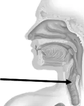
The structure indicated by the arrow in the picture to the right is the
larynx
esophagus
pharynx
trachea
Explain how the unique shape of erythrocytes enables them to best carry out their function.
Their biconcave shape increases surface area and flexibility, allowing efficient gas exchange and passage through narrow capillaries.
Their spherical shape decreases surface area, reducing gas exchange and preventing water loss.
Their elongated shape allows them to contract to push oxygen into tissues.
Their irregular shape enables them to engulf pathogens by phagocytosis.
You are a doctor and are treating a patient with liver failure due to chronic alcoholism. One of the results of this is reduced synthesis of plasma proteins, like fibrin. What issue can you expect to arise because of this?
Impaired blood clotting leading to excessive bleeding and bruising
Increased oxygen-carrying capacity of the blood
Enhanced immune memory and faster vaccination response
Hyperglycemia due to reduced insulin production
Which option best defines hematopoiesis and correctly identifies two other organ systems that support this process?
Formation of blood cells in bone marrow; supported by the urinary/endocrine system (kidneys release erythropoietin) and the skeletal system (marrow housing stem cells)
Clotting of blood in damaged vessels; supported by the digestive and respiratory systems
Destruction of old blood cells in the spleen; supported by the integumentary and muscular systems
Circulation of blood through vessels; supported by the lymphatic and reproductive systems
Which sequence correctly lists the major steps of hemostasis from first to last?
Platelet plug formation → Vascular spasm → Coagulation
Vascular spasm → Platelet plug formation → Coagulation
Coagulation → Platelet plug formation → Vascular spasm
Vascular spasm → Coagulation → Platelet plug formation
Assume you are blood type A. What agglutinogens are present on your red blood cells?
Antigen A
Antigen B
Antigens A and B
No ABO antigens
Assume you are blood type A. What antibodies are present in your plasma?
Anti-A antibodies
Anti-B antibodies
Anti-A and anti-B antibodies
No ABO antibodies
Assume you are blood type A. What would happen if you received a transfusion of type B blood, and why?
No reaction; type A plasma lacks antibodies
Mild fever; incompatible Rh factor only
Agglutination and possible hemolysis because anti-B antibodies bind B antigens
Improved oxygen delivery because B cells carry more hemoglobin
Assume you are blood type A. To whom can you donate blood, and why?
Only to type O; it lacks antigens
To types A and AB because recipients lack anti-A antibodies against donor A antigen
Only to type A because AB has anti-A antibodies
To all ABO types because A is a universal donor
Which option correctly identifies the three blood flow circuits of the heart and their primary roles?
Pulmonary (heart to lungs and back), Systemic (heart to body tissues and back), Coronary (blood supply to the heart muscle itself)
Systemic (oxygenates blood), Lymphatic (returns interstitial fluid), Pulmonary (feeds the myocardium)
Coronary (gas exchange in lungs), Hepatic portal (feeds myocardium), Systemic (returns blood to lungs)
Pulmonary (to body tissues), Coronary (to lungs), Systemic (to myocardium)
What is the function of the myocardium, and in which ventricle is it thicker and why?
Contracts to pump blood; thicker in the right ventricle to send blood to the lungs at high pressure
Provides electrical insulation; thicker in the left ventricle to slow conduction
Contracts to pump blood; thicker in the left ventricle to generate higher pressure for systemic circulation
Stores nutrients; equal thickness in both ventricles for balanced output
Choose the option that best describes the pathway of blood through the heart, starting where oxygen-poor blood enters the heart and ending where oxygen-rich blood leaves to the body.
Superior and inferior vena cava → Right atrium → Tricuspid valve → Right ventricle → Pulmonary valve → Pulmonary trunk → Lungs → Pulmonary veins → Left atrium → Mitral valve → Left ventricle → Aortic valve → Aorta
Pulmonary veins → Left atrium → Mitral valve → Left ventricle → Aortic valve → Aorta → Lungs → Pulmonary trunk → Right ventricle → Tricuspid valve → Right atrium → Venae cavae
Aorta → Left ventricle → Mitral valve → Left atrium → Pulmonary veins → Lungs → Pulmonary trunk → Right ventricle → Tricuspid valve → Right atrium → Venae cavae
Superior and inferior vena cava → Right ventricle → Tricuspid valve → Right atrium → Pulmonary valve → Lungs → Pulmonary veins → Left ventricle → Mitral valve → Left atrium → Aorta
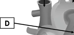
In the heart diagram, what structure is labeled A? Use the same word bank as in the pathway item: Aorta, Aortic valve, Inferior vena cava, Left atrium, Left ventricle, Lungs, Mitral valve, Pulmonary trunk, Pulmonary valve, Pulmonary veins, Right atrium, Right ventricle, Superior vena cava, Tricuspid valve.
Superior vena cava
Pulmonary trunk
Aorta
Right atrium
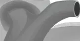
In the heart diagram, what structure is labeled B? Use the same word bank as in the pathway item: Aorta, Aortic valve, Inferior vena cava, Left atrium, Left ventricle, Lungs, Mitral valve, Pulmonary trunk, Pulmonary valve, Pulmonary veins, Right atrium, Right ventricle, Superior vena cava, Tricuspid valve.
Pulmonary trunk
Aorta
Pulmonary valve
Superior vena cava
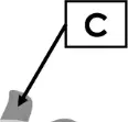
In the heart diagram, what structure is labeled C? Use the same word bank as in the pathway item: Aorta, Aortic valve, Inferior vena cava, Left atrium, Left ventricle, Lungs, Mitral valve, Pulmonary trunk, Pulmonary valve, Pulmonary veins, Right atrium, Right ventricle, Superior vena cava, Tricuspid valve.
Aorta
Left atrium
Pulmonary veins
Pulmonary trunk
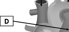
In the heart diagram, what structure is labeled D? Use the same word bank as in the pathway item: Aorta, Aortic valve, Inferior vena cava, Left atrium, Left ventricle, Lungs, Mitral valve, Pulmonary trunk, Pulmonary valve, Pulmonary veins, Right atrium, Right ventricle, Superior vena cava, Tricuspid valve.
Right atrium
Left atrium
Right ventricle
Left ventricle
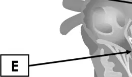
In the heart diagram, what structure is labeled E? Use the same word bank as in the pathway item: Aorta, Aortic valve, Inferior vena cava, Left atrium, Left ventricle, Lungs, Mitral valve, Pulmonary trunk, Pulmonary valve, Pulmonary veins, Right atrium, Right ventricle, Superior vena cava, Tricuspid valve.
Right ventricle
Left ventricle
Left atrium
Pulmonary veins

In the heart diagram, what structure is labeled F? Use the same word bank as in the pathway item: Aorta, Aortic valve, Inferior vena cava, Left atrium, Left ventricle, Lungs, Mitral valve, Pulmonary trunk, Pulmonary valve, Pulmonary veins, Right atrium, Right ventricle, Superior vena cava, Tricuspid valve.
Left ventricle
Right ventricle
Right atrium
Aortic valve
Which set lists three primary factors that determine resistance to blood flow in vessels and identifies the one with the greatest impact?
Blood viscosity, vessel length, and vessel radius (radius has the greatest impact)
Cardiac output, capillary density, and stroke volume (stroke volume has the greatest impact)
Blood type, hematocrit, and blood pressure (blood pressure has the greatest impact)
Valve number, heart rate, and venous return (heart rate has the greatest impact)
An electrocardiogram (ECG) is used in clinical settings. Which option best states its purpose and correctly describes how the ECG’s trace would differ for a heart beating at a high frequency, a low frequency, and one that is not beating?
It measures the heart’s electrical activity; high frequency shows closely spaced repeating waves, low frequency shows widely spaced repeating waves, and no heartbeat shows a flat line.
It measures blood pressure; high frequency shows tall waves, low frequency shows short waves, and no heartbeat shows a flat line.
It measures heart sounds; high frequency shows louder spikes, low frequency shows quieter spikes, and no heartbeat shows random noise.
It measures oxygen levels; high frequency shows a rising baseline, low frequency shows a falling baseline, and no heartbeat shows a zigzag line.
The respiratory tract is divided into conducting and respiratory zones. Which option correctly states the primary function of each zone and places each listed structure into the correct zone? Structures: Nose, Pharynx, Larynx, Trachea, Bronchi, Bronchioles, Alveolar ducts, Alveoli.
Conducting zone transports, warms, and filters air: Nose, Pharynx, Larynx, Trachea, Bronchi, Bronchioles. Respiratory zone is the site of gas exchange: Alveolar ducts, Alveoli.
Conducting zone is the site of gas exchange: Alveolar ducts, Alveoli. Respiratory zone transports, warms, and filters air: Nose, Pharynx, Larynx, Trachea, Bronchi, Bronchioles.
Conducting zone transports, warms, and filters air: Nose, Pharynx, Larynx only. Respiratory zone is the site of gas exchange: Trachea, Bronchi, Bronchioles, Alveolar ducts, Alveoli.
Conducting zone is the site of mucus secretion: Alveoli, Alveolar ducts, Bronchioles. Respiratory zone moves air: Nose, Pharynx, Larynx, Trachea, Bronchi.
The overall function of the respiratory system is to supply oxygen to our cells for what critical process?
Cellular respiration (ATP production)
Photosynthesis
Blood clotting
Protein translation
Which statement best explains the pressure–volume relationship that causes air to flow into and out of the lungs during breathing?
Inhalation: thoracic and lung volume increase, intrapulmonary pressure falls below atmospheric pressure, and air flows in; exhalation: volume decreases, intrapulmonary pressure rises above atmospheric pressure, and air flows out.
Inhalation: thoracic volume decreases, intrapulmonary pressure rises above atmospheric pressure, and air flows in; exhalation: volume increases, intrapulmonary pressure falls below atmospheric pressure, and air flows out.
Inhalation and exhalation occur when intrapulmonary pressure equals atmospheric pressure, so no volume change is required.
Airflow depends only on blood pressure changes in the pulmonary vessels and is independent of thoracic volume.
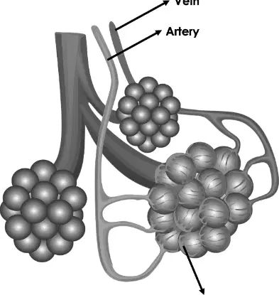
Referring to the pictured alveoli with labeled artery, vein, and capillary network, what is the purpose of these different blood vessels that connect to the alveoli?
Pulmonary arteries deliver deoxygenated blood to the capillary network for gas exchange, and pulmonary veins carry oxygenated blood away from the alveoli back to the heart.
Pulmonary arteries deliver oxygenated blood to the capillary network, and pulmonary veins remove carbon dioxide–rich blood from the body to the alveoli.
Both arteries and veins carry only oxygenated blood near the alveoli; capillaries remove oxygen from the lungs to the air.
Arteries and veins near the alveoli function mainly to regulate airway pressure, not gas transport.

