
Font size
 Worksheets
Lab Practical 2
Total questions: 35
Worksheet time: 26mins
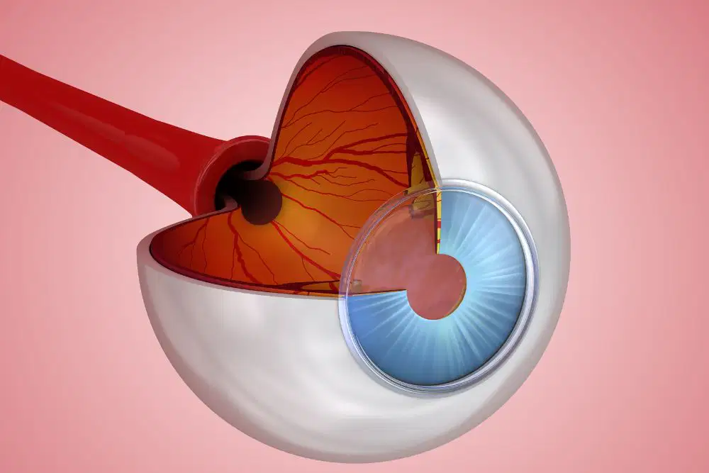
Name the structure. (Is a highly vascular layer)
Platysma
Choroid
Retina
Iris

Name the structure.
Cornea
Pupil
Iris
Sarcomere
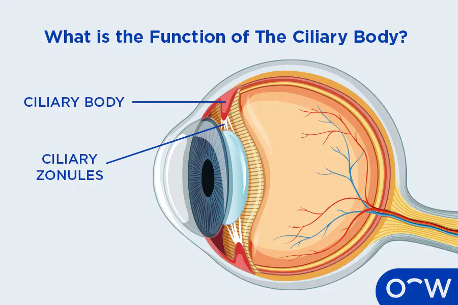
Name the structure. (Also known as suspensory ligaments)
Ciliary Body
Ciliary Zonule
Lens
Cornea

Name the structure. (The colored part)
Pupil
Lens
Choroid
Iris
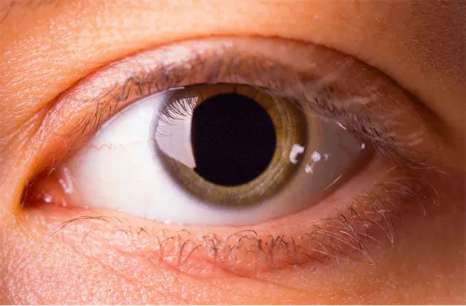
Name the structure. (The black part)
Iris
Pupil
Lens
Sclera
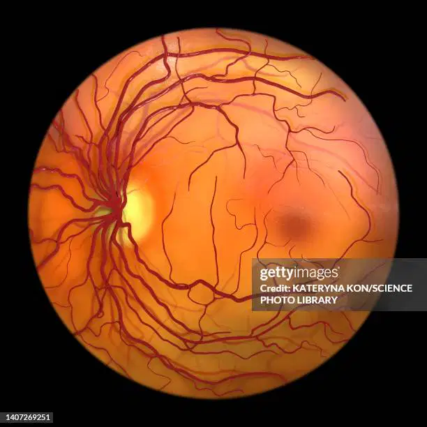
Name the structure. (Also known as the neural layer)
Cornea
Pituitary Gland
Retina
Pupil

Name the structure. (White part of the eye)
Ciliary Zonule
Retina
Pupil
Sclera
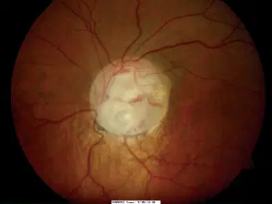
Name the structure. (White highlighted part)
Ulnar Nerve
Temporal Nerve
Cornea
Optic Disc
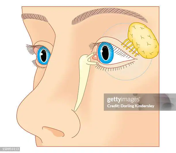
Name the structure. (Yellow part)
Lacrimal Gland
Pituitary Gland
Medulla
Pons

Name the structure.
Pupil
Thalamus
Hypothalamus
Lens
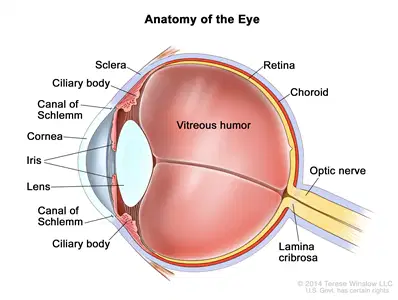
The canal of schlemm is also known as:
Scleral venous sinus
Sclera
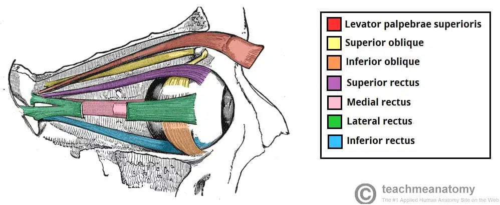
Which eye muscle pulls the eyeball towards the nose?
medial rectus extrinsic eye muscle
superior rectus extrinsic eye muscle
Lateral rectus extrinsic eye muscle
inferior rectus extrinsic eye muscle

Name the structure. (Also known as the ear drum)
External auditory canal
Auditory ossicles
Cochlea
Tympanic membrane
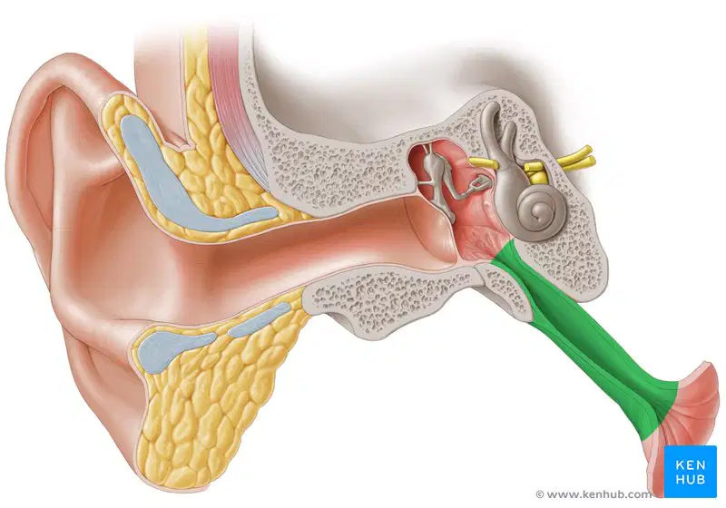
Name the structure.
Eustachian Tube
Pharyngotympanic Tube
Auditory Tube
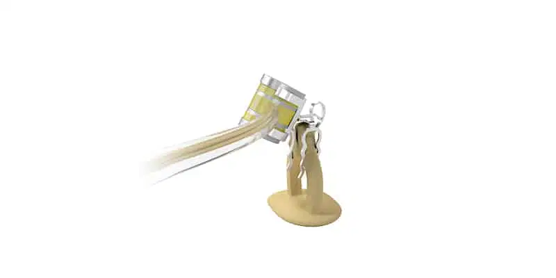
Name the structure.
Anvil
Stapes
Cochlea
Malleus
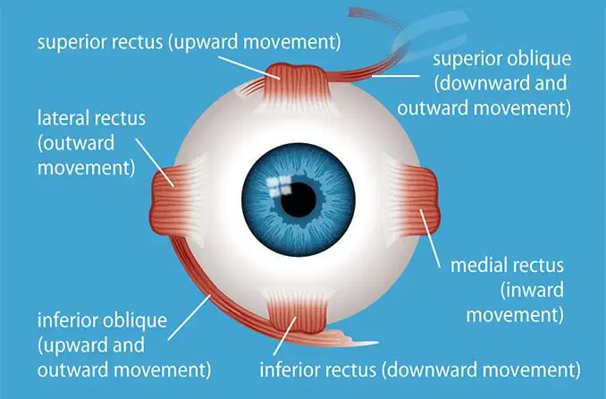
Name the structure. (Pulls the eye downward)
medial rectus extrinsic eye muscle
superior rectus extrinsic eye muscle
superior oblique extrinsic eye muscle
inferior rectus extrinsic eye muscle
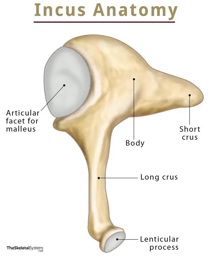
This structure is also known as:
Anvil
Hammer
Malleus
Stirrup

Name the structure.
Malleus
Incus
Cochlea
Stapes
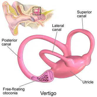
Name the structure.
(a)
Name the structure in the brain that has a X shape and the optic nerves from the two eyes cross over.
(a)
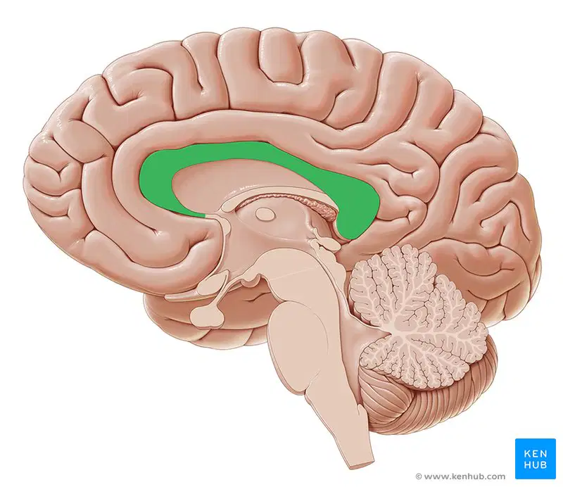
Name the structure.
(a)
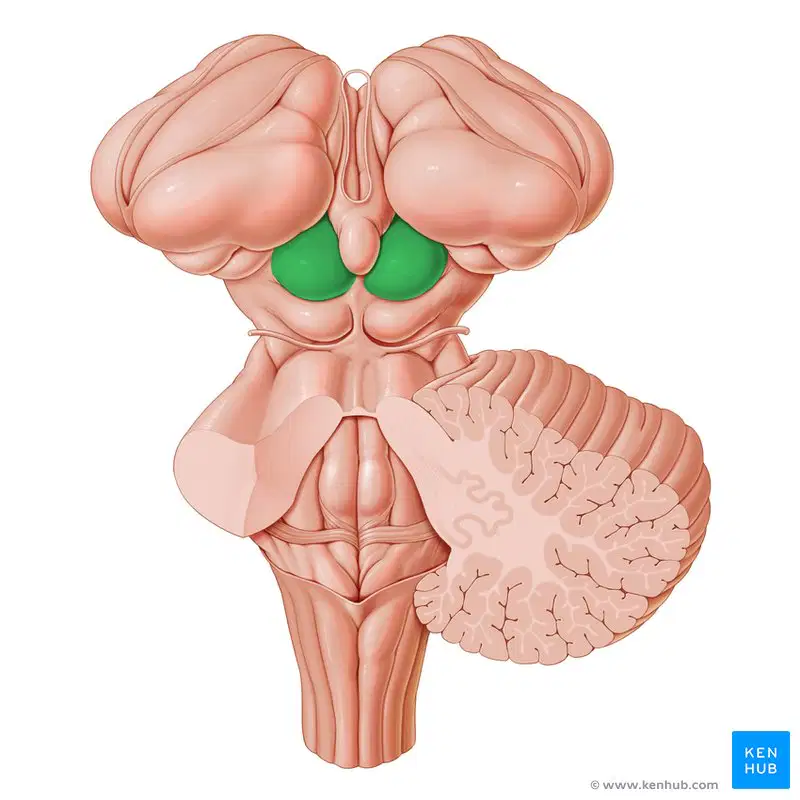
Name the structure and its two lobes.
(a)

Name the structure.
(a)
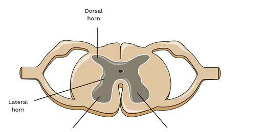
What is the horn that is on the bottom left called?
(a)
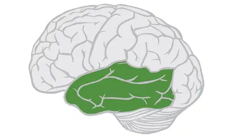
Name the structure.
(a)

What is the bottom line pointing to?
(a)
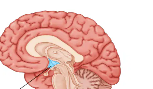
Name the structure.
(a)

Name the structure highlighted in pink.
(a)
What is the cranial nerve responsible for smell. (Name and CN#)
(a)
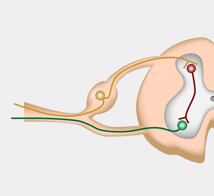
What is the bulge in this picture called?
(a)
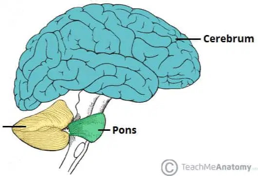
Name the structure highlighted in yellow.
(a)

Name the structure.
(a)
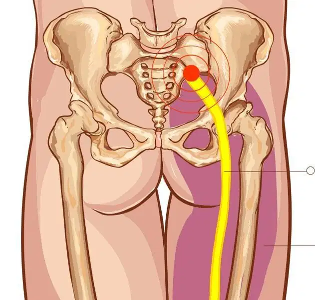
Name the structure.
(a)
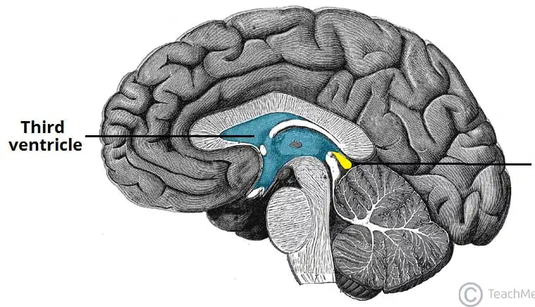
Name the structure.
(a)

Name the structure.
(a)

