
Font size
 Worksheets
Introduction to Histology and Tissue Structure
Total questions: 41
Worksheet time: 21mins
According to the overview, tissues are described as consisting of two key components. Which pair correctly identifies these components?
Cells and extracellular matrix (ECM)
Organs and organelles
Cells and intracellular matrix (ICM)
Fibers and membranes only
Which statement best characterizes the extracellular matrix (ECM) as presented in the summary?
A fluid containing only electrolytes and water
A network of macromolecules such as collagen fibrils and basement membranes that supports cells
A compartment inside the nucleus that stores DNA
A set of ribosomes that synthesize proteins for secretion
Select the functions attributed to the ECM in the provided material.
Providing structural support to cells
Facilitating nutrient transport to cells
Mediating waste removal
Generating ATP for cellular metabolism
Which statement describes the interaction between cells and the ECM in this section?
Cells produce ECM and are influenced by ECM molecules that interact with cell surface receptors
Cells are isolated from ECM and never contact it
ECM components act only as barriers without signaling roles
Cells cannot recognize ECM components due to lack of receptors

In the labeled connective tissue diagram, which structure is identified as providing tensile strength within the ECM?
Elastic fibers
Collagen fibers
Ground substance
Adipocyte (fat cell)

Based on the diagram, which option correctly pairs a cell type with its likely role?
Adipocyte — lipid storage
Macrophage — forming basement membrane
Mesenchymal cell — transporting oxygen
Blood vessel endothelial cell — synthesizing elastic fibers

Which combination of ECM components shown in the diagram forms the non-fibrous portion that fills space between cells?
Ground substance
Collagen fibers
Elastic fibers
Basement membrane only
Which statements about cell–ECM interactions from the introduction are accurate? Select all that apply.
Cells produce ECM molecules
ECM components are recognized and attach to cell receptors
Cells and ECM interact through cell surface receptors
ECM functions exclusively in energy production

Refer to the leukocyte rolling diagram. Which step immediately follows selectin-mediated rolling to promote firm adhesion?
Proteoglycan shedding from endothelium
Integrin activation by chemokines leading to high-affinity binding
Macrophage phagocytosis of microbes in the vessel lumen
Cytokine release (TNF, IL-1) causing endothelial detachment

In the leukocyte migration sequence shown, which molecules present on the endothelial surface initially mediate the slow rolling of leukocytes under blood flow?
Integrin ligands
Selectins and their ligands
Chemokines bound to fibrin
Fibronectin receptors

Based on the diagram, which statements correctly describe the role of chemokines in leukocyte recruitment? Select all that apply.
Chemokines displayed on proteoglycans signal leukocyte integrins to switch from low- to high-affinity states
Chemokines are released by macrophages stimulated by microbes
Chemokines directly form the integrin ligand on the endothelium
Chemokines guide leukocyte migration into fibrin and fibronectin of the extracellular matrix
According to the introduction slide on tissues, what enables organs to perform overall organism function?
Exclusive presence of a single tissue type
Random association of cells with ECM
An organized combination of multiple tissues
Absence of molecular methods in histology
From the tissue introduction slide, which statements are accurate regarding histology? Select all that apply.
Histology relies on microscopes due to small cell and matrix sizes
Histology primarily studies only organ-level macroscopic structures
Histology uses molecular methods to study tissue structure and function
Histology is essential for identifying tissue types formed by specific cell–ECM associations
Which statement best describes the relationship between fundamental tissues and organs in the human body?
Organs are single tissues specialized for one function.
Fundamental tissues combine in specific arrangements to form organs.
Organs are collections of cells without tissue organization.
Tissues only exist within organs and have no independent structure.
Histology primarily studies tissues using which approaches? Select all that apply.
Light and electron microscopy to visualize structure
Molecular methods to analyze proteins and genes
Astronomical imaging to map large-scale patterns
Seismography to detect mechanical waves
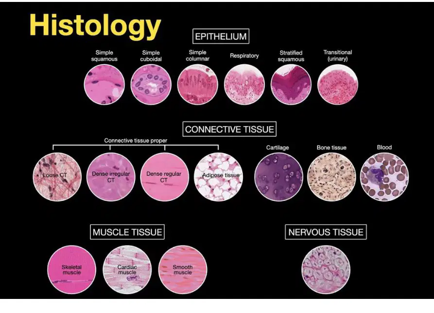
Based on the labeled images of tissue types, which pairing correctly matches a tissue category with an example subtype?
Epithelial tissue — skeletal muscle
Connective tissue — adipose tissue
Muscle tissue — stratified squamous
Nervous tissue — dense regular connective tissue
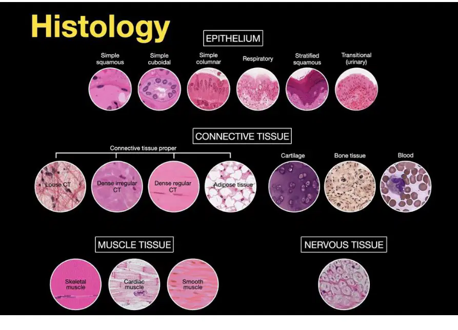
Which set lists the four fundamental tissue types that underpin organ composition?
Epithelial, connective, muscle, nervous
Epithelial, cartilage, bone, blood
Muscle, tendon, ligament, nerve
Connective, epithelial, endocrine, exocrine
Refer to the diagram titled "Overview of Tissue Types". Which category is primarily responsible for covering surfaces and lining cavities of the body?
Connective tissue
Muscle tissue
Epithelial tissue
Nervous tissue
In the same overview diagram, which tissue type is specialized for rapid communication and signal processing? Select the best answer.
Epithelial tissue
Connective tissue
Muscle tissue
Nervous tissue
Which of the following functions most closely aligns with connective tissue in the tissue overview diagram?
Generating force for movement
Forming protective coverings and linings
Providing support, binding, and structural integrity
Conducting electrical impulses
Based on the diagram, which pair correctly matches a tissue type to a characteristic function? Select all that apply.
Epithelial tissue — secretion and absorption
Muscle tissue — force generation and movement
Nervous tissue — cushioning and energy storage
Connective tissue — transport of neural signals
Which statement best differentiates muscle tissue from epithelial tissue in the diagram-based overview?
Muscle tissue primarily covers surfaces, whereas epithelial tissue contracts to produce movement.
Muscle tissue contracts to generate force, whereas epithelial tissue forms linings and surfaces.
Both primarily conduct electrical impulses for communication.
Both primarily provide structural support and bind other tissues.
Select the tissue type that most directly underpins rapid reflexes and information integration, as summarized in the overview visual.
Connective tissue
Nervous tissue
Epithelial tissue
Muscle tissue
Histology relies on multiple scientific advances to study cells and tissues. Which statement best captures the role of microscopic approaches introduced in this chapter?
They enable visualization and analysis of tissue structure at scales beyond the unaided eye.
They replace all biochemical methods, making molecular assays unnecessary.
They are limited to gross anatomy and cannot resolve cellular details.
They focus only on tissue staining and exclude sample preparation steps.
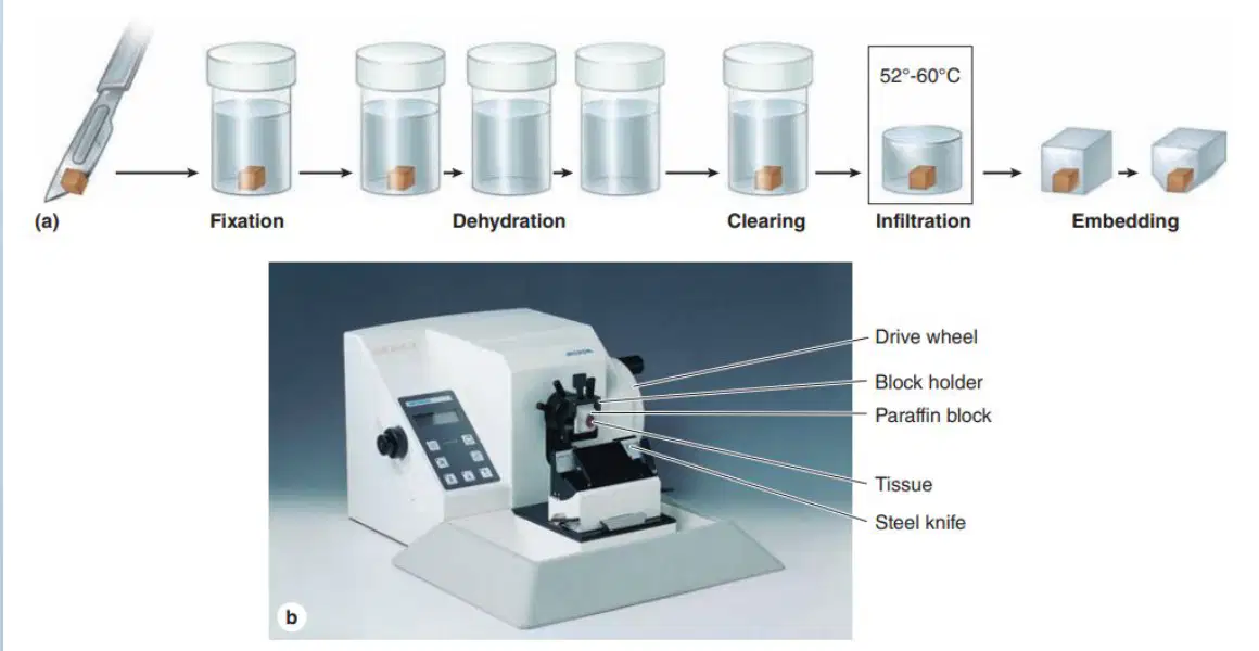
In the illustrated workflow for preparing tissues for study, place emphasis on the purpose of embedding after fixation, dehydration, clearing, and infiltration. What is the primary reason embedding is performed?
To stabilize the infiltrated tissue within a supportive medium for thin sectioning.
To increase the water content of tissue for better staining contrast.
To remove all residual fixative by prolonged washing at room temperature.
To enlarge tissues so features appear bigger under low magnification.
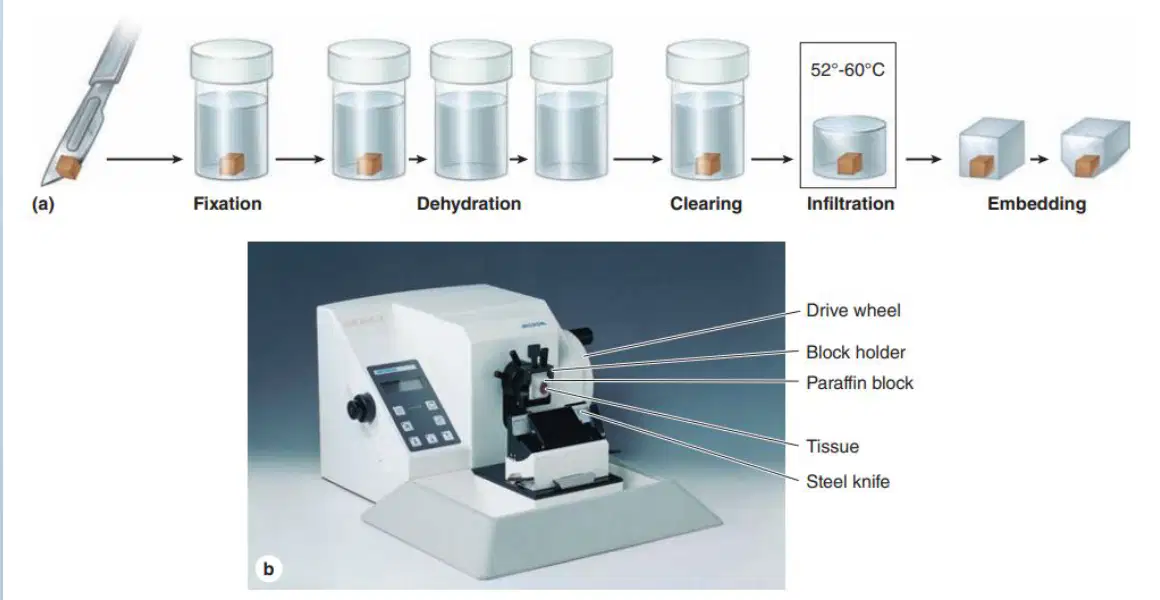
Considering modern tools in histology, which set correctly pairs a device component with its function in the sectioning instrument shown? Select all that apply.
Drive wheel—advances the block against the knife for controlled slicing.
Block holder—securely clamps the paraffin block containing tissue.
Steel knife—cuts thin sections from the paraffin-embedded tissue.
Paraffin block—houses tissues during fixation to prevent shrinkage.
In histologic research, what is the common procedure used to make tissues suitable for light microscopy with transmitted light?
Preparation of whole organs without alteration
Preparation of tissue sections or slices
Immersion of tissues in opaque dye to block light
Observation of tissues under reflected light microscopy
Why must tissues and organs be sliced into thin, translucent sections before microscopic examination with transmitted light?
They are usually too thick for light to pass through
Translucent sections prevent dehydration
Thin sections enhance reflected light imaging
Thick samples improve resolution in light microscopy
Which statement best describes how tissue sections are handled for examination after slicing?
They are suspended in saline and viewed directly
They are mounted on glass slides for examination
They are placed in metal cassettes for observation
They are glued to coverslips without slides
Select the challenges commonly encountered during preparation that can affect interpretation of tissue sections.
Artifacts
Distortions
Loss of components
Uniform enhancement of contrast
Which goal defines an ideal tissue preparation outcome?
Maximizing color intensity regardless of structure
Maintaining the tissue's original structure and composition
Reducing the sample thickness to below 1 nm
Eliminating all cellular components to simplify imaging
Light microscopy with transmitted light is used after which preparatory step in histology?
Cryofracture of intact organs
Preparation of thin tissue sections
Application of electron-dense stains
Mounting tissues in opaque resin blocks
Which combination of events accurately represents the minimal workflow for tissue sectioning prior to examination?
Slice tissues into thin sections, mount on glass slides
Stain tissues heavily, view without sectioning
Freeze tissues, observe under reflected light
Compress tissues to increase opacity, then mount
In the context of histologic research, which microscopy mode is explicitly mentioned for examining prepared tissues?
Polarized light microscopy with reflected light
Light microscopy with transmitted light
Scanning electron microscopy
Confocal fluorescence microscopy
Which statement is most consistent with the rationale behind sectioning for microscopy?
Sectioning reduces sample transparency
Sectioning allows light to pass through previously opaque samples
Sectioning increases tissue thickness for better focus
Sectioning eliminates the need for glass slides
Which of the following are direct consequences of imperfect preparation that investigators must be cautious about when interpreting images?
Preparation artifacts that mimic structures
Distortions that alter spatial relationships
Loss of components that changes composition
Guaranteed preservation of original morphology
What is the primary reason glass slides are used in the workflow described?
They enable sectioning by acting as cutting surfaces
They serve as supports to mount sections for examination
They replace coverslips to enhance contrast
They prevent artifacts and distortions during preparation
Which option best contrasts ideal preparation with common real-world outcomes?
Ideal: exaggerates features; Real: removes artifacts
Ideal: maintains original structure and composition; Real: artifacts and loss often occur
Ideal: relies on reflected light; Real: depends on transmitted light
Ideal: uses thick samples; Real: uses ultrathin samples
Identify the step that directly addresses the problem of tissues being too thick for light transmission.
Fixation in strong acids
Slicing tissues into thin, translucent sections
Dehydration to increase opacity
Embedding in opaque resin for support
Which practice would most likely compromise the goal of ideal preparation as stated?
Maintaining structural integrity during processing
Introducing distortions during sectioning
Avoiding loss of components through gentle handling
Mounting sections carefully on glass slides
When assessing images from prepared tissue sections, which awareness improves scientific rigor?
Assuming all features are artifact-free
Recognizing that artifacts, distortions, and loss of components may influence observations
Believing thicker sections yield better transmitted light images
Ignoring the role of mounting on glass slides

