
Font size
 Worksheets
Lecture Objectives
Total questions: 101
Worksheet time: 51mins
Which pairing correctly matches a heart landmark with its level or relation?
Base—extends to the level of the 2nd rib
Apex—located above the base
Base—positioned at the 5th intercostal space
Apex—found at the sternal angle
Select all statements that correctly characterize the apex of the heart as presented in the lecture objectives.
It lies at the 5th intercostal space
It is slightly below the nipple on the left side
It is aligned with the Left mid clavicular line (LMCL)
It is superior to the base
Which statement about the base and apex relationship is accurate in this lecture overview?
The base lies below and the apex above
Both base and apex are at the same level
The base lies above and the apex below
The apex lies above the 2nd rib
In clinical surface anatomy terms given, where would you palpate to locate the heart’s apex beat?
Right 5th intercostal space at RMCL
Left 5th intercostal space slightly below the nipple at LMCL
Left 2nd intercostal space at LMCL
Midline at the sternal angle
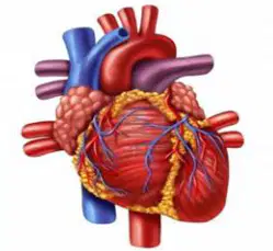
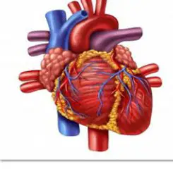
Refer to the diagram showing a stylized heart and bullet list of positional facts. Which bullet identifies the rib level associated with the heart’s base?
1st rib
2nd rib
4th rib
5th rib
Which statement best describes the heart’s primary role in circulation as presented in the lesson?
A skeletal muscle that stores blood for later use
A complex muscular pump that maintains blood pressure and flow through the lungs and the rest of the body
A passive reservoir that equalizes blood volume between organs
A neural regulator that initiates vasoconstriction without moving blood
Approximately how many times does the heart pump in a typical day according to the material?
10,000 times
50,000 times
100,000 times
200,000 times
What is the approximate daily volume of blood moved by the heart as stated in the content?
720 liters (190 gallons)
2,400 liters (630 gallons)
7,200 liters (1,900 gallons)
12,000 liters (3,170 gallons)
From where does the heart receive its own blood supply?
Pulmonary veins
Systemic capillaries
Coronary arteries
Aortic arch
According to the functions listed, which materials are transported by the cardiovascular system to and from cells? Select all that apply.
Oxygenated blood to cells
Deoxygenated blood into the heart
Nutrients
Hormones
Exhaled air to the lungs
Which statement accurately captures the bidirectional role of the cardiovascular system described?
It only delivers oxygenated blood to tissues.
It only returns deoxygenated blood to the lungs.
It transports oxygenated blood to cells and brings deoxygenated blood into the heart from the body.
It stores nutrients until cells request them.
Based on the slide, which list correctly identifies additional substances carried by the cardiovascular system besides blood gases?
Minerals and enzymes
Nutrients, hormones, and waste products
Vitamins and antibodies only
Water and electrolytes only
Which layer is explicitly named as the visceral layer of the pericardium in the material?
Endocardium
Epicardium
Myocardium
Parietal pericardium
Identify the correct pairing of heart structure and description from the lesson.
Pericardium: inner lining of heart chambers
Myocardium: muscular layer responsible for pumping action
Endocardium: outer surface of the heart
Epicardium: fibrous sac surrounding the lungs
Which components are listed under “Heart Muscles” in the instructional content? Select all that apply.
Pericardium
Myocardium
Endocardium
Valvular apparatus
According to the material, which of the following is part of the pericardium?
Parietal layer
Myocardial layer
Endocardial layer
Coronary layer
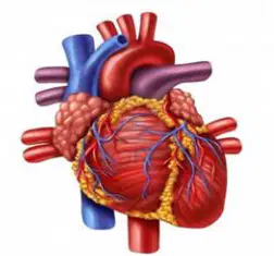
Consider the small heart diagram shown. Which vessel type is specifically noted as supplying the heart tissue itself?
Pulmonary arteries
Coronary arteries
Portal veins
Systemic arterioles
Which statement best describes a primary function of the cardiovascular system?
It transports oxygen from the lungs to body tissues.
It produces hormones within endocrine glands.
It digests nutrients in the small intestine.
It generates electrical impulses in neurons.
Select all roles that the cardiovascular system performs in material transport throughout the body.
Delivery of oxygen to tissues
Return of deoxygenated blood to the lungs
Transport of nutrients absorbed from digestion
Distribution of hormones to target organs
Elimination of solid food waste via the colon
Which pairing correctly matches the substance with its transport direction or destination in the cardiovascular system?
Deoxygenated blood—carried from tissues back to the lungs
Hormones—stored permanently in blood vessels
Nutrients—moved from the liver only to the kidneys
Metabolic wastes—retained in tissues to conserve energy
In the context of cardiovascular transport, which statement about wastes is accurate?
Waste products are circulated to organs that process and remove them.
Wastes are kept in the bloodstream indefinitely to maintain pressure.
Only gaseous wastes are transported; dissolved wastes are not moved.
Wastes are neutralized exclusively in skeletal muscle.
Which set lists materials the cardiovascular system distributes to coordinate body functions?
Hormones and nutrients
Bile and digestive enzymes
Synaptic neurotransmitters and axons
Keratin and melanin
Refer to the diagram of the heart wall layers. Which layer directly lines the heart chambers and valves, providing a smooth surface for blood flow?
Pericardium
Myocardium
Endocardium
Epicardium
In the heart wall diagram, which layer consists primarily of cardiac muscle responsible for the force of contraction?
Endocardium
Myocardium
Fibrous pericardium
Serous pericardium
Which statement correctly describes the pericardium as shown in the heart wall visual? Select all that apply.
It is a protective sac surrounding the heart.
It forms the inner endothelial lining of the chambers.
It has fibrous and serous components.
It generates the heartbeat through contractile cells.
Examine the micrograph-style diagram of cardiac muscle. Which feature is unique to cardiac muscle cells and facilitates synchronized contraction?
Sarcomeres arranged in myofibrils
Intercalated discs connecting adjacent cells
Multiple peripheral nuclei per fiber
Voluntary innervation via somatic motor neurons
In the cardiac muscle structure image, what is the primary functional role of intercalated discs?
Store calcium for contraction
Provide electrical and mechanical coupling between cells
Anchor valves to papillary muscles
Line the interior of the heart chambers
Based on the heart wall illustration, match each layer to its description.
Pericardium — outer protective sac
Myocardium — muscular middle layer
Endocardium — inner endothelial lining
Epicardium — internal valve tissue
According to the diagram focusing on cardiac muscle cells, which characteristic best differentiates them from skeletal muscle fibers?
Lack of striations
Long multinucleated cylindrical fibers
Branched cells with single central nucleus
Absence of intercellular junctions
From the heart wall layers figure, which statement accurately links a layer to its clinical relevance?
Inflammation of the endocardium is called pericarditis.
Damage to the myocardium primarily affects the heart’s pumping capacity.
Epicardial thickening directly obstructs blood flow inside chambers.
Pericardium forms valve cusps that ensure one-way flow.
In the cardiac muscle micro-structure diagram, which cell-to-cell junction within intercalated discs permits rapid ionic current spread for coordinated depolarization?
Tight junctions
Gap junctions
Hemidesmosomes
Adherens junctions
Which statement best describes the primary function of heart valves in the human heart?
To propel blood through muscular contraction
To ensure one-way blood flow by opening and closing with pressure changes
To generate the heart’s electrical impulses
To exchange gases directly with circulating blood
Which pair correctly identifies the atrioventricular (AV) valves?
Aortic and Pulmonic
Mitral (Bicuspid) and Tricuspid
Semilunar and Atrioventricular
Chordae tendineae and Papillary
What is the structural distinction between the two AV valves?
Both have three cusps supported by chordae tendineae
Mitral valve has two cusps (bicuspid) while tricuspid valve has three cusps
Mitral valve is semilunar; tricuspid valve is atrioventricular
Mitral valve opens to the right ventricle; tricuspid opens to the left ventricle
Which statement about chordae tendineae in relation to AV valves is accurate?
They prevent valve cusps from prolapsing into the atria during ventricular contraction
They initiate valve closure by generating action potentials
They attach semilunar valves to the arterial wall
They open the valves by pulling cusps apart during diastole
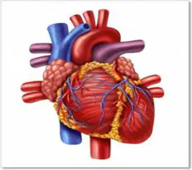
According to the slide on normal heart sounds, what produces the S2 (dub) sound?
Closure of atrioventricular valves
Opening of semilunar valves
Closure of aortic and pulmonic (semilunar) valves
Contraction of atrial myocardium
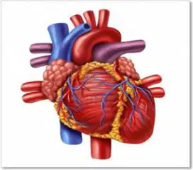
Where is the S2 (dub) sound best auscultated based on standard cardiac landmarks?
Apex over the mitral area
Base area over aortic and pulmonic areas
Left lower sternal border at the tricuspid area
Midpoint known as Erb’s point
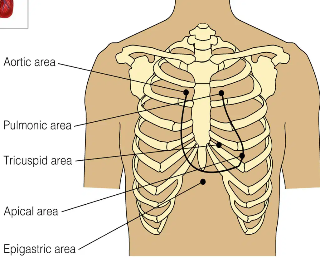
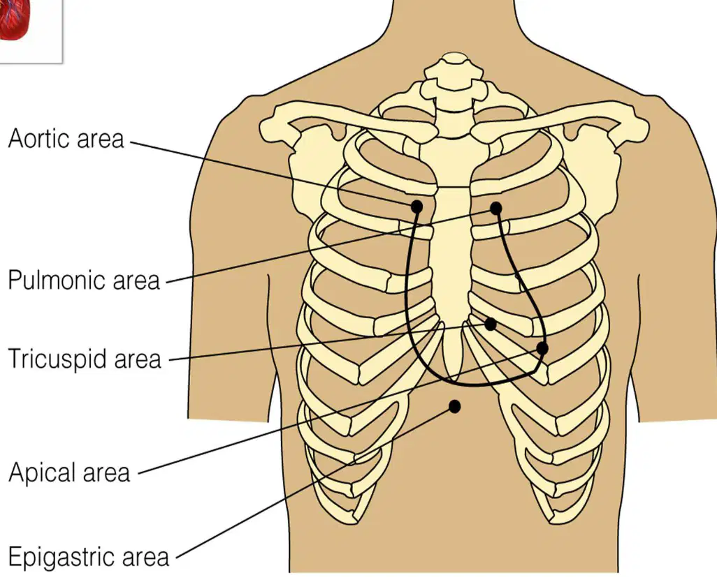
Match each auscultation area with its location along the chest wall.
Aortic area: 2nd intercostal space, right sternal border
Pulmonic area: 2nd intercostal space, left sternal border
Tricuspid area: 5th intercostal space, left sternal border
Mitral area: 5th intercostal space, 1 cm medial to midclavicular line
Which landmark corresponds to the point midway between the pulmonic and tricuspid areas?
Erb’s point
Point of maximal impulse (PMI)
Left midclavicular line (LMCL)
Apex beat

Refer to the diagram showing labeled cardiac listening landmarks. Which area is most associated with auscultating the tricuspid valve mechanics, including observing AV valve closure during ventricular contraction?
Aortic area
Pulmonic area
Tricuspid area
Apical area
Based on the visual landmarks, which observation best fits relaxed ventricles with AV valves open?
Blood flows from atria to ventricles while the semilunar valves are open
Atrioventricular valves permit filling; semilunar valves remain closed
Semilunar valves open to allow ejection as AV valves close
Both AV and semilunar valves open simultaneously to equalize pressure
Which combination of valve states characterizes contracting ventricles in the cardiac cycle? Select all that apply.
AV valves closed to prevent backflow into atria
Semilunar valves open to permit ejection into great arteries
AV valves open to enhance ventricular filling
Semilunar valves closed to protect arterial pressure during diastole

When assessing semilunar valve function using the cardiac landmarks diagram, which area would primarily reflect aortic valve ejection sounds during ventricular contraction?
Pulmonic area
Aortic area
Tricuspid area
Apical area
Which statement best explains why AV valves close at the onset of ventricular systole?
Atrial pressure suddenly exceeds ventricular pressure
Chordae tendineae actively pull the cusps together
Rising ventricular pressure forces the leaflets to coapt, preventing regurgitation into atria
Semilunar valves closing transmit force that shuts the AV valves
Considering the labeled apical area on the diagram, which valve event is most clearly appreciated at the apex during diastole?
Opening snap of semilunar valves as ventricles relax
AV valve opening that permits ventricular filling
Semilunar valve closure initiating isovolumetric relaxation
Tricuspid regurgitation murmur during ejection
Which statement correctly identifies the semilunar valves based on their locations and roles in the cardiac cycle?
They are the tricuspid and mitral valves that sit between atria and ventricles.
They are the aortic and pulmonary valves that guard the exits of the left and right ventricles.
They are valves found only in veins that prevent backflow into the atria.
They are specialized muscular rings located in the aorta and pulmonary artery but not connected to the heart.

Select all correct statements about the aortic and pulmonary valves as shown in the circulation diagram of the heart.
The aortic valve controls blood leaving the left ventricle into the aorta.
The pulmonary valve controls blood leaving the right ventricle into the pulmonary artery.
Both valves prevent backflow into their respective ventricles after ejection.
Both valves regulate blood entering the atria from the venae cavae.

In the depicted flow sequence, which structure is immediately downstream of the right ventricle when blood exits through a semilunar valve?
Pulmonary artery
Left atrium
Aorta
Superior vena cava

According to the heart diagram, which path correctly traces blood ejected by the left ventricle through its semilunar valve?
Left ventricle → mitral valve → left atrium → lungs
Left ventricle → aortic valve → aorta → rest of body
Left ventricle → tricuspid valve → right atrium → venae cavae
Left ventricle → pulmonary valve → pulmonary artery → lungs
Refer to the diagram showing the four main heart valves. Which valve controls blood flow from the left ventricle into the systemic circulation via the aorta?
Mitral valve
Aortic valve
Pulmonary valve
Tricuspid valve
Based on the heart valve overview diagram, which valves are atrioventricular (AV) valves separating atria from ventricles? Select all that apply.
Aortic valve
Mitral valve
Pulmonary valve
Tricuspid valve
In the illustrated heart valve layout, which valve permits blood to exit the right ventricle toward the lungs?
Pulmonary valve
Aortic valve
Mitral valve
Tricuspid valve
Using the heart valve diagram, which statement accurately pairs each valve with its adjoining chambers or vessels?
Mitral valve: between right atrium and right ventricle
Tricuspid valve: between left atrium and left ventricle
Aortic valve: between left ventricle and aorta
Pulmonary valve: between right ventricle and pulmonary artery
Which event primarily produces the S1 ("Lub") heart sound during cardiac auscultation?
Opening of the aortic and pulmonic valves
Closure of the mitral and tricuspid valves at the onset of ventricular systole
Rapid ventricular filling in early diastole
Closure of the aortic and pulmonic valves at end-systole
Where is the S1 heart sound best appreciated with the stethoscope?
At the left lower sternal border and apex, over the mitral/tricuspid areas
At the right upper sternal border, over the aortic area
At the left upper sternal border, over the pulmonic area
Over the carotid arteries during inspiration
Which valve closures are responsible for the S2 ("Dub") heart sound?
Mitral and tricuspid valve closure
Aortic and pulmonic valve closure
Mitral and aortic valve closure
Tricuspid and pulmonic valve closure
Select all statements that correctly characterize normal S1 and S2 timing within the cardiac cycle.
S1 marks the start of ventricular systole
S2 marks the end of ventricular systole and onset of diastole
S1 occurs when semilunar valves close
S2 occurs when atrioventricular valves open
During normal respiration, how does the physiologic splitting of S2 typically present?
Single, unchanging component throughout inspiration and expiration
Wider during inspiration due to delayed pulmonic valve closure and earlier aortic valve closure
Narrower during inspiration due to earlier pulmonic valve closure
Absent in healthy individuals
A patient’s heart rate is 75 beats/min. Approximately how long does one cardiac cycle last, and which heart sound marks its transition from systole to diastole?
About 0.4 seconds; S1
About 0.8 seconds; S2
About 1.0 second; S1
About 0.8 seconds; S1
Refer to the diagram of chest auscultation landmarks. Which landmark is classically used to listen for aortic valve sounds?
Left second intercostal space at the sternal border
Right second intercostal space at the sternal border
Left third intercostal space at the sternal border (Erb’s point)
Left fifth intercostal space at the midclavicular line
Using the labeled chest diagram, which site best corresponds to the pulmonic area for auscultation?
Right second intercostal space, parasternal
Left second intercostal space, parasternal
Left fifth intercostal space, midclavicular
Lower left sternal border at the fourth intercostal space
In the illustrated landmarks, Erb’s point is typically located at which position?
Right third intercostal space along the sternal border
Left third intercostal space along the sternal border
Left fourth intercostal space at the midclavicular line
Left fifth intercostal space at the anterior axillary line
Based on the diagram, which areas are found along the left sternal border? Select all that apply.
Pulmonic area
Aortic area
Erb’s point
Tricuspid area
Which landmark is most appropriate for auscultating the tricuspid valve according to the diagram?
Right second intercostal space at the sternal border
Left lower sternal border near the fourth or fifth intercostal space
Left fifth intercostal space at the midclavicular line
Left third intercostal space at the sternal border
The mitral (apical) area in the diagram is best described as located at which position?
Right fifth intercostal space at the midclavicular line
Left fifth intercostal space at the midclavicular line
Left second intercostal space at the sternal border
Left third intercostal space at the sternal border
Which pairing of landmark and typical heart sound focus is correctly matched according to standard auscultation positions shown in the diagram?
Aortic area — best for S1 intensity
Pulmonic area — best for aortic murmurs
Erb’s point — best for hearing S2 and mixed murmurs
Mitral area — best for pulmonic valve sounds
Refer to the heart conduction diagram. Which structure normally initiates the electrical impulse that sets the heart rate?
Atrioventricular (AV) node
Sinoatrial (SA) node
Bundle of His
Purkinje fibers
In the depicted conduction pathway, which statement best describes the role of the AV node?
It directly depolarizes ventricular myocardium to cause contraction.
It delays the impulse from the atria before it reaches the ventricles.
It serves as the primary pacemaker of the heart under resting conditions.
It carries impulses across the interventricular septum via Purkinje fibers.
Which sequence correctly traces the normal conduction of the heartbeat as shown in the diagram?
SA node → AV node → Purkinje fibers → Bundle of His
AV node → SA node → Bundle of His → Purkinje fibers
SA node → AV node → Bundle of His → Purkinje fibers
SA node → Purkinje fibers → AV node → Bundle of His
Which structures are part of the ventricular conduction network illustrated? Select all that apply.
Bundle of His
Purkinje fibers
Bachmann’s bundle
SA node
According to the diagram, what is the primary function of Bachmann’s bundle?
To conduct impulses from the AV node into the ventricles
To synchronize depolarization between the left and right atria
To increase heart rate by overriding the SA node
To provide a backup pacemaker in the ventricles
Which of the following best explains how heart rate is normally regulated in the conduction system shown?
The AV node sets the fastest intrinsic rate, pacing the heart.
The Purkinje fibers generate impulses that determine resting heart rate.
The SA node acts as the pacemaker, with its spontaneous depolarization controlling rate.
Bachmann’s bundle modulates ventricular rate through septal delay.
Select the statements that accurately compare SA node and AV node behavior illustrated in the diagram. (Multi-select)
SA node initiates impulses; AV node delays them before ventricular conduction.
Both nodes are located within the ventricles.
AV node receives impulses from the atria; SA node is located near the superior right atrium.
SA node transmits directly to Purkinje fibers without intermediate pathways.
A block in the Bundle of His would most directly affect which part of the conduction pathway shown?
Propagation from the SA node to the AV node
Interatrial conduction via Bachmann’s bundle
Transmission of impulses from the AV node into the ventricular Purkinje network
Autonomic modulation of SA node firing
Refer to the heart-circulation diagram. Which vessel brings deoxygenated blood from the upper body directly into the right atrium?
Pulmonary vein
Superior vena cava
Aorta
Pulmonary artery
According to the diagram of cardiac blood flow, what is the immediate destination of blood after leaving the right ventricle?
Pulmonary artery
Left atrium
Aorta
Inferior vena cava
In the depicted pathway, which structure carries oxygenated blood from the lungs back to the heart?
Pulmonary vein
Superior vena cava
Pulmonary artery
Inferior vena cava
Based on the diagram, arrange the following structures to show the path of oxygenated blood from the heart to the body: Left ventricle → ____ → systemic tissues → systemic veins → right atrium.
Pulmonary artery
Aorta
Pulmonary vein
Inferior vena cava
Which pairing correctly matches chamber or vessel with the oxygenation status of the blood shown in the diagram? Select all that apply.
Right atrium — deoxygenated
Left ventricle — deoxygenated
Pulmonary artery — oxygenated
Pulmonary vein — oxygenated
Aorta — deoxygenated
According to the visual sequence, where does blood go immediately after entering the left atrium?
Aorta
Left ventricle
Pulmonary artery
Right ventricle
From the diagram, which vessels deliver deoxygenated blood to the right atrium from the lower body?
Inferior vena cava
Pulmonary vein
Aorta
Left coronary artery
In the illustrated circulation, which vessel distributes oxygenated blood from the left ventricle to the entire systemic circuit?
Pulmonary artery
Superior vena cava
Aorta
Right coronary artery
Which statement best defines the cardiac cycle in terms of heart activity and timing?
A single heartbeat consisting of coordinated atrial and ventricular events that repeat with each beat
Only the period of ventricular contraction (systole) measured per minute
Only the period of ventricular relaxation (diastole) measured per second
A random sequence of heart sounds with no relation to pressure or volume changes
During systole, which change primarily occurs within the ventricles?
Pressure rises and volume decreases as blood is ejected
Pressure falls and volume increases as chambers fill
Both pressure and volume rise due to valve closure
Neither pressure nor volume changes because the heart is at rest
Select the events that characterize diastole in a normal cardiac cycle.
Atrial contraction completes ventricular filling
Ventricular pressure falls and AV valves open
Semilunar valves open to allow ejection
Ventricular volume increases as blood enters
Heart rate is the number of cardiac cycles per minute. Which scenario would directly increase heart rate?
Shortening of diastole while systole duration remains similar
Lengthening of diastole with unchanged systole
Elimination of atrial contraction from the cycle
Maintaining equal durations of systole and diastole
Which sequence correctly orders the major mechanical phases within one cardiac cycle?
Atrial contraction → Ventricular contraction → Ventricular ejection → Ventricular relaxation and filling
Ventricular contraction → Atrial contraction → Ventricular relaxation → Ventricular ejection
Atrial relaxation → Ventricular relaxation → Ventricular ejection → Atrial contraction
Ventricular ejection → Atrial contraction → Ventricular contraction → Ventricular relaxation
In a pressure–volume framework of the cardiac cycle, which valve actions align with the start and end of ventricular systole?
AV valves close at the start; semilunar valves close at the end
Semilunar valves open at the start; AV valves open at the end
AV valves open at the start; semilunar valves open at the end
No valve changes occur at phase boundaries
In pulmonary circulation, which vessel carries deoxygenated blood away from the right ventricle toward the lungs?
Pulmonary vein
Pulmonary artery
Aorta
Coronary artery
Which statement best describes the role of the pulmonary veins in pulmonary circulation?
They transport deoxygenated blood from the lungs to the right atrium.
They return oxygenated blood from the lungs to the left atrium.
They deliver oxygenated blood from the left ventricle to systemic tissues.
They shunt blood from arterioles to venules within the lungs.
Place the following in the correct sequence for pulmonary circulation starting at the right ventricle: 1) Pulmonary trunk, 2) Pulmonary arteries, 3) Pulmonary capillaries in the lungs, 4) Pulmonary veins, 5) Left atrium.
1 → 2 → 3 → 4 → 5
2 → 1 → 3 → 4 → 5
1 → 3 → 2 → 4 → 5
1 → 2 → 4 → 3 → 5
Which pair correctly matches blood oxygen status with vessel type in the pulmonary circuit?
Pulmonary artery—oxygenated; Pulmonary vein—deoxygenated
Pulmonary artery—deoxygenated; Pulmonary vein—oxygenated
Both pulmonary artery and pulmonary vein carry oxygenated blood
Both pulmonary artery and pulmonary vein carry deoxygenated blood
The pulmonary trunk is best defined as:
A branch of the aorta supplying systemic tissues
The large vessel leaving the right ventricle that bifurcates into the pulmonary arteries
A vein that drains oxygenated blood from the lungs into the left ventricle
A microscopic vessel connecting arterioles to venules
Which statements are true of deoxygenated blood in pulmonary circulation? Select all that apply.
It exits the heart via the pulmonary trunk.
It enters the lungs through the pulmonary arteries.
It returns to the heart through the pulmonary veins.
It is pumped from the left ventricle to the lungs.
Oxygenated blood produced by gas exchange in the lungs travels back to the heart through which structure and empties into which chamber?
Pulmonary artery → right atrium
Pulmonary vein → left atrium
Aorta → right ventricle
Superior vena cava → left ventricle
Which description correctly contrasts systemic and pulmonary roles of arteries and veins?
In both circuits, arteries carry oxygenated blood and veins carry deoxygenated blood.
In the pulmonary circuit, arteries carry deoxygenated blood to the lungs, while veins carry oxygenated blood back to the heart.
In the pulmonary circuit, veins carry deoxygenated blood to the lungs, while arteries carry oxygenated blood back to the heart.
Arteries and veins swap roles randomly depending on heart rate.
Consider gas exchange in pulmonary capillaries. Which outcome directly results from this exchange for the blood leaving the lungs?
Blood remains deoxygenated and enters the right atrium.
Blood becomes oxygenated and returns via pulmonary veins to the left atrium.
Blood becomes oxygenated and flows through the aorta to systemic tissues without reentering the heart.
Blood becomes deoxygenated and is sent through the pulmonary arteries to the left ventricle.
Which combination accurately names structures and their direction of flow in pulmonary circulation? Select all that apply.
Pulmonary trunk—carries blood from right ventricle toward lungs
Pulmonary veins—carry blood from lungs toward left atrium
Pulmonary arteries—carry blood from left ventricle toward systemic tissues
Aorta—returns blood from lungs toward the right atrium
According to the instructional text, what is the immediate branching pattern of the pulmonary trunk within the pulmonary circulation?
It divides into anterior and posterior pulmonary arteries
It splits into left and right pulmonary arteries to supply each lung
It bifurcates into superior and inferior lobar arteries directly
It continues as a single vessel into the left lung only
Select all statements that correctly describe the role of the left and right pulmonary arteries in lung blood supply.
Each carries deoxygenated blood from the heart to its respective lung
Both arise from the pulmonary trunk before entering the lungs
They primarily drain oxygenated blood from the lungs back to the heart
They deliver blood to the lung tissue for gas exchange
Which sequence best represents blood flow from the right ventricle to the lungs based on the section’s explanation?
Right ventricle → aorta → pulmonary trunk → lungs
Right ventricle → pulmonary trunk → left and right pulmonary arteries → lungs
Right ventricle → superior vena cava → pulmonary trunk → lungs
Right ventricle → coronary arteries → pulmonary trunk → lungs

