
 Worksheets
Heart and Circulation REVIEW
Total questions: 121
Worksheet time: 1hrs 6mins
Which statement best explains why the muscle wall of the left ventricle is about twice as thick as the wall of the right ventricle?
It pumps blood a short distance to the lungs only.
It must generate higher pressure to send blood through the systemic circulation.
It receives oxygenated blood directly from the lungs.
It has more valves than the right ventricle.
Pulmonary circulation is defined in this unit as which pathway?
Blood from the heart to the lungs to be oxygenated and back to the heart.
Blood from the heart muscle to the coronary sinus and into the vena cava.
Blood from the aorta to all body tissues and back via veins.
Blood from the right atrium to the left ventricle through the mitral valve.
Which circulation specifically supplies nutrients and oxygen to the heart muscle itself?
Pulmonary circulation
Systemic circulation
Cardiac circulation
Portal circulation
According to the text, blood from cardiac veins gathers in the coronary sinus and then empties where?
Into the superior vena cava
Directly through separate openings in the right atrium
Into the left atrium via the pulmonary veins
Into the inferior vena cava
Systemic circulation in this unit refers to blood leaving the heart through which vessel to supply the body?
Pulmonary artery
Coronary artery
Aorta
Superior vena cava
Which pair correctly matches a heart valve with its position between chambers?
Tricuspid valve between left atrium and left ventricle
Mitral (bicuspid) valve between right atrium and right ventricle
Tricuspid valve between right atrium and right ventricle
Pulmonary valve between left atrium and left ventricle
Heart valves have small flaps commonly called what?
Cusps
Papillae
Chordae
Septae
Which valves are located between the atria and the ventricles and are held in place by cord-like tissues called chordae tendineae?
Aortic and pulmonary valves
Tricuspid and mitral valves
Mitral and aortic valves
Pulmonary and tricuspid valves
What is another commonly used name for the mitral valve mentioned in the material?
Tricuspid valve
Bicuspid valve
Semilunar valve
Coronary valve
According to the summary, what is the heart’s basic task?
To filter blood plasma
To pump blood to the lungs and then to the rest of the body
To store oxygen for later use
To control nerve impulses to muscles
Blood returning from the body’s systemic circulation enters which heart chamber before passing through the tricuspid valve?
Left atrium
Right atrium
Left ventricle
Right ventricle
After passing through the tricuspid valve from the right atrium, blood moves into which structure and then heads to the lungs to be oxygenated?
Left ventricle and aorta
Right ventricle and pulmonary artery
Left atrium and pulmonary vein
Right ventricle and coronary sinus
Where is the coronary artery located relative to the aortic valve, and why is it described as the freshest blood?
Behind the mitral valve; it has just been filtered by the kidneys
At the location of the aortic valve; it is the first blood vessel that comes off the aorta
Within the pulmonary trunk; it just left the lungs
Inside the right atrium; it mixes with venous blood
Which statement about the aortic and pulmonary valves’ cusps aligns with the material?
They usually have one cusp each.
They usually have two cusps each.
They usually have three cusps each.
They have variable numbers of cusps without a common pattern.
If any branch of a coronary artery becomes blocked, what is the most likely immediate effect on heart muscle supplied by that branch?
The muscle will continue normally due to multiple alternate pathways
The muscle may receive extra blood from the pulmonary arteries
The muscle can become ischemic because the artery is an end artery
The muscle automatically switches to nutrients from lymphatic vessels
Which option correctly identifies the pacemaker of the heart and its location?
Atrioventricular node in the interventricular septum
Bundle of His in the right atrium
Sinoatrial node in the right atrium
Purkinje fibers in the left ventricle
A special branch of the right coronary artery supplies the sinoatrial node. What is this artery commonly called?
SA nodal artery
AV nodal artery
Left marginal artery
Posterior descending artery
Which description best explains the coronary sinus?
A large venous structure in the left atrium that collects cardiac veins and opens to the right atrium
An arterial channel that delivers oxygen-rich blood to the ventricles
A lymphatic duct draining excess fluid from the myocardium into subclavian veins
A valve regulating flow between the left ventricle and aorta
Where does the coronary sinus open into and what structure may guard its opening?
Left atrium; bicuspid valve
Right atrium; Thebesian valve
Right ventricle; tricuspid valve
Pulmonary trunk; semilunar valve
Which statement best distinguishes systole from diastole?
Systole is ventricular contraction and diastole is ventricular relaxation
Systole is atrial filling and diastole is atrial emptying
Systole occurs when valves close and diastole when valves open
Systole is the heart’s rest phase; diastole is the work phase
What is the main role of the atrioventricular (AV) node in the heart’s electrical system?
It initiates the heartbeat and sets the overall pace
It delays the electrical signal before it passes to the ventricles
It contracts the left ventricle directly
It returns deoxygenated blood to the right atrium
Which structure carries the electrical impulse from the AV node into the ventricular septum?
Purkinje fibers
Bundle of His
Coronary sinus
SA nodal artery
Which statement correctly describes Purkinje fibers?
They originate in the right atrium and act as the heart’s pacemaker
They are specialized conduction fibers that distribute impulses through the ventricles
They are venous tributaries draining into the coronary sinus
They are lymphatic vessels that return excess fluid to circulation
Which statement about the lymphatic system aligns with the section’s guidance?
It returns excess tissue fluid to circulation via subclavian veins through main lymphatic ducts
It pumps oxygenated blood to the myocardium through coronary arteries
It stores electrical impulses for the SA node
It drains directly into the coronary sinus
Which layer directly covers the heart muscle and is considered the outermost layer of the heart wall?
Pericardium
Epicardium
Myocardium
Endocardium
What is the primary function of the myocardium in the heart?
Forms a protective sac around the heart
Lines the inner chambers and valves
Provides the muscular contractions that pump blood
Separates the left and right sides of the heart
Which statement best distinguishes the left heart from the right heart?
The left heart pumps oxygen-poor blood to the lungs; the right heart pumps oxygen-rich blood to the body.
The left heart pumps oxygen-rich blood to the body; the right heart pumps oxygen-poor blood to the lungs.
Both left and right hearts pump the same blood to all tissues.
The left heart only receives blood while the right heart only sends blood.
The endocardium is best described as which of the following?
The muscular wall responsible for contraction
The inner lining of the heart chambers and valves
The outer sac anchoring the heart in the chest
The partition separating atria and ventricles
Which pair correctly matches the septum with the regions it separates?
Interatrial septum separates the ventricles; interventricular septum separates the atria.
Interatrial septum separates the left and right atria; interventricular septum separates the left and right ventricles.
Interatrial septum separates the atria from the ventricles; interventricular septum separates the ventricles from great vessels.
Both septa separate the left heart from the right heart without specifying chambers.
A student claims the epicardium and pericardium are the same structure with the same role. Which is the most accurate evaluation of this claim?
Accurate: both are the inner lining of the heart chambers.
Partly accurate: epicardium is the outer heart wall layer; pericardium is the surrounding sac with protective function.
Inaccurate: epicardium is the muscular pumping layer; pericardium is the inner lining.
Accurate: both are septa that divide the heart.
Which statement best describes the role of the coronary arteries?
They supply oxygenated blood and nutrients to the heart muscle itself.
They carry deoxygenated blood from the heart to the lungs.
They transport hormones from the adrenal glands to the heart.
They drain blood from the myocardium directly into the aorta.
Which branch is correctly matched with a region it supplies?
Left anterior descending (LAD) — anterior interventricular septum and anterior walls of both ventricles
Right marginal branch — left atrium and pulmonary veins
Circumflex branch — right ventricle outflow tract
Posterior descending artery — left atrial appendage
Which list includes four major coronary artery branches?
Left anterior descending, circumflex, right coronary artery, posterior descending
Superior vena cava, pulmonary trunk, aorta, coronary sinus
Tricuspid, bicuspid, aortic, pulmonary
Papillary, chordae tendineae, septal, marginal veins
Which pairing correctly identifies a major coronary artery?
Left coronary artery and right coronary artery
Pulmonary artery and aorta
Superior vena cava and inferior vena cava
Coronary sinus and pulmonary vein
Which vessels are the major coronary veins that drain the myocardium?
Great cardiac vein, middle cardiac vein, small cardiac vein
Pulmonary veins, superior vena cava, inferior vena cava
Aorta, pulmonary trunk, brachiocephalic vein
Internal jugular vein, subclavian vein, femoral vein
What is the coronary sinus?
A large venous channel on the posterior heart that collects coronary venous blood and empties into the right atrium
A muscular ridge separating the ventricles
An arterial branch that supplies the left atrium
A valve between the left atrium and left ventricle
Which coronary veins flow directly into the coronary sinus?
Great cardiac vein, middle cardiac vein, and small cardiac vein
Pulmonary veins only
Superior and inferior venae cavae
Anterior cardiac veins draining into the right ventricle
When blood leaves the coronary sinus, where does it enter next?
Right atrium
Left atrium
Pulmonary trunk
Left ventricle
Which are the two atria of the heart?
Right atrium and left atrium
Right ventricle and left ventricle
Aorta and pulmonary trunk
Mitral and tricuspid
Where does blood in the right atrium come from just before it enters?
Superior and inferior venae cavae and coronary sinus
Pulmonary veins
Left ventricle via the aortic valve
Pulmonary trunk
Where does blood in the left atrium come from just before it enters?
Pulmonary veins
Inferior vena cava
Coronary sinus
Right ventricle
Name the two ventricles of the heart.
Right ventricle and left ventricle
Right atrium and left atrium
Aorta and pulmonary trunk
Mitral and tricuspid
Where does the right ventricle pump blood to?
Pulmonary trunk leading to the lungs
Aorta supplying systemic circulation
Coronary sinus
Left atrium
Where does the left ventricle pump blood to?
Aorta for systemic circulation
Pulmonary trunk
Coronary sinus
Right atrium
What structure separates the right and left ventricles internally?
Interventricular septum
Chordae tendineae
Coronary sinus
Atrioventricular groove
Which valve controls blood flow from the right atrium to the right ventricle?
Tricuspid valve
Bicuspid (mitral) valve
Aortic semilunar valve
Pulmonary semilunar valve
Which valve controls blood flow from the left atrium to the left ventricle?
Bicuspid (mitral) valve
Tricuspid valve
Pulmonary semilunar valve
Coronary valve
Which description best matches the chordae tendineae?
Tendinous cords anchoring atrioventricular valve leaflets to papillary muscles
Muscular ridges separating ventricles
Arterial branches supplying the myocardium
Venous channels draining into the coronary sinus
What is the role of papillary muscles during ventricular contraction?
They tighten the chordae tendineae to prevent AV valve prolapse into the atria.
They open the semilunar valves directly.
They pump blood into the coronary sinus.
They generate electrical impulses of the SA node.
Skill/Concept: An impulse travels from the atria to the ventricles after a brief delay that allows the ventricles to fill. Which structure is primarily responsible for this delay and relay?
Sino-Atrial (SA) Node
Atrio-Ventricular (AV) Node
Purkinje fibers
Aortic semilunar valve
Skill/Concept: After leaving the AV node, the electrical signal moves down the interventricular septum before spreading through the ventricles. Which pathway carries the signal down the septum?
Bundle of His
Pulmonary semilunar valve
Sino-Atrial (SA) Node
Atrial myocardium
Strategic Thinking: A patient’s ECG shows ventricles contracting in an uncoordinated fashion despite normal atrial activity. Which part of the conduction system is most likely failing to distribute the impulse efficiently through the ventricular walls?
Sino-Atrial (SA) Node
Atrio-Ventricular (AV) Node
Bundle of His
Purkinje fibers
Recall: What does an Electrocardiogram (ECG) provide to healthcare providers?
A mechanical measurement of blood pressure only
An electrical readout that reflects how the heart is functioning
A visual image of heart valves opening and closing
A test that directly measures oxygen levels in the blood
Which statement best defines an ECG for a patient? It is a noninvasive method that measures the heart’s electrical activity during the cardiac cycle to assess whether the heart rate is normal or abnormal and whether parts of the heart are overworked.
An invasive test that measures blood flow by inserting a catheter into the heart
A noninvasive test that measures the heart’s electrical activity over time
A scan that uses X-rays to image heart structures only
A breathing test that records lung capacity during exercise
What is a common reason doctors use the results of an ECG? Choose the best answer based on the description.
To determine if the heart’s electrical signals show normal rhythm or signs of overwork
To calculate the exact amount of blood in each heart chamber
To diagnose lung infections by measuring oxygen levels
To measure bone density around the ribcage
Which placement and count correctly describe a modern 12-lead ECG?
12 electrodes placed on the chest to produce 12 views
10 electrodes used to produce 12 different electrical views: six on the chest and one on each limb
8 electrodes used to produce 10 views: four on the chest and four on the limbs
10 electrodes used to produce 10 views: five on the chest and five on the limbs
Which ECG feature represents the initial electrical signal generated at the sinoatrial (SA) node and passing to the atrioventricular (AV) node?
P wave
QRS complex
T wave
ST interval
What does the PR interval specifically measure on an ECG?
Time from the start of the P wave to the start (first deflection) of the QRS complex
Time from the end of the QRS complex to the start of the T wave
Time from the start of the T wave to the next P wave
Time from the end of the P wave to the end of the QRS complex
Which statement best describes the QRS complex?
It is the big spike reflecting electrical signals that lead to ventricular contraction
It represents atrial depolarization and relaxation
It is the period of zero potential between depolarization and repolarization
It shows ventricular repolarization returning to baseline
According to the text, the electrical signal occurs slightly before the actual muscle movement. Which example illustrates this timing?
The P wave appears just before the atria contract to push blood into the ventricles
The T wave appears just before the SA node fires
The ST segment appears just before the AV node receives the signal
The QRS complex appears just before the atria relax
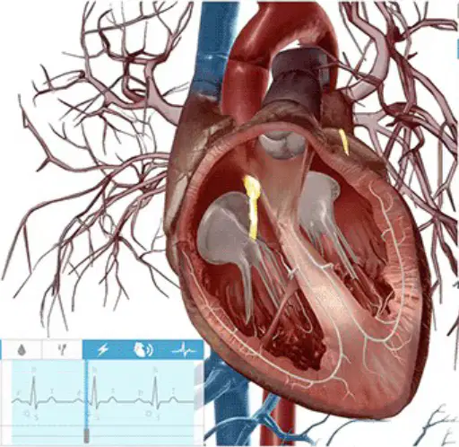
Which pathway correctly traces the electrical signal after reaching the AV node?
AV node → atrioventricular bundle (bundle of His) → bundle fibers → Purkinje fibers
AV node → SA node → Purkinje fibers → bundle of His
AV node → pulmonary artery → Purkinje fibers → ventricles
AV node → aorta → SA node → bundle fibers
Which ECG component corresponds to ventricular repolarization, when electrical activity returns to baseline and the ventricles relax?
T wave
P wave
QRS complex
PR interval
Which pair correctly matches an ECG feature with the cardiac event it reflects?
P wave — signal from SA node leading to atrial contraction
ST interval — atrial contraction and relaxation
QRS complex — atrial repolarization clearly visible
T wave — ventricular depolarization
PR interval — time from end of P wave to end of QRS complex
Which statement best describes the main purpose of an ECG in medical practice?
To measure blood pressure in the arteries
To record the heart’s electrical activity to help diagnose conditions
To check lung capacity during exercise
To determine a person’s body temperature after illness
A patient reports chest pain and shortness of breath. According to the material, which test is commonly used to investigate these symptoms?
MRI of the brain
ECG to assess heart electrical activity
Ultrasound of the abdomen
Vision screening for eye strain
Which consequence is associated with untreated atrial fibrillation?
Greater risk of stroke compared with people without AFib
Improved exercise tolerance
Lower chance of heart-related death
No change in blood flow to the body
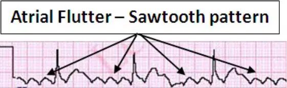
Which ECG finding is characteristic of atrial flutter?
Sawtooth appearance of the P waves
Complete absence of QRS complexes
Prolonged flat T waves
Regular, tall P waves before each QRS
Which application of ECG technology beyond diagnosing arrhythmias is mentioned?
Noninvasive detection of hypoglycemia episodes
Measuring bone density in older adults
Testing lung diffusion capacity
Determining hydration level using skin electrodes
Which structure acts as the heart’s primary pacemaker by initiating rhythmic electrical impulses?
Atrioventricular (AV) node
Purkinje fibers
Sinoatrial (SA) node
Bundle of His
If the primary pacemaker is damaged, can other specialized heart cells assume pacemaker duties? Choose the best answer.
No; only the primary pacemaker can generate impulses
Yes; secondary pacemaker regions can take over at slower rates
Yes; skeletal muscle cells can replace cardiac pacemaker cells
No; the heart stops without the primary pacemaker
What is the main advantage of gap junctions in cardiac muscle tissue?
They store calcium for contraction
They electrically couple cells to allow rapid impulse spread
They increase membrane thickness to prevent depolarization
They slow signal transmission to lengthen diastole
In typical skeletal muscle, the resting potential is about −70 mV and threshold is −55 mV. In cardiac cells, there is no true resting potential; instead, the lower cell potential is approximately −60 mV and the threshold potential for cardiac nerve cells is around −40 mV. Based on this information, which pair best represents cardiac values?
Lower potential −70 mV; threshold −55 mV
Lower potential −60 mV; threshold −40 mV
Lower potential −50 mV; threshold −30 mV
Lower potential −80 mV; threshold −60 mV
The sarcoplasmic reticulum in heart tissue is very high in which ion needed to maintain the plateau phase of heart muscle contraction?
Sodium (Na+)
Potassium (K+)
Calcium (Ca2+)
Chloride (Cl−)
Why is the plateau phase of the cardiac action potential critical for the heart’s function?
It shortens contraction so more beats can occur
It prevents synchronized contraction of cells
It sustains contraction long enough for effective pumping
It eliminates the need for calcium influx
The total refractory period for typical skeletal muscle is about 1 millisecond. What is the approximate refractory period for heart muscle tissue?
1 millisecond
5 milliseconds
200 milliseconds
1000 milliseconds
Which statement best defines venipuncture?
Insertion of a needle into an artery to measure pressure
Insertion of a needle into a vein to draw blood or give medication
Compression of a vein to stop bleeding
Surgical removal of a varicose vein
Which term describes a localized bulge in an artery wall caused by weakening of the vessel?
Atherosclerosis
Aneurism
Phlebitis
Ischemia
Arteriosclerosis versus atherosclerosis—what is the most accurate distinction?
Arteriosclerosis is hardening of arteries generally; atherosclerosis is plaque buildup of fats within arterial walls
Arteriosclerosis is venous inflammation; atherosclerosis is arterial spasm
Arteriosclerosis is low blood pressure; atherosclerosis is high blood pressure
Arteriosclerosis is clot formation; atherosclerosis is vessel dilation
Which term refers to abnormally low blood pressure?
Hypertension
Hypotension
Ischemia
Phlebitis
Deep vein thrombosis (DVT) most nearly means which of the following?
Inflammation of a superficial vein
Formation of a blood clot in a deep vein, often in the legs
Bursting of an artery with internal bleeding
Spasm of coronary arteries
Peripheral Vascular Disease (PVD) primarily involves which system and effect?
Central nervous system and plaque in cerebral arteries
Peripheral circulation with narrowed or blocked vessels reducing blood flow to limbs
Respiratory system and alveolar collapse
Endocrine system and hormone imbalance
Intermittent claudication is most closely associated with which symptom pattern?
Constant chest pain at rest
Cramping pain in a limb during activity that improves with rest due to reduced blood flow
Sudden numbness on one side of the body lasting seconds
Diffuse joint pain unrelated to movement
Phlebitis refers to what condition?
Inflammation of a vein
Inflammation of the heart muscle
Inflammation of an artery
Inflammation of lymph nodes
Which statement correctly describes a Transient Ischemic Attack (TIA)?
Permanent loss of brain function from prolonged ischemia
Short-term neurological deficits caused by temporary reduced blood flow to the brain
Cardiac event due to coronary plaque rupture
Pulmonary event due to embolism in the lungs only
A cerebrovascular incident (stroke) most directly results from which event?
Overproduction of red blood cells
Interruption of blood supply to brain tissue, either by blockage or bleeding
Infection of the meninges by bacteria
Failure of venous valves in the legs
Ischemia is best defined as which of the following?
Complete absence of oxygen in the blood
Reduced blood flow to tissues leading to oxygen shortage
Excessive blood flow causing hypertension
Inflammation of arterial walls
Which pairing of vessels correctly states the direction of blood flow relative to the heart?
Arteries carry blood toward the heart; veins carry blood away from the heart
Arteries carry blood away from the heart; veins carry blood toward the heart
Both arteries and veins carry blood away from the heart
Capillaries carry blood directly from the heart to the lungs
Normal blood pressure is often approximated as 120/80 mmHg. What do these two numbers represent?
Systolic and diastolic pressures, respectively
Diastolic and systolic pressures, respectively
Pulse pressure and mean arterial pressure
Venous pressure and capillary pressure
Which arteries are the first branches coming off the aorta to supply the heart itself?
Left and right coronary arteries
Left and right carotid arteries
Left and right subclavian arteries
Left and right iliac arteries
Which set lists the typical three major branches of the aortic arch and their general destinations?
Left coronary, right coronary, celiac—heart and abdomen
Brachiocephalic trunk, left common carotid, left subclavian—head and upper limbs
Renal, superior mesenteric, inferior mesenteric—kidneys and intestines
Common iliac, femoral, popliteal—pelvis and leg
From the abdominal aorta, which arteries primarily supply the stomach, spleen, liver, and kidneys?
Coronary arteries supply stomach and spleen; carotids supply kidneys
Celiac trunk branches supply stomach, spleen, and liver; renal arteries supply kidneys
Brachiocephalic trunk supplies liver; femoral arteries supply kidneys
Superior mesenteric supplies liver; inferior mesenteric supplies spleen
Starting at the common iliac arteries, which pathway best describes how blood reaches the feet?
Common iliac → renal → tibial → plantar
Common iliac → external iliac → femoral → popliteal → tibial/plantar arteries
Common iliac → internal carotid → brachial → radial → digital
Common iliac → hepatic → splenic → gastric → pedal
Which major vein returns blood from the lower body to the heart by traveling through the diaphragm into the abdominal cavity?
Inferior vena cava
Superior vena cava
Portal vein
Azygous vein
The inferior vena cava enters the abdominal cavity at the level of which type of vertebra?
Cervical vertebra
Thoracic vertebra
Sacral vertebra
Coccygeal vertebra
Which set of veins primarily returns blood from abdominal organs to the inferior vena cava?
Hepatic, renal, and gonadal veins
Carotid, jugular, and subclavian veins
Pulmonary, coronary, and portal veins
Femoral, popliteal, and tibial veins
Blood from the lower limbs mostly returns via which vein, while blood from the pelvic cavity itself returns via the internal veins of the pelvis?
Femoral vein; internal iliac vein
Great saphenous vein; external iliac vein
Popliteal vein; common iliac vein
Anterior tibial vein; lumbar vein
Which veins drain the kidneys directly into the inferior vena cava?
Renal veins
Hepatic veins
Lumbar veins
Jugular veins
Which statement best describes the gonadal veins?
They drain reproductive organs and empty into larger abdominal veins
They carry oxygenated blood to the gonads
They originate in the thoracic cavity
They connect the heart directly to the pelvic cavity
Below a named ligament near the groin, which artery begins its course down the leg before becoming the popliteal vein behind the knee?
Femoral artery
Tibial artery
Fibular artery
Axillary artery
In the lower leg, which two deep veins return blood from the area around the tibia?
Anterior tibial and posterior tibial veins
Great saphenous and small saphenous veins
Femoral and popliteal veins
Hepatic and renal veins
Which vein returns blood from the fibula region and is named after the associated artery?
Fibular (peroneal) vein
Anterior tibial vein
Posterior tibial vein
Popliteal vein
Which pair of large superficial veins drain the upper and lower leg and are commonly involved in varicose veins?
Greater saphenous and lesser saphenous veins
Femoral and popliteal veins
Anterior tibial and posterior tibial veins
Renal and hepatic veins
Which major vein collects blood from the head, neck, and upper extremities and drains into the heart?
Superior vena cava
Inferior vena cava
Portal vein
Coronary sinus
The left and right brachiocephalic trunks drain directly into which vessel?
Superior vena cava
Inferior vena cava
Aorta
Pulmonary trunk
Which venous system connects lumbar veins to the superior vena cava via the thoracic vertebrae?
Azygous venous system
Portal venous system
Coronary venous system
Pulmonary venous system
As lumbar veins ascend and change names at the thoracic level, which set of veins do they form to drain the thoracic and abdominal cavity walls?
Azygous, hemi-azygous, and accessory-azygous veins
Renal, hepatic, and portal veins
Jugular, subclavian, and vertebral veins
Femoral, popliteal, and tibial veins
Which veins receive blood from the cephalic vein in the arm pit and continue downward as the axillary vein?
Subclavian veins
Jugular veins
Renal veins
Saphenous veins
Which pathway best traces venous return from the upper limb to the heart?
Cephalic vein → subclavian vein → brachiocephalic trunk → superior vena cava
Tibial vein → popliteal vein → femoral vein → inferior vena cava
Hepatic vein → portal vein → aorta → heart
Renal vein → lumbar vein → azygous vein → inferior vena cava
Which system provides a collateral connection between the inferior and superior vena cavae through veins that run alongside the vertebral column?
Azygous venous system
Pulmonary arterial system
Coronary arterial system
Portal hepatic system
Choose the correct responses.
Ventricular Depolarization
Atrial Depolarization
Ventricular Repolarization

Identify this rhythm
Sinus tachycardia
Normal Sinus Rhythm
Supraventricular Tachycardia
Atrial fibrillation

Identify this rhythm
Sinus Tachycardia
Atrial fibrillation
Atrial flutter
Normal Sinus Rhythm

Identify this rhythm
Ventricular Fibrillation
Ventricular Tachycardia
Pulseless Electrical Activity
Atrial fibrillation
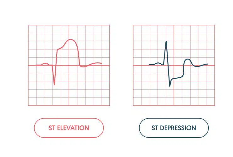
What does the depressed ST wave in the image possibly indicate
A heart block leading to ischemia
Rapid hear beat
low blood pressure
heart attack (myocardial infarction)
What does an elevated ST wave in the image possibly indicate
A heart block leading to ischemia
Rapid hear beat
low blood pressure
heart attack (myocardial infarction)
There are (a) pulmonary veins that return to the left atrium of the heart.
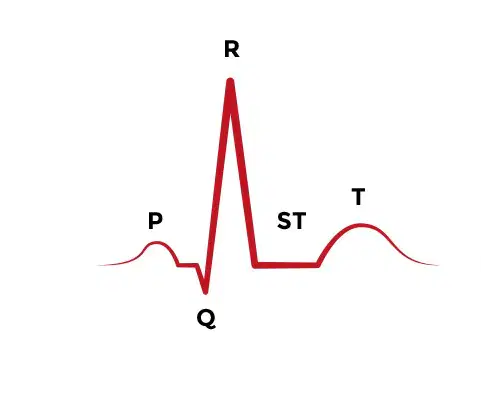
Look at the image. Why is the line labeled ST flat?
The heart is resting
The atria are repolarizing
The person has damage from a heart attack
They have a heart block
Label the heart
right atrium
left atrium
left ventricle
right ventricle
Label the heart
right atrium
left atrium
left ventricle
right ventricle
Correctly identify the following areas of the heart.

Match the following terms to their description
When blood leaves this chamber of the heart, it heads to the lungs.
Right ventricle
The main artery which distributes oxygenated blood to the entire body
Aorta
This blood vessel splits in two (L/R) and sends deoxygenated blood to the lungs to be refreshed.
Pulmonary artery
The chamber in the heart that receives oxygenated blood from the lungs.
Left atrium

