
 Worksheets
Lecture 11 oral pathology
Total questions: 55
Worksheet time: 28mins
Cysts of the jaws are broadly classified according to the origin of the epithelium lining the cavity. Which of the following best describes an odontogenic cyst?
A cyst arising from epithelial remnants of the nasolacrimal duct
A cyst arising from odontogenic epithelium derived from the dental lamina
A cyst arising from embryonic remnants of the thyroglossal duct
A cyst arising from respiratory epithelium of the maxillary sinus
A cyst formed from sequestrated surface epithelium of the oral mucosa
A 32-year-old patient presents with a well-defined, unilocular radiolucency in the posterior mandible. The lesion is lined by parakeratinised stratified squamous epithelium with palisaded basal cells. Histopathological diagnosis confirms an odontogenic keratocyst. Based on the current WHO classification principles, how should this cyst be correctly categorised?
Developmental non-odontogenic cyst
Inflammatory odontogenic cyst
Developmental odontogenic cyst
Pseudocyst of the jaws
Soft tissue cyst of the oral mucosa
A 35-year-old patient presents with a well-defined radiolucency near the apex of a non-vital upper incisor. Histology reveals epithelial lining with Rushton bodies. Based on classification, to which group does this cyst most likely belong?
Non-odontogenic cyst
Developmental odontogenic cyst
Inflammatory odontogenic cyst
Soft tissue cyst of the gingiva
Cystic odontogenic tumour
Which of the following mechanisms primarily accounts for the enlargement of most jaw cysts?
Cellular proliferation of the cyst wall
Accumulation of keratin within the lumen
Expansion due to internal hydrostatic pressure
Invasion of adjacent bone by inflammatory cells
Deposition of new bone within the cyst cavity
Why do odontogenic keratocysts typically fail to cause significant jaw expansion until they become very large?
They contain inflammatory exudate that prevents expansion
They enlarge by epithelial proliferation rather than osmotic expansion
They lack significant internal hydrostatic pressure
They develop only within the cortical bone
They arise exclusively in soft tissue sites
A 40-year-old man presents with a painless, slow-growing swelling of the mandible. On palpation, a crackling sensation is noted, and radiographs show a well-defined radiolucency with a thin sclerotic border. Which clinical feature best explains this finding?
Infection of the cyst wall
New bone formation by periosteum
Degeneration of cyst lining epithelium
Resorption of adjacent tooth roots
Deposition of keratin within the cyst cavity
Which of the following radiological features is most typical of a slowly enlarging jaw cyst?
Ill-defined radiolucent margins
Mixed radiolucent and radiopaque appearance
Sharply defined unilocular radiolucency with corticated margin
Irregular sclerotic lesion with internal septa
Generalised widening of the periodontal ligament space
What is the most common and effective method of treating most jaw cysts?
Aspiration and antibiotic therapy
Enucleation with primary closure
Curettage followed by irrigation
Marsupialisation only
Resection of affected bone segment
Which of the following best explains the rationale for performing marsupialisation in selected cysts?
To prevent loss of adjacent teeth
To promote epithelial transformation of cyst lining
To decompress large cysts and prevent jaw fracture
To obtain complete cyst lining for histopathology
To allow keratinisation of the cystic epithelium
A 30-year-old patient presents with a large unilocular radiolucency in the mandible. Due to the risk of fracture, the surgeon opens the cyst to the oral cavity, sutures the lining to mucosa, and maintains a gauze dressing for decompression. Which procedure has been performed?
Enucleation
Marsupialisation
Apicectomy
Curettage
Partial mandibulectomy
Which of the following is a key diagnostic feature of a radicular cyst?
Associated with a vital tooth
Associated with an impacted tooth
Arises from the reduced enamel epithelium
Originates from a non-vital tooth
Arises from the cell rests of Serres
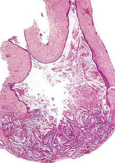
Which of the following histological findings most strongly supports the diagnosis of a radicular cyst?
Keratinised stratified squamous epithelium
Non-keratinising epithelial lining with cholesterol clefts and mural nodules
Pseudostratified ciliated columnar epithelium with goblet cells
Fibrous capsule without epithelial lining
Orthokeratinised squamous epithelium with basal palisading
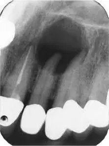
A 45-year-old man presents with a painless swelling above an upper lateral incisor. The tooth is non-vital, and radiographs show a sharply defined unilocular radiolucency with a corticated margin. What is the most likely diagnosis?
Residual cyst
Dentigerous cyst
Radicular cyst
Lateral periodontal cyst
Odontogenic keratocyst
What is the main distinguishing feature of a lateral radicular cyst?
It develops adjacent to a vital tooth root
It arises at the opening of a lateral accessory canal of a non-vital tooth
It originates from remnants of the dental lamina
It is always associated with pericoronitis
It occurs exclusively in deciduous teeth
Which of the following best differentiates a residual radicular cyst from other radicular cysts?
It is associated with a vital tooth
It forms following removal of the associated non-vital tooth
It shows keratinisation of the epithelial lining
It commonly arises in the deciduous dentition
It always contains Rushton bodies
A 10-year-old presents with a buccal swelling adjacent to an erupting mandibular first molar. The tooth is vital but surrounded by mild pericoronitis. Radiography reveals a cystic lesion in the buccal bifurcation area. What is the most likely diagnosis?
Dentigerous cyst
Lateral periodontal cyst
Mandibular buccal infected cyst
Residual radicular cyst
Odontogenic keratocyst
Which of the following best defines a dentigerous cyst?
A cyst arising at the apex of a non-vital tooth
A cyst surrounding the crown of an unerupted tooth with lining attached at the cementoenamel junction
A cyst associated with the lateral root surface of a vital tooth
A cyst forming within the nasopalatine canal
A cyst related to a developing tooth germ before enamel formation
Which radiographic feature most reliably distinguishes a dentigerous cyst from an enlarged dental follicle?
Multilocular appearance with septa
Corticated outline with radiolucency exceeding 2–3 mm around the crown
Association with a vital tooth
Irregular radiopaque margin
Root resorption of adjacent teeth only
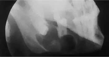
A 20-year-old male presents with an unerupted lower third molar displaced towards the inferior border of the mandible. Radiographs show a unilocular radiolucency enclosing the crown with a well-defined corticated margin. What is the most likely diagnosis?
Odontogenic keratocyst
Radicular cyst
Dentigerous cyst
Lateral periodontal cyst
Ameloblastoma
Which of the following statements correctly describes an eruption cyst?
It occurs in adults and causes cortical bone expansion
It develops around the roots of erupted teeth
It is a superficial form of dentigerous cyst associated with an erupting tooth in children
It is lined by keratinised squamous epithelium
It always requires surgical enucleation
Which of the following features best defines an odontogenic keratocyst (OKC)?
Cyst arising from the reduced enamel epithelium of an unerupted tooth
Developmental odontogenic cyst lined by parakeratinised stratified squamous epithelium with palisaded basal cells
Inflammatory cyst arising from the epithelial rests of Malassez
Cystic lesion of the maxilla associated with a vital tooth
Odontogenic cyst containing mucous-secreting cells and duct-like structures
Which radiographic feature is most characteristic of an odontogenic keratocyst?
Unilocular radiolucency with displacement and resorption of adjacent teeth
Poorly defined radiolucent area without cortication
Multilocular or scalloped radiolucency extending along the medullary cavity with minimal cortical expansion
Radiopaque lesion involving the tooth crown
Heart-shaped radiolucency at the midline of the palate
A 25-year-old male presents with a large asymptomatic radiolucent lesion in the posterior mandible extending into the ramus. The lesion is multilocular, with no cortical expansion or root resorption. Biopsy reveals parakeratinised epithelium with a palisaded basal cell layer. What is the most appropriate management approach?
Observation only
Enucleation with primary closure
Marsupialisation followed by secondary enucleation
Simple aspiration and curettage
Resection and reconstruction in all cases
Which of the following factors contributes most significantly to recurrence of odontogenic keratocysts?
Thick fibrous cyst wall that prevents enucleation
Poor blood supply to the cyst lining
Residual epithelial remnants and daughter cysts left after incomplete removal
Excessive bone formation around the cyst
Infection causing cyst wall fibrosis
Which gene mutation is primarily responsible for Basal Cell Naevus Syndrome (Gorlin-Goltz syndrome)?
SHH gene on chromosome 7p
PTCH gene on chromosome 9q
TP53 gene on chromosome 17p
APC gene on chromosome 5q
SMO gene on chromosome 7q
Which of the following clinical features is most characteristic of Basal Cell Naevus Syndrome?
Multiple epidermoid cysts on the scalp
Multiple odontogenic keratocysts and palmar pits
Jaw expansion due to dentigerous cysts
Bilateral parotid gland cysts
Periapical radiolucencies involving non-vital teeth
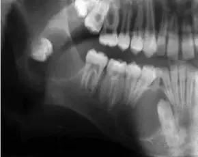
A 15-year-old patient presents with multiple mandibular cysts resembling odontogenic keratocysts on radiographs. His father had a history of recurrent jaw cysts and multiple skin lesions on the face. Clinical examination reveals small dark pits on the palms. What is the most likely diagnosis?
Orthokeratinised odontogenic cyst
Multiple dentigerous cysts
Basal cell naevus (Gorlin-Goltz) syndrome
Langerhans cell histiocytosis
Cleidocranial dysplasia
Which of the following best differentiates an orthokeratinised odontogenic cyst from an odontogenic keratocyst?
Orthokeratinised cysts are more aggressive and recur frequently
Orthokeratinised cysts are linked with PTCH mutations and syndromic forms
Orthokeratinised cysts have orthokeratinised linings and are not associated with Basal Cell Naevus Syndrome
Orthokeratinised cysts are inflammatory in origin
Orthokeratinised cysts contain Rushton bodies in the lining epithelium
Which of the following best describes the lateral periodontal cyst?
Inflammatory cyst arising from a non-vital tooth apex
Developmental odontogenic cyst located beside the root of a vital tooth
Cyst associated with unerupted teeth in the follicular space
Soft tissue cyst forming above the alveolar ridge in children
Multilocular cyst arising exclusively in the maxilla
Which histological feature is most characteristic of a lateral periodontal cyst?
Parakeratinised epithelial lining with palisaded basal cells
Respiratory-type epithelium with goblet cells
Thin squamous or cuboidal lining with focal epithelial plaques showing clear cells
Pseudostratified columnar lining with cilia
Orthokeratinised lining with prominent rete ridges
A 60-year-old patient presents with a painless swelling in the anterior mandible. Radiographs reveal a multilocular, well-circumscribed radiolucency between the roots of vital lower premolars. Histology shows a thin epithelial lining with focal plaques identical to those seen in lateral periodontal cysts. What is the most likely diagnosis?
Odontogenic keratocyst
Botryoid odontogenic cyst
Lateral radicular cyst
Glandular odontogenic cyst
Central giant cell granuloma
Which feature is diagnostic of a calcifying odontogenic cyst?
Orthokeratinised epithelial lining
Presence of ghost cells that may calcify
Pseudostratified columnar epithelium with mucous cells
Parakeratinised lining with palisaded basal cells
Thick fibrous wall with Rushton bodies
In a calcifying odontogenic cyst, what is the usual histological appearance of the lining epithelium?
Resembles reduced enamel epithelium
Stratified squamous epithelium with rete pegs
Ameloblastoma-like epithelium with cuboidal basal cells and stellate reticulum
Pseudostratified columnar respiratory epithelium
Parakeratinised lining with reverse polarity
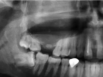
A 25-year-old patient presents with a painless swelling in the anterior maxilla. Radiographs reveal a unilocular radiolucency containing small flecks of radiopacity. Histology shows ghost cells that have undergone calcification and induction of dentinoid formation. What is the most likely diagnosis?
Ameloblastoma
Odontogenic keratocyst
Calcifying odontogenic cyst
Lateral periodontal cyst
Dentigerous cyst
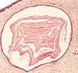
Which of the following statements about gingival cysts of the newborn is correct?
They are lined by keratinised epithelium and persist into childhood
They are developmental cysts arising from the rests of Malassez
They are small keratin-filled cysts derived from the rests of Serres
They occur exclusively on the midpalatine raphe
They require surgical removal to prevent recurrence
Epstein’s pearls differ from gingival cysts of the newborn primarily in which aspect?
They arise from odontogenic epithelium
They are found along the midpalatine raphe
They contain mucous cells instead of keratin
They are larger and require excision
They are associated with unerupted teeth
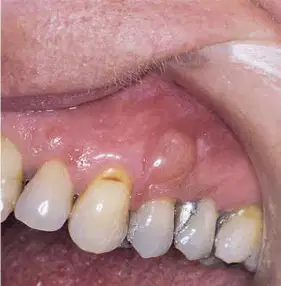
A 50-year-old patient presents with a painless, dome-shaped swelling in the upper canine region. The lesion measures 0.8 cm and has caused slight pressure erosion of the underlying bone. The overlying tooth is vital. What is the most likely diagnosis?
Lateral radicular cyst
Lateral periodontal cyst
Gingival cyst of adult
Residual radicular cyst
Odontogenic keratocyst
Which of the following statements best describes a nasopalatine duct cyst?
An odontogenic cyst arising from rests of Malassez
A developmental cyst of vestigial nasopalatine duct origin
A cyst derived from periapical inflammation of a non-vital tooth
A cyst arising from mucous retention in the minor salivary glands
An epithelial-lined cavity associated with an unerupted tooth
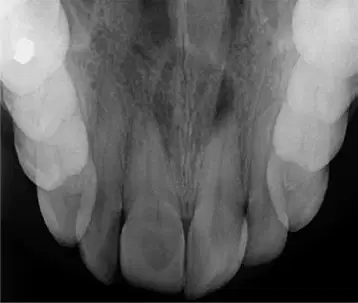
What is the characteristic radiographic feature of a nasopalatine duct cyst in the anterior occlusal or periapical films?
Ill-defined radiolucency
Heart-shaped or pear-shaped radiolucency
Unilocular, well-defined radiolucent lesion with sclerotic border
Unilocular radiolucency with root resorption
Well-defined radiolucent, radiopaque or mixed opacity
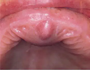
A 42-year-old man presents with a swelling in the anterior palate between the central incisors. The teeth are vital, and radiography reveals a corticated, heart-shaped radiolucency in the midline. Histology shows ciliated columnar and stratified squamous epithelium with mucous glands. What is the most likely diagnosis?
Radicular cyst
Nasopalatine duct cyst
Residual cyst
Lateral periodontal cyst
Odontogenic keratocyst
Which of the following statements best describes a nasolabial cyst?
It is an odontogenic cyst arising from rests of Malassez
It is a developmental soft tissue cyst derived from the nasolacrimal duct
It is a cyst arising from the nasopalatine duct
It is a cystic degeneration of the maxillary sinus lining
It is a cyst associated with an unerupted canine tooth
Which of the following clinical features is most characteristic of a nasolabial cyst?
Swelling in the anterior palate between central incisors
Intraosseous swelling causing egg-shell crackling
Extraosseous swelling beneath the nasolabial fold distorting the nostril
Midline swelling in the neck that moves with swallowing
Dome-shaped swelling in the canine–premolar gingival region
Which of the following statements best describes a sublingual dermoid cyst?
It is an odontogenic cyst arising from rests of Malassez
It is a developmental cyst lined by keratinising epithelium with skin adnexae
It is a salivary retention cyst related to the sublingual gland
It is a cystic lesion arising from the thyroglossal duct
It is a cystic degeneration of lymphatic tissue
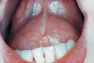
A 25-year-old patient presents with a firm, midline swelling beneath the tongue that elevates the floor of the mouth and interferes with speech. The lesion is non-translucent and not related to meals. What is the most likely diagnosis?
Ranula
Sublingual dermoid cyst
Thyroglossal duct cyst
Epidermoid cyst of the neck
Nasolabial cyst
Which of the following statements best describes a thyroglossal duct cyst?
It develops from remnants of the first branchial cleft
It arises from epithelial remnants of the embryonic thyroglossal duct
It is a cystic degeneration of cervical lymph nodes
It results from obstruction of the thyroglossal gland duct
It originates from the nasolacrimal duct
A 17-year-old presents with a painless, fluctuant swelling in the midline of the neck just below the hyoid bone. The swelling moves upward when he protrudes his tongue. Histology shows a cyst lined by squamous epithelium containing thyroid follicles. What is the most likely diagnosis?
Thyroglossal duct cyst
Branchial cyst
Dermoid cyst
Epidermoid cyst
Cystic hygroma
A 28-year-old woman presents with a soft, fluctuant swelling located at the anterior border of the sternocleidomastoid, just above the angle of the mandible. The swelling has gradually enlarged over months and is not tender. There is no movement with swallowing or tongue protrusion. Which of the following is the most likely diagnosis?
Thyroglossal duct cyst
Branchial cyst
Cystic hygroma
Dermoid cyst
Lymphadenitis
Branchial cysts most commonly arise from which pharyngeal arch?
First arch
Third arch
Fourth arch
Second arch
Fifth arch
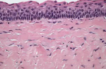
A 27-year-old patient presents with a painless swelling in the posterior mandible. Radiographic examination reveals a multilocular radiolucency extending along the medullary cavity with minimal bone expansion. Histology shows a parakeratinized lining with a palisaded basal layer. What is the most likely diagnosis?
Dentigerous cyst
Radicular cyst
Ameloblastoma
Odontogenic keratocyst
Lateral periodontal cyst
A 15-year-old presents with a painless midline neck swelling that moves upward during swallowing. Histology reveals stratified squamous epithelium with clusters of ectopic thyroid tissue. What is the best management option?
Marsupialization
Simple excision
Sistrunk procedure
Enucleation
Observation
Histological examination of a thyroglossal duct cyst often reveals:
Ghost cells
Parakeratinized epithelial lining
Stratified squamous epithelium or respiratory epithelium with ectopic thyroid tissue
Lymphoid aggregates with germinal centers
Mucous metaplasia
Which of the following teeth is most commonly associated with dentigerous cysts?
Lower premolars
Lower third molars
Upper central incisors
Upper first molars
Lower canines
Odontogenic keratocysts are most commonly found in the:
Anterior maxilla
Posterior maxilla
Posterior mandible
Anterior mandible
Midline palate
A 25-year-old male presents with an unerupted lower third molar associated with a large unilocular radiolucency that envelopes the crown. The cyst is attached at the cementoenamel junction. What is the most likely diagnosis?
Odontogenic keratocyst
Radicular cyst
Dentigerous cyst
Calcifying odontogenic cyst
Ameloblastoma
A 48-year-old patient presents with a painless, slowly enlarging swelling in the anterior mandible. Radiographs show a well-defined unilocular radiolucency
displacing adjacent teeth. Histology reveals epithelial thickenings with mucous cells forming gland-like structures that secrete mucin. Which is the most likely
diagnosis?
Botryoid odontogenic cyst
Lateral radicular cyst
Glandular odontogenic
Odontogenic keratocyst
Residual radicular cyst

