
 Worksheets
Muscle Structure and Function Quiz
Total questions: 191
Worksheet time: 2hrs 36mins
What is the detailed structure of skeletal muscle, myofibrils, and myofilaments referred to as?
Ultrastructure
Molecular structure
Sliding filament theory
Ratchet mechanism
Which two proteins are primarily involved in the molecular structure of muscle contraction?
Myosin and actin
Tropomyosin and troponin
Collagen and elastin
Keratin and fibrin
What is the basic repeating unit of muscle?
Myofibril
Sarcomere
Actin
Fascia
What does the term "muscle ultrastructure" refer to?
The arrangement of blood vessels in muscles
A complex arrangement of many protein-protein interactions
The structure of muscle fibers only
The organization of fascia in muscles
What is the role of fascia in muscle structure?
It is the basic repeating unit of muscle
It surrounds and supports muscle fibers
It is a type of protein involved in muscle contraction
It is a blood vessel supplying nutrients to muscles
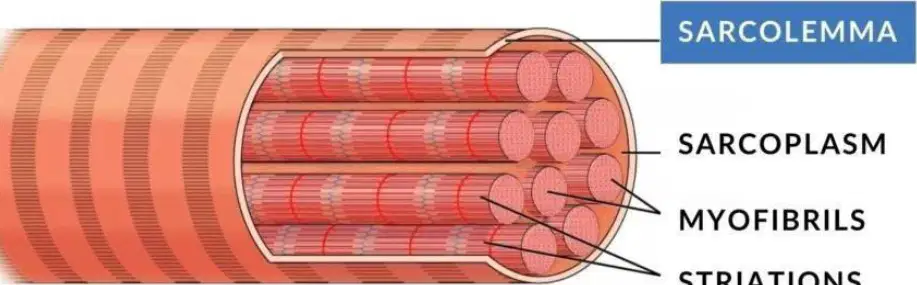
What is the cytoplasmic matrix of a muscle fiber called?
Sarcoplasm
Sarcolemma
Myofibril
Striation
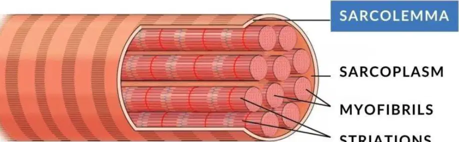
What smaller structures are embedded in the cytoplasmic matrix of a muscle fiber?
Sarcolemma
Sarcoplasm
Myofibrils
Striations
What is the term used for cytosol in muscle fibers?
Myofibril
Sarcoplasm
Sarcolemma
Mitochondria
What is the primary function of transverse tubules (T-tubules) in muscle fibers?
Store calcium
Transmit electrical signals
Produce ATP
Surround myofibrils
What are transverse tubules (T-tubules) made of?
Flattened vesicles
Series of membranous folds extending from the plasma membrane
Glycogen-rich cytosol
Thick and thin filaments
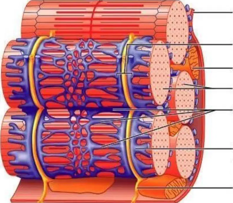
Which structure in the muscle fiber is responsible for storing calcium ions?
Myofibril
Sarcolemma
Sarcoplasmic reticulum
Mitochondria
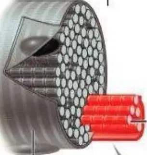
What is the function of the mitochondria in muscle fibers?
To store calcium ions
To produce energy for muscle contraction
To transmit electrical signals
To form the structural framework of the muscle fiber
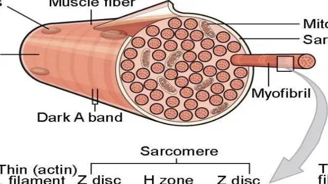
What is the diameter of myofibrils?
1 mm
1 μm
10 μm
0.1 μm
What is the basic contractile unit of a muscle fiber?
Myofibril
Sarcomere
Actin
Myosin
Which two main protein filaments are responsible for muscular contraction?
Actin and Myosin
Myosin and Tropomyosin
Actin and Troponin
Myosin and Titin
What is the function of actin and myosin in the sarcomere?
They provide energy for the muscle
They are responsible for muscular contraction
They store calcium ions
They transport oxygen to the muscles

What is the role of the M-line in the sarcomere?
It anchors the thin filaments.
It is the boundary of the sarcomere.
It holds the thick filaments in place.
It contains both thick and thin filaments.
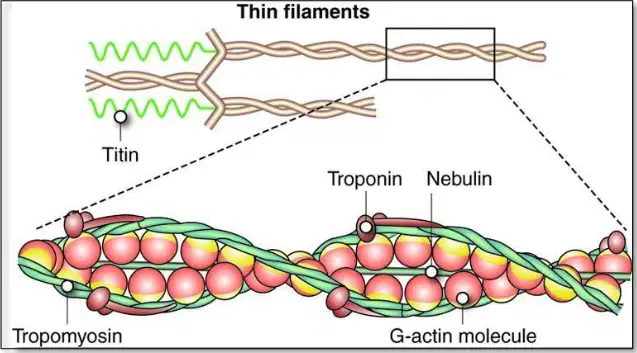
What are the three components that make up the thin filament in muscle structure?
F-actin, tropomyosin, and troponin
Myosin, titin, and nebulin
Tropomyosin, myosin, and titin
Actin, nebulin, and myosin
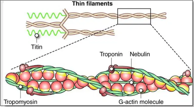
Which protein molecule in the thin filament is wound into a double helix?
Tropomyosin
F-actin
Troponin
Myosin
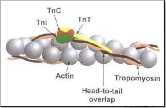
What is the role of troponin in the thin filament structure?
It forms the double helix structure of actin.
It lies in the groove of the F-actin helix.
It consists of globular proteins attached to the tropomyosin strand.
It connects actin to myosin filaments.
Which proteins regulate actin in thin filament arrangement?
Tropomyosin and troponin
Myosin and actin
Tropomyosin and myosin
Actin and troponin
What does the TnC protein bind to in the thin filament arrangement?
Tropomyosin
F-actin
Calcium (Ca²⁺) ions
Myosin
What is the approximate number of myosin polypeptides in thick filaments?
100
200
400
600
What do the heads of myosin filaments form when they stick out?
Thick central portion
Cross bridges with actin filaments
Light meromyosin
A-band
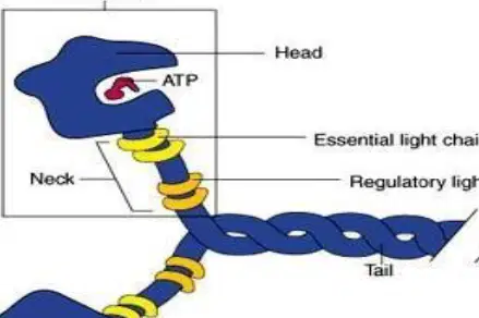
What is the function of each myosin head?
Binding to actin and storing energy
Binding to ATP and hydrolyzing it
Forming the tail domain
Regulating light chains
Who independently proposed the sliding filament theory in 1950 to explain how muscle fibers contract?
Albert Einstein and Isaac Newton
A. F. Huxley and H. E. Huxley
Charles Darwin and Gregor Mendel
James Watson and Francis Crick
What happens when the actin and myosin filaments slide between each other?
The muscle relaxes
The muscle contracts
The muscle elongates
The muscle remains stationary
What is the role of cross bridges in the sliding filament theory?
They prevent the actin and myosin filaments from interacting
They facilitate the sliding of actin and myosin filaments past each other
They provide structural support to the sarcomere
They are responsible for muscle relaxation
What is formed between the myosin head and the myosin binding site on the actin filament during contraction?
ATP molecules
Cross-bridges
Actin filaments
Myosin heads
Which step in the powerstroke mechanism involves the movement of the myosin head?
Attachment of myosin
Breaking of actin/myosin interaction
Contraction of the myosin head
Re-attachment further along the actin filament
What is the difference between isometric and isotonic contraction?
Isometric contraction involves movement, while isotonic contraction does not
Isometric contraction involves no change in muscle length, while isotonic contraction involves muscle length change
Isometric contraction is voluntary, while isotonic contraction is involuntary
Isometric contraction occurs in the heart, while isotonic contraction occurs in the skeletal muscles
Which bone is located in the upper arm?
Radius
Ulna
Humerus
Tibia
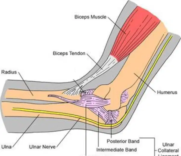
What is the name of the nerve shown in the diagram?
Ulnar nerve
Biceps nerve
Radius nerve
Ligament nerve
How many bones are muscles usually attached to?
One bone
Two different bones
Three bones
No bones
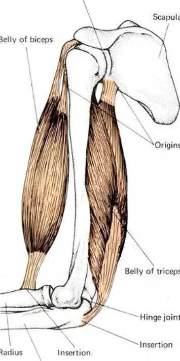
Which part of the muscle is labeled as "Belly of biceps" in the diagram?
The fixed attachment of the muscle
The middle part of the muscle
The moving attachment of the muscle
The joint of the muscle
How is any movement in the body brought about?
By the relaxation of muscles
By the contraction of a number of muscles working together
By the stretching of muscles
By the expansion of muscles
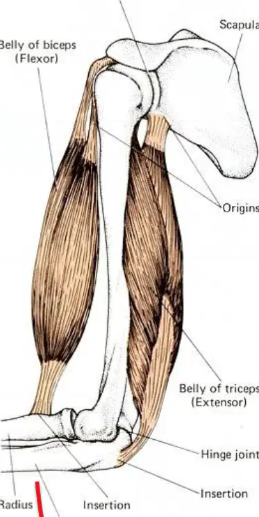
What is the function of the triceps brachii muscle?
Flexes the elbow
Extends the arm
Rotates the wrist
Moves the fingers
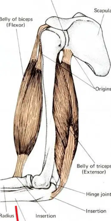
Which joint is located at the shoulder?
Ball and socket joint
Hinge joint
Elbow joint
Wrist joint
What does the term "action is complex" mean in the context of muscle movement?
Only one muscle is involved in movement.
Movement involves many muscles working together.
Flexion and extension are the only actions possible.
Muscles do not play a role in movement.
What is the name of the loose connective tissue sheath that binds each muscle?
Perimysium
Epimysium
Endomysium
Myofibril

What do myofibrils contain?
Tendons
Sarcomeres
Bones
Nerves
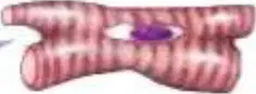
Where can cardiac muscles be found in the human body?
Attached to bones
In the walls of the myocardium
In the organs
In the skin

Which type of muscle is found in organs?
Skeletal muscle
Cardiac muscle
Smooth muscle
Bone muscle
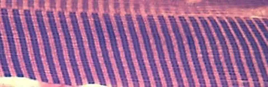
What is another name for striated muscle?
Smooth muscle
Cardiac muscle
Striped or skeletal muscle
Involuntary muscle
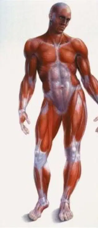
Which type of muscle is under direct nervous control?
Voluntary muscle
Involuntary muscle
Cardiac muscle
Smooth muscle
What is removed during a muscle biopsy?
A small 'tube' of muscle
A piece of skin
A bone fragment
A blood sample
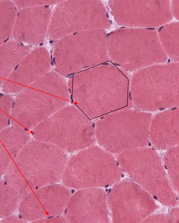
What shape are the fibers in striated muscle?
Circular
Hexagonal
Triangular
Square
What type of muscle is a cardiac muscle?
Smooth muscle
Striated muscle related to smooth muscle
Skeletal muscle
Voluntary muscle
Is the cardiac muscle voluntary or involuntary?
Voluntary
Involuntary
Both voluntary and involuntary
Neither voluntary nor involuntary

Where are the nuclei located in cardiac muscle cells?
At the edges of the cell
Centrally placed
Scattered randomly
Outside the cell
What is one characteristic of smooth muscle?
It is found in the heart.
It is a high endurance muscle.
It is controlled voluntarily.
It is attached to bones.
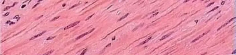
Which characteristic is true for smooth muscle tissue?
It has striations.
It has sustained contraction.
It has multiple nuclei per cell.
It is voluntary.
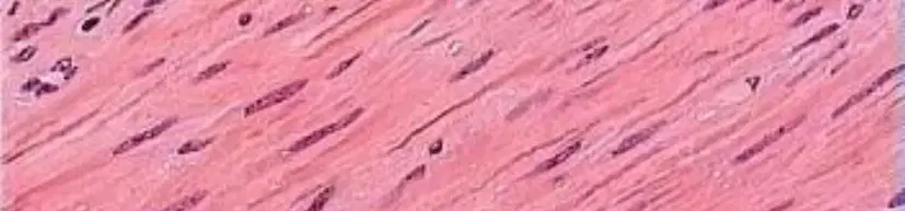
What is a key difference between smooth muscle and skeletal muscle?
Smooth muscle has striations, while skeletal muscle does not.
Smooth muscle has one nucleus per cell, while skeletal muscle has multiple nuclei per cell.
Smooth muscle is voluntary, while skeletal muscle is involuntary.
Smooth muscle is found in the heart, while skeletal muscle is found in the stomach.
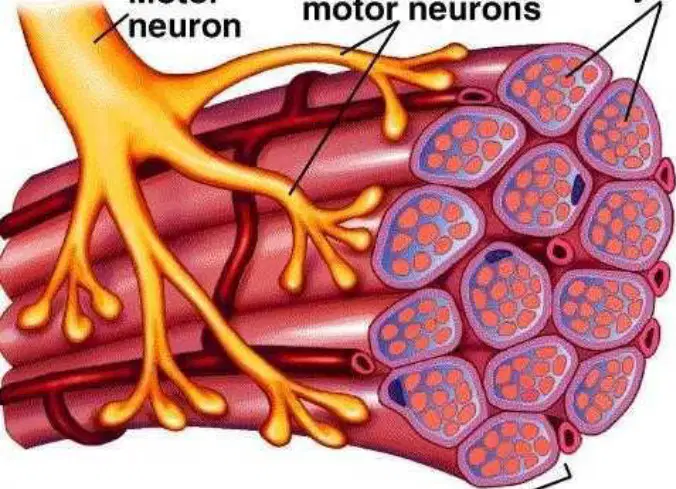
What is a motor unit composed of?
A single motor neuron and the group of muscle fibers it innervates
Multiple motor neurons and one muscle fiber
Only muscle fibers
Only motor neurons
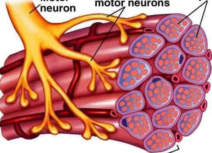
Why do some motor neurons innervate more than one muscle fiber?
Because there are fewer muscle fibers than motor neurons
Because there are considerably more muscle fibers than motor neurons
Because motor neurons are larger than muscle fibers
Because muscle fibers are not connected to motor neurons
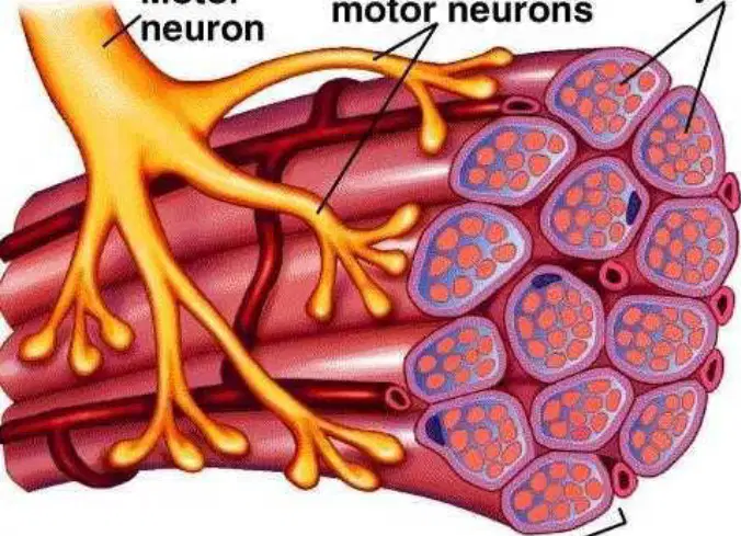
What part of the motor unit connects to the muscle fibers?
Myofibrils
Branches of motor neurons
Muscle fiber nucleus
Skeletal muscle
Which muscle has the fewest fibres per motor unit?
Eye muscles
Biceps muscles
Larynx muscle
Average body muscles
How many muscle fibres per motor unit are found in the eye muscles?
2-3 fibres/MU
10 fibres/MU
1000+ fibres/MU
150 fibres/MU
What is the average number of muscle fibres per motor unit in the human body?
10 fibres/MU
150 fibres/MU
1000+ fibres/MU
2-3 fibres/MU
What happens when there are fewer muscle fibres per motor unit?
Increased strength
Increased dexterity
Decreased control
Decreased flexibility
Which muscle type is voluntary?
Skeletal
Cardiac
Smooth
Visceral
Which muscle type has high endurance and is involuntary?
Skeletal
Cardiac
Striated
None
Smooth muscle is primarily located in:
Bones
Heart
Digestive tract & vessels
Skin
Skeletal muscles make up approximately what percentage of body mass?
20%
40%
60%
70%
How many skeletal muscles exist in the human body?
150
300
650
1000
What attaches muscle to bone?
Ligament
Cartilage
Tendon
Fascia
What attaches bone to bone?
Tendon
Ligament
Fascia
Epimysium
The fixed attachment of a muscle is known as the:
Insertion
Origin
Anchor
Muscles can only:
Push
Pull
Twist
Vibrate
Flexors function to:
Open a joint
Close a joint
Rotate a joint
Immobilize a joint
Extensors function to:
Open a joint
Close a joint
Rotate a joint
Lock a joint
12. Biceps brachii primarily:
Extends the elbow
Flexes the elbow
Rotates the wrist
Flexes the shoulder
Triceps brachii primarily:
Flexes elbow
Extends elbow
Flexes wrist
Supinates hand
The connective tissue surrounding an entire muscle is:
Endomysium
Perimysium
Epimysium
Sarcolemma
A fascicle is a bundle of:
Myofibrils
Fibers
Filaments
Cells only
Myofibrils contain the contractile unit known as:
Actin
Sarcomere
Myosin
Endomysium
Cardiac muscle contains:
Peripheral nuclei
Multiple nuclei
1-2 centrally located nuclei
No nuclei
Cardiac muscle contains unique structures called:
Nodes
Intercalated discs
Sarcomeres only
Gap fibers
Smooth muscle cells contain how many nuclei?
Many
None
1 central nucleus
2 peripheral nuclei
Smooth muscle contraction is generally:
Short and weak
Sustained
Voluntary
Uncoordinated
The functional unit of muscle activity is the:
Muscle
Sarcomere
Motor unit
Myofibril
A motor unit includes:
Motor neuron only
Muscle fibers only
Motor neuron + fibers it innervates
Spinal cord
Smaller motor units allow:
Less control
More precise control
Random contraction
Which muscle has the fewest fibers per motor unit?
Biceps
Larynx
Quadriceps
Gastrocnemius
The average number of fibers per motor unit is:
10
100
150
1000
The neuromuscular junction is also called:
Motor plate
Motor endplate
Myoneural disc
Muscle receptor
The muscle response to a single stimulus is a:
Spasm
Twitch
Tetany
Cramp
The latent period of a twitch lasts approximately:
0.005 sec
0.04 sec
0.05 sec
1 sec
The contraction period lasts:
0.005 sec
0.04 sec
0.05 sec
1 sec
The relaxation period lasts:
0.005 sec
0.04 sec
0.05 sec
1 sec
A stimulus during the refractory period produces:
Stronger contraction
Weaker contraction
No response
Tetany
Summation of contraction occurs when:
Muscle relaxes
Second stimulus arrives before relaxation
Low-frequency firing
Fiber fatigues
Increasing the number of motor units firing is:
Twitching
Recruitment
Tetany
Fatigue
Increasing the stimulus frequency leads to:
Wave summation
Relaxation
Cramping
Inhibition
Sustained maximal contraction is called:
Wave summation
Tetany
Twitching
Fatigue
Isometric contraction means:
Same tension
Same length
Changing tension
Changing length
In isometric contraction, the muscle:
Shortens
Lengthens
Stays same length
Melts
Isotonic contraction means:
Same tension
Same length
Changing tension
Changing length
Concentric contraction means muscle:
Lengthens
Shortens
Stays same
Trembles
Eccentric contraction means muscle:
Shortens
Remains still
Lengthens
Contracts isometrically
Eccentric contractions often cause:
Less damage
More muscle damage
No effect
Instant fatigue
During eccentric contraction, force is:
Greater than load
Less than load
Same as load
Not measurable
Muscles are stronger in which contraction?
Concentric
Isometric
Eccentric
None
Tetanus occurs at frequencies above:
5 pulses/sec
10 pulses/sec
20 pulses/sec
40 pulses/sec
Which muscle type is voluntary?
Skeletal
Cardiac
Smooth
Visceral
Which membrane encloses an individual muscle fiber?
Endomysium
Sarcolemma
Sarcoplasm
Perimysium
Plasmalemma
Which of the following is rich in glycogen, ATP and creatine phosphate within muscle?
Sarcoplasmic reticulum
Sarcolemma
Sarcoplasm
Endomysium
T-tubule
What is the primary role of T-tubules in muscle fibers?
Store calcium
Synthesize ATP
Anchor myofilaments
Transmit electrical signals inward
Produce acetylcholine
What is the main function of the sarcoplasmic reticulum (SR)?
Sequester (store) calcium
Produce acetylcholine
Generate action potentials
Form the Z-line
Synthesize tropomyosin
What structural unit is formed by a T-tubule plus two adjacent terminal cisternae?
A. Sarcomere
B. Myofibril
C. Triad
D. Motor unit
E. Neuromuscular junction
Approximately how wide are myofibrils?
1 μm
10 μm
0.1 μm
5 μm
100 nm
Which band is the light (isotropic) band of the sarcomere?
A-band
I-band
H-zone
M-line
Z-disc
Which band corresponds to the dark (anisotropic) region of the sarcomere?
A-band
I-band
Z-line
H-zone
M-line
What is found in the middle of the I-band?
A-band
H-zone
Z-line (Z-disc)
M-line
T-tubule
Which band corresponds to the region containing only thick filaments when muscle is relaxed?
A. I-band
B. A-band
C. H-zone
D. Z-line
E. M-line
Which band contains both thin and thick filaments overlapping?
I-band
H-zone
A-band
Z-line
T-tubule
What structural feature runs between Z-lines defining the sarcomere boundary?
M-line
H-zone
A-band
Sarcomere extends between Z-lines
Triad
What is the basic repeating unit of a muscle fiber?
Sarcomere
Myofibril
Myofilament
Sarcoplasm
Sarcolemma
What is the basic repeating unit of a muscle fiber?
Sarcomere
Myofibril
Myofilament
Sarcoplasm
Which filament is referred to as the thin filament?
Myosin
Actin
Titin
Nebulin
Myomesin
What two filaments give skeletal muscle its striated appearance?
A. Titin and nebulin
B. Troponin and tropomyosin
C. T-tubules and SR
D. Actin (thin) and myosin (thick)
E. Myomesin and desmin
Which three components make up the thin filament?
A. F-actin, tropomyosin, troponin
B. Myosin, titin, nebulin
C. Actinin, myomesin, desmin
D. TnC, TnI, TnT only
E. Actin, myosin, tropomyosin
Which component of the thin filament is an α-helical protein lying in the groove of F-actin?
Troponin
Tropomyosin
TnC
Nebulin
Titin
Which proteins are mentioned as associated with thin filament structure in the images (besides actin)?
Myomesin and desmin
Titin only
Titin and nebulin
Dystrophin and dystroglycan
Myosin and actinin
Which structure contains the actin-binding sites covered by tropomyosin?
Myosin head
F-actin (thin filament)
Sarcoplasmic reticulum
T-tubule
M-line
E. Sarcolemma
Sarcolemma
Myofibril
Endomysium
Perimysium
Which troponin subunit binds calcium?
TnT
TnI
TnM
TnC
TnA
In the resting state, what covers the myosin-binding sites on actin?
Troponin C
Tropomyosin
Myosin heads
Titin
Nebulin
Which protein binds to tropomyosin as part of the troponin complex?
TnC
TnT
TnI
TnX
TnM
Which troponin subunit binds directly to actin in the thin filament?
TnI
TnT
TnC
TnA
TnB
What is the role of troponin I (TnI) in the thin filament?
Bind Ca2+
Bind tropomyosin
Bind myosin
Bind to F-actin and help inhibit interaction in resting state
Hydrolyse ATP
Which protein is primarily responsible for blocking myosin binding sites on actin at rest?
TnC
Tropomyosin
Myosin regulatory light chain
Titin
Myomesin
What event exposes the myosin binding site on actin during contraction?
ATP binding to myosin
Phosphate release from myosin
Ca2+ binding to TnC
Phosphate release from actin
Which molecule is the direct trigger for conformational change in tropomyosin to expose actin sites?
ATP
ADP
Ca2+
Na+
Acetylcholine
Which of the following is an effect of troponin TnC binding Ca2+?
A. Inhibition of ATP hydrolysis
B. Blocking of actin-myosin binding
C. Collapse of the sarcomere
D. Conformational change in troponin-tropomyosin exposing active sites
E. Breakdown of tropomyosin
What happens to tropomyosin when Ca2+ concentration rises in the sarcoplasm?
It binds more tightly to actin and prevents binding
It shifts position to uncover myosin-binding sites
It hydrolyses ATP
It forms cross-bridges with myosin
It degrades
Approximately how many myosin polypeptides make up a myosin filament?
About 400
About 40
About 4,000
About 40,000
About 100
Which region of myosin packs together to form the thick central portion of the filament?
Head region
Tail (coiled-coil) region
S1 fragment only
Neck light chain
ATP-binding loop
Which protein forms most of the tail region (light meromyosin) of myosin?
Light meromyosin composes most of the tail
Heavy meromyosin composes most of the tail
S1 fragment is the tail
TnT forms the tail
Which part of the myosin molecule binds and hydrolyses ATP?
A. Tail
B. Light meromyosin
C. Head
D. Titin-binding domain
E. Tail coiled-coil
What energy form is stored on the myosin head after ATP hydrolysis?
ATP
ADP + inorganic phosphate (Pi)
cAMP
Creatine phosphate
Glucose-6-phosphate
Where are the myosin heads located on the molecule?
In the tail region only
Evenly along the tail
At the N-terminal parts of the heavy chains (head region)
At the C-terminal tail only
On titin
Which fragment corresponds to the active head portion of myosin often studied in experiments?
LMM (light meromyosin)
Tail fragment
Neck fragment
S1 fragment
C-terminal fragment
Which light chains are found on the myosin neck region?
Essential and regulatory light chains
Actin and myosin light chains
Troponin light chains
Titin light chains
Myomesin light chains
What structural feature gives the myosin tail its rod-shaped character?
Beta-sheet stacking
Coiled-coil α-helical rod formed by heavy chains
Triple helix collagen-like motif
Microtubule binding
Actin-binding repeats
Which of the following best describes the functional role of the myosin head?
Only structural support
Binds actin and hydrolyses ATP to generate force
Stores calcium
Forms the Z-line
Forms the sarcolemma
Who proposed the sliding filament theory around 1950?
Huxley only
Huxley and Hill
Debrunner and Huxley
A. F. Huxley and H. E. Huxley
Bernard and Huxley
According to the sliding filament theory, how do myosin filaments produce contraction?
Myosin heads walk along actin using ATP
Actin filaments shorten chemically
Sarcolemma constriction pulls filaments
Calcium breaks down actin
ATP polymerizes actin
Which of the following best describes the force of contraction according to the sliding filament theory?
Caused by movement of cross-bridges (myosin heads)
Generated by ATP hydrolysis in mitochondria exclusively
Result of calcium binding to ACh receptors
Due to the elongation of actin filaments
Due to increased sarcoplasmic volume
What is the first step in the sliding filament contraction cycle once a nerve signal arrives?
ATP binds myosin causing detachment
Calcium is released from the SR exposing binding sites
ADP is released from myosin
Tropomyosin binds more tightly
Actin polymerizes
Release of which molecule from myosin initiates the power stroke?
ATP
Water (H2O)
ADP
Calcium
Creatine phosphate
What causes the separation (detachment) of the actin-myosin cross-bridge?
Binding of a new ATP molecule to the myosin head
Calcium binding to troponin
ADP release from myosin
Actin polymerization
Sarcolemma depolarization
Select the correct answer.
Binding of Ca2+ to TnC
Release of inorganic phosphate
Hydrolysis of ATP to ADP + Pi
Oxidation of ADP
Which event describes the 'cocking' of the myosin head?
ADP release
Powerstroke
Hydrolysis of ATP to ADP + Pi which reorients the head
Binding of actin to tropomyosin
Calcium uptake by SR
What is the immediate consequence of ATP binding to the myosin head in the cross-bridge cycle?
Powerstroke
Release of Pi only
Calcium release
Tropomyosin movement
Detachment of myosin from actin
Which of these is a correct sequence in the contraction cycle?
ATP binds → Pi release → Ca2+ release
ADP release → Pi release → Ca2+ binding
Ca2+ binding → cross-bridge formation → Pi release → ADP release (powerstroke)
Tropomyosin covers site → ATP binds → muscle contracts
Acetylcholine reuptake → contraction → Ca2+ release
Which event directly follows the powerstroke in the cross-bridge cycle?
Ca2+ reuptake into SR
Formation of new actin monomers
TnC releases Ca2+
Binding of ATP to myosin causing detachment from actin
Hydrolysis of ATP to ADP + Pi without detachment
Which statement correctly describes the sequence when ATP is hydrolyzed on the myosin head?
Hydrolysis immediately detaches myosin from actin
Hydrolysis to ADP + Pi energizes (cocks) the head ready to bind actin
Hydrolysis occurs after detachment of tropomyosin
Hydrolysis causes permanent binding to actin
Hydrolysis produces glucose
Which molecule must be present for cross-bridge cycling to continue?
ATP
Glucose only
What happens to filament overlap during contraction?
Overlap between actin and myosin increases
Overlap decreases
Overlap stays the same
Myosin dissociates completely
Actin shortens chemically
Which statement about the sliding filament theory diagrams is correct?
They show actin shortening chemically
They show A-band length decreasing
They show increased overlap between filaments during contraction
They depict the sarcolemma as the force source
They indicate myosin dissolving during contraction
Which sarcomere band remains constant in length during contraction?
I-band
A-band
H-zone
Z-line
M-line
Which sarcomere region shortens during muscle contraction?
A. I-band
B. A-band
C. M-line
D. T-tubule
E. SR terminal cisternae
What happens to the H-zone during contraction?
It lengthens
It doubles in width
It is reduced or disappears
It becomes more electron-dense
It forms new Z-lines
Which statement about the A-band during contraction is correct?
A. A-band length remains constant
B. A-band shortens
C. A-band disappears
D. A-band doubles in length
E. A-band becomes the I-band
Which of the following is TRUE about the sarcomere during contraction?
Both A-band and I-band lengthen
A-band shortens while I-band remains same
I-band shortens while A-band remains constant
H-zone enlarges
Z-lines move apart
What does the ratchet mechanism of contraction describe?
Action potential propagation along nerves
Biochemical and biophysical events converting ATP energy into displacement of filaments
Passive stretching of muscle
Mitochondrial ATP production only
Neurotransmitter recycling
Which of these is NOT a step of the cross-bridge (powerstroke) action listed in the slides?
A. Attachment of myosin
B. Contraction of myosin head
C. Breaking of actin/myosin interaction
D. Synthesis of actin monomers
E. Re-attachment further along actin
What chemical reaction in the myosin head provides the energy for conformational change?
ATP → ADP + Pi + Energy
ADP → ATP + Pi
Glucose → Pyruvate
Creatine → Creatine phosphate
GTP hydrolysis
Approximately how far does the powerstroke move the actin filament towards the M-line?
0.5–1 nm
5–12 nm
50–100 nm
100–200 nm
20–30 nm
Which of the following best characterizes the powerstroke?
Change in myosin head conformation that pulls actin toward the M-line
Synthesis of ATP on the myosin head
Release of Ca2+ from SR
Polymerization of actin
Uptake of Na+ into the SR
How many ATP molecules are used per myosin head for each powerstroke?
1
2
3
4
5
How many ATP molecules are used per myosin head for each powerstroke?
One ATP molecule
Two ATP molecules
Zero ATP molecules
Variable: 0-3 ATP
Four ATP molecules
What is true about ADP + Pi release from myosin when actin is absent?
Release is faster without actin
ADP + Pi cannot be released at all
Release is slow without actin
Release is independent of actin
ADP is immediately converted to ATP
What describes the cyclical depiction of events in the myosin head?
A linear one-time reaction
A repetitive cycle of binding, powerstroke, detachment and re-cocking
An irreversible collapse
A single-step detachment
A process independent of ATP
In resting muscle, what form are most myosin heads in?
Myosin-ATP
Myosin-ADP-Pi
Myosin-ADP only
Free myosin monomers
Actomyosin complex
Which event is the first listed step in the detailed ratchet mechanism on the slide?
A. Actin is uncovered when Ca2+ binds troponin
B. ATP binds myosin and causes detachment
C. ADP is released from myosin
D. Actin is cleaved
E. Myosin dissociates into monomers
What forms when ATP replaces ADP on the myosin head while it is bound to actin?
Myosin-ADP only
Myosin-Pi
Myosin-actin-ADP
Actomyosin-ATP (A-M-ATP) complex
Titin-actin complex
What occurs immediately after phosphate (Pi) is released from myosin bound to actin?
ATP binds and detaches myosin
Calcium is pumped back into SR
The powerstroke occurs as the myosin head changes conformation
Which term describes the complex formed when ATP replaces ADP in the myosin head while still associated with actin?
Myosin-ADP
A-M-ATP (actomyosin-ATP) complex
Myosin-Pi complex
Tropomyosin-actin complex
Crosslink complex
What best defines excitation-contraction coupling (ECC)?
Synthesis of actin and myosin
Rapid communication between membrane electrical events and Ca2+ release leading to contraction
The sliding of filaments past each other
Reuptake of calcium into the SR
Neurotransmitter synthesis in the motor neuron
How does the depolarization travel into the interior of the muscle fiber?
Via the T-tubule system
Via the SR only
By diffusion of calcium
Through gap junctions
Along collagen fibers
Which structure releases Ca2+ into the sarcoplasm in response to the T-tubule depolarization?
Mitochondria
Golgi apparatus
Sarcoplasmic reticulum (terminal cisternae)
Nucleus
Endoplasmic reticulum of adjacent cells
Which structure transmits action potentials into the interior of the muscle fiber to trigger Ca2+ release?
T-tubules
SR terminal cisternae
Mitochondria
Endomysium
Sarcomere
Which structure is directly adjacent to T-tubules and releases calcium during ECC?
Terminal cisternae of the sarcoplasmic reticulum
Nucleus
Mitochondrion
Golgi apparatus
Endomysium
What is the initial chemical released at the neuromuscular junction to trigger muscle contraction?
Dopamine
Acetylcholine (ACh)
Serotonin
Glutamate
GABA
What role does acetylcholine (ACh) play at the neuromuscular junction?
It is released from the nerve terminal to depolarize the sarcolemma
It stores calcium in the SR
It binds troponin directly
It hydrolyzes ATP
It blocks T-tubules
What effect does Ca2+ binding to TnC have on TnI and tropomyosin?
TnI disengages from actin causing tropomyosin to uncover active sites
TnI binds more tightly to actin preventing contraction
Tropomyosin binds to myosin
TnT releases ATP
TnC hydrolyzes ATP
How is Ca2+ removed from the myofiber to allow muscle relaxation?
Diffusion into extracellular space
Binding permanently to troponin
Active pumping back into the sarcoplasmic reticulum
Conversion into ATP
Sequestration by mitochondria permanently
What is the consequence of pumping Ca2+ back into the SR?
Muscle relaxes
Muscle contracts further
More cross-bridges form
A-band shortens
ATP is synthesized
What is the fate of Ca2+ after muscle contraction to restore the resting state?
It diffuses out through the sarcolemma
It is bound permanently to troponin
It is actively pumped back into the SR
It is converted to Mg2+
It is exocytosed

