
 Worksheets
Special studies
Total questions: 90
Worksheet time: 1hrs 25mins
Amniotic fluid
Ascites
Pleural fluid
Pericardial fluid
(a)
Pleural effusions (thoracentesis)
Ascites (paracentesis
(a)
Removal and examination of living tissue from the body
(a)
Obtains cells from tissue
Ensures sufficient cell collection while maintaining patient comfort
(a)
Disorganized cells lose spatial arrangement as seen in the lesion
(a)
Pathologist evaluates the specimen during the procedure to assess the need for additional (a)
Which tool is used for biopsy and fine needle aspiration?
18-25 gauge needles
19-20 gauge needles
30-50 gauge needles

This shows a
(a)
Needle choice should be made with respect to area being sampled
True
False
Spinal
Franseen
Chiba
Bard
Biopince
(a)
Real-time visualization of needle is a strength of sonography and can be challenging
True
False
Sonographic appearance of needle is either a (a) line or dot depending on imaging plane used
A specialized ultrasound technique that uses microbubble contrast agents to enhance vascular and tissue imaging.
(a)
Microbubbles (1–10 μm) act as strong reflectors of ultrasound waves.
(a)
They remain within the vascular system, allowing real-time visualization of blood flow
(a)
(a) imaging preserves microbubble integrity for dynamic assessment.
Liver: Characterization of focal liver lesions (benign vs malignant).
Kidneys: Evaluation of cystic or solid masses
(a)
Cardiology: Myocardial perfusion and left ventricular opacification.
Oncology: Tumor perfusion and therapy response monitoring
(a)
Real-time perfusion imaging
No ionizing radiation
(a)
Safe for patients with renal impairment
High spatial and temporal resolution
(a)
Operator-dependent
Limited penetration in deep or obese patients
(a)
Contraindicated in patients with known hypersensitivity to contrast agents
(a)
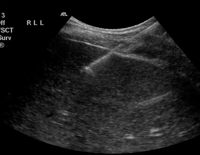
This shows
(a)
Which procedure removes ascites?
Thoracentesis
Paracentesis
Biopsy
FNA
Fluid is drained for comfort and there is a known cause
(a)
New onset of ascites
Unknown etiology
(a)
Which abdominal quadrant is best for paracentesis?
RUQ
LUQ
LLQ
RLQ
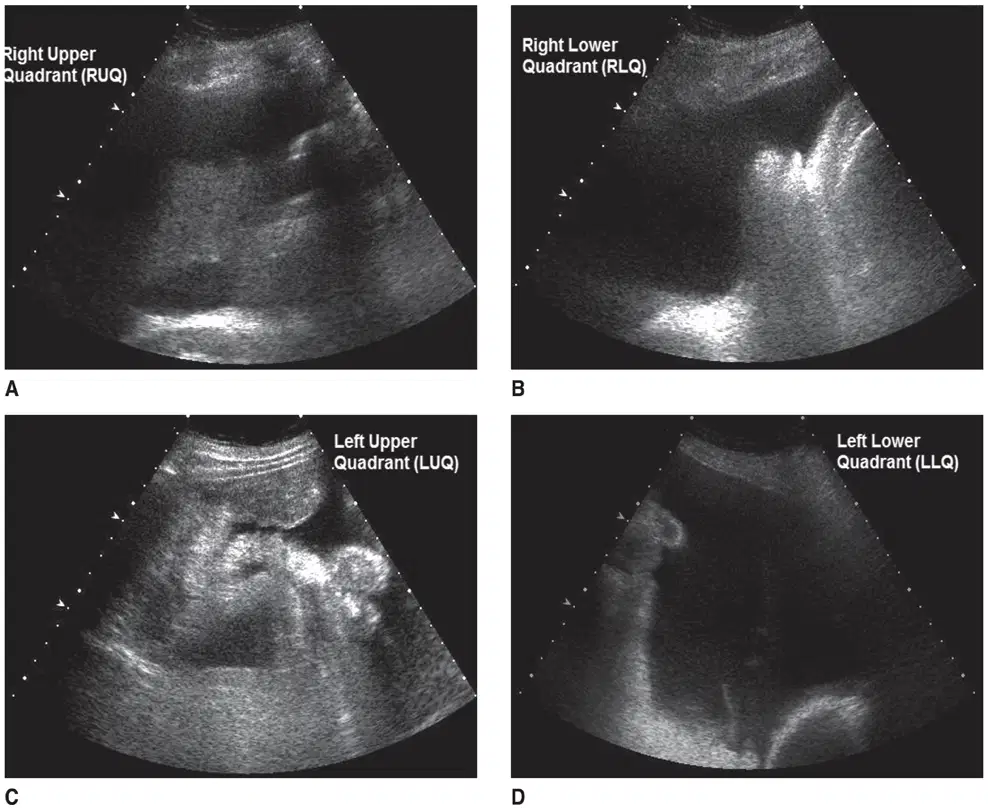
This shows (a)
Which procedure is the removal of pleural effusion
Parecentesis
Thoracentesis
Biopsy
FMA
Diagnostic thoracentesis is done when cause is not known
True
False
Performed when cause is already known
Fluid is removed for comfort
(a)
Pain
Bleeding
Pneumothorax
Infection
Spleen or liver puncture
Vasovegal events
(a)
Chest ultrasound for pleural effusion should be done in the same position as during the thoracentesis
True
False
Sitting up with legs over the side of the bed is ideal position for (a)
Above 9th rib to avoid subdiaphragmatic puncture
(a)
Midway between spine and posterior axillary line where ribs are easily palpated
(a)
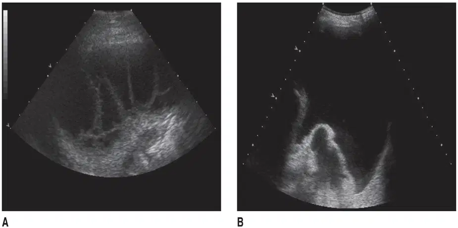
This shows
(a)
Removal of amniotic fluid for:
Genetic testing
Assessment of fetal lung maturity
(a)
Used to relieve hydronephrosis
Catheter is inserted through the back into the kidney and extends into the ureter, allowing urine to drain to the bladder
(a)
Catheter is left in place to drain the abscess and manage infection
(a)
Which of the following following is not a contraindication?
Bleeding disorder
Unable to give consent
Unable to cooperate
Able to give consent
Platelet therapy
Fresh frozen plasma (FFP)
Anticoagulant therapy
(a)
Alpraxolam (Xanax)
Lorezepam (Ativan)
Diazepam (Valium)
Midazolam (Versed)
(a)
Drowsiness
Restlessness
Cardiac irregularities
Nausea
Skin rash
Safer than deep sedation
(a)
Most common
Requires patient cooperation during procedure
Local tissue anesthetized
(a)
Lidocaine (Xyelocaine)
Sodium bicarbonate
(a)
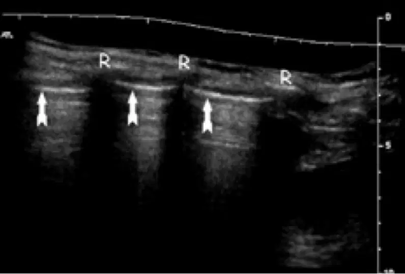
This shows
(a)
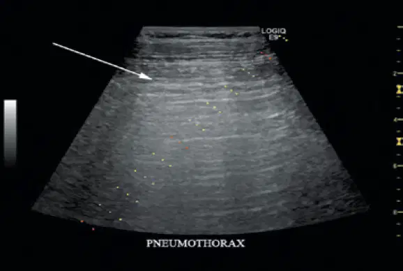
This shows
(a)

This shows
(a)

this shows
(a)
Non-ionizing radiation
Fast exam (can be done in 5-10 minutes)
(a)
Need patient cooperation
Need patient to take in a deep breath
(a)
Normal exam with quiet respiration and sniff
(a)
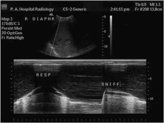
This shows
(a)
No movement with respiration and cranial or absent movement with sniff.
(a)
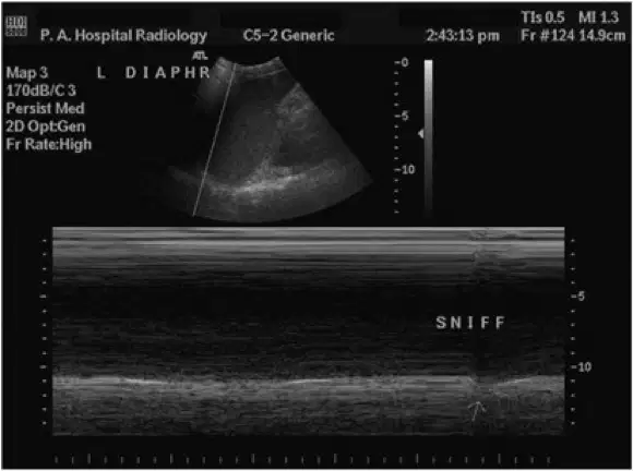
This shows
(a)
allows users to quickly distinguish between normal and abnormal anatomical findings at the bedside and various clinical settings
(a)
Diagnostic
Monitoring
Procedural guidance
Resuscitation
Screening
(a)
In trauma setting can be used during resuscitation for cardiac arrest
(a)
Cardiac standstill or clotting within cardiac ventricles and atrii or decreased contractions
(a)
Screening can rapidly assess patients to avoid the use of radiation
Originally used to screen for abdominal aortic aneurysm
(a)
has evolved into an integral extension of the clinical examination, providing real-time diagnostic insights at the bedside.
(a)
Goal directed focused exam that answers brief and important clinical questions in an organ system or for clinical signs and symptoms involving multiple organ system
True
False
focused assessment with sonography in trauma
(a)
Provides rapid evaluation tool for trauma, and/ or acute patients (ICU)
(a)
Pericardium
Thorax
Abdomen
Pelvis
(a)
children and pregnant-no ionizing radiation
Hemodynamically unstable
Penetrating trauma with multiple wounds
(a)
Inability to differentiate blood from other fluids (ascites, urine)
Obese patients
(a)
FAST exam position
(a)
Trendelenburg to shift fluid to dependent areas
Sagittal oblique and coronal scanning
Use liver and spleen as acoustic windows
(a)
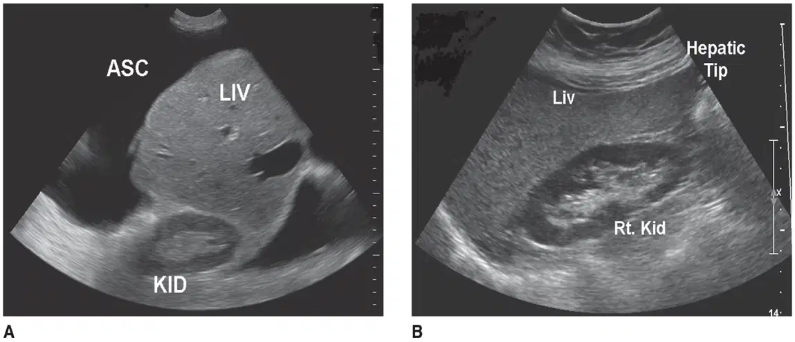
This shows
(a)
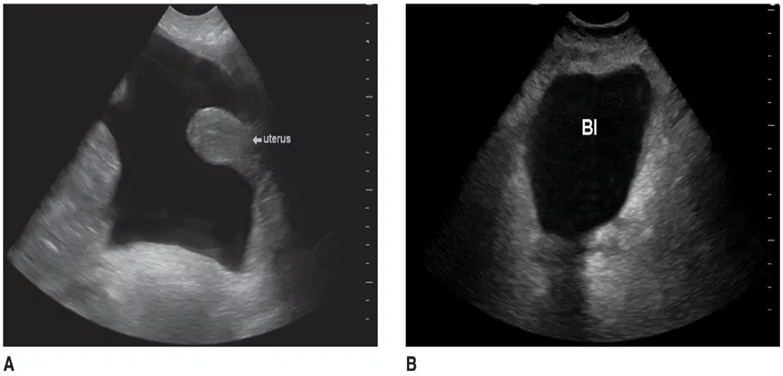
This shows
(a)
Sonography of the pericardial space
Small pericardial effusion can become tamponade
(a)
Patient must receive immediate treatment to avoid life threatening
Pericardiocentesis
(a)

This shows
(a)
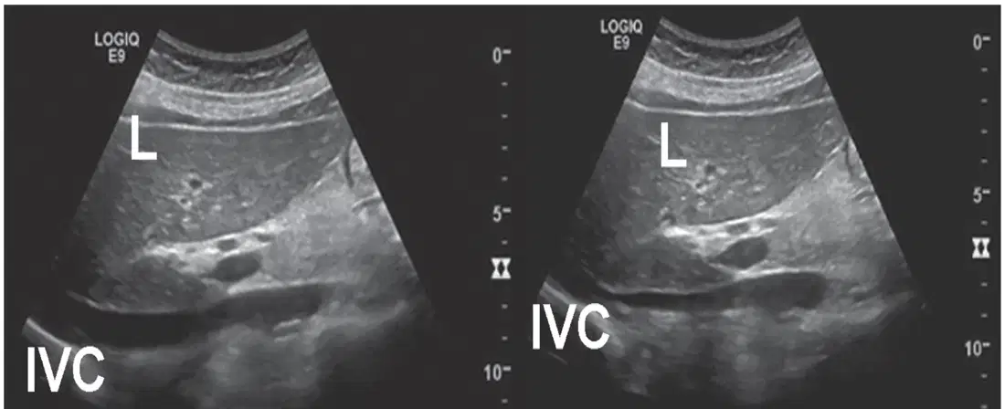
This shows
(a)
visceral pleura slides back and forth under the parietal pleura between the ribs during respiration
(a)
Absence of sliding indicates
(a)
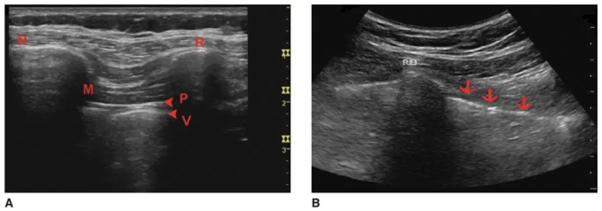
This shows
(a)
Surgical transfer of a healthy organ (allograft) from a donor to a recipient with organ failure.
(a)
Positioned in iliac fossa; connected to iliac vessels and bladder
(a)
Placed in right iliac fossa; drainage may be enteric or bladder-based
(a)
Implanted orthotopically with vascular and biliary anastomoses
(a)
Superficial; normal corticomedullary differentiation; assess ureter and vessels
(a)
Lobulated, homogeneous; located near iliac vessels; perfusion visible with Doppler
(a)
Normal echotexture; evaluate hepatic artery, portal vein, and bile ducts
(a)
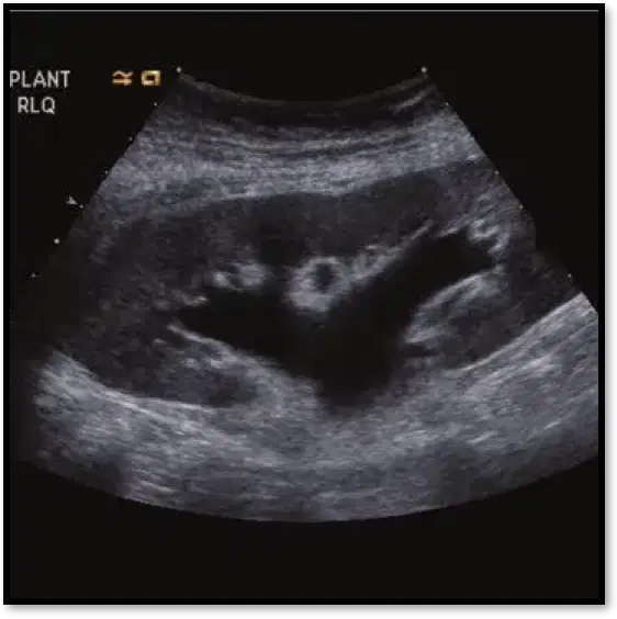
This shows
(a)
Evaluate the Inferior Vena Cava (IVC) during inspiration and expiration
(a)
Rule out cardiac tamponade if the IVC collapses by (a) % or more
Ultrasound guides catheter insertion into arterial and venous structures.
Provides real-time visualization of the needle for accurate placement.
(a)

