
Font size
 Worksheets
Cardiovascular System - Worksheet Extracted Questions
Total questions: 88
Worksheet time: 44mins
According to the analogy provided, fluids flow through a pipe only when they are forced to do so. What produces the force that makes the fluids flow?
A pump
A filter
A valve
A thermometer
In the analogy, what does the pump in the body represent?
The heart
The lungs
The liver
The kidney
The heart is a single organ composed of two pumps in one. What does the right side of the heart pump blood to?
The lungs
The brain
The liver
The kidneys
Which side of the heart is responsible for systemic circulation?
The left side
The right side
Both sides
Neither side
What is the main function of the heart in generating blood pressure?
To increase body temperature
To contract and force blood through the vessels
To digest food
To filter waste
The heart separates the pulmonary and systemic circulation.
True
False
What is the function of the left atrium in the heart?
Receives deoxygenated blood
Receives oxygenated blood
Pumps blood to the lungs
Pumps blood to the stomach
Which valve ensures one-way blood flow from the left ventricle to the aorta, preventing back flow of oxygenated blood?
Mitral valve
Tricuspid valve
Aortic semilunar valve
Pulmonary valve
Fill in the blank: During short periods of vigorous exercise, the amount of blood pumped per minute increases seven-fold (approx. ___ per min at rest) to increase the supply of blood and O₂ to the skeletal muscles.
5L
2L
10L
8L
The heart is a muscular organ, shaped like a blunt cone, approximately the size of a ________ and weighs less than 1 lb.
closed fist
tennis ball
football
apple
What is the blunt, rounded point of the heart called?
Apex
Base
Septum
Valve
In the posterior view of the heart, what is the direction of the base?
A) Anteriorly and slightly inferiorly
B) Posteriorly and slightly superiorly
C) Laterally and slightly inferiorly
D) Medially and slightly superiorly
The heart is surrounded by its own cavity called the pericardial cavity, formed by the pericardium.
True
False
The importance of knowing the location and shape of the heart for health professionals is:
It helps in accurate diagnosis and treatment of heart-related conditions.
It is only useful for academic purposes.
It is mainly important for athletes.
It is not relevant to patient care.
Match the following anatomical terms with their descriptions:
Apex
The blunt, rounded point of the heart
Base
The larger, flat part at the opposite end
Pericardial cavity
The cavity formed by the pericardium surrounding the heart
What is the protective fibrous sac that encloses the heart called?
Pericardial cavity
Pericardium or pericardial sac
Myocardium
Endocardium
The pericardium consists of two layers: the outer layer is called the ________ and is composed of tough, fibrous connective tissue.
Fibrous pericardium
Serous pericardium
Epicardium
Endocardium
The serous pericardium is composed of two parts: the parietal pericardium, which lines the fibrous pericardium, and the ________, which covers the heart surface.
Visceral pericardium (Epicardium)
Myocardium
Endocardium
Fibrous pericardium
What is the function of pericardial fluid?
Increases friction
Reduces friction as the heart moves within the pericardium
Anchors the heart within the mediastinum
Pumps blood
Pericarditis is the ________ of the serous pericardium.
inflammation
infection
degeneration
rupture
Cardiac tamponade occurs when fluid or blood ________ within the pericardial cavity and compresses the heart from the outside.
accumulates
dissipates
evaporates
decreases
The right and left atria are known as the ________ chambers of the heart.
receiving
pumping
contracting
distributing
Which groove separates the atria from the ventricles?
Anterior interventricular sulcus
Coronary sulcus
Posterior interventricular sulcus
Atrioventricular groove
Which layer of the pericardium is in direct contact with the heart surface?
Visceral pericardium (Epicardium)
Parietal pericardium
Fibrous pericardium
Serous pericardium
Which vein carries deoxygenated blood from the upper limbs before entering the heart to the right atrium?
Inferior Vena Cava
Superior Vena Cava
Pulmonary Vein
Coronary Sinus
Fill in the blank: The pulmonary vein carries oxygenated blood from the lungs to the _____
left atrium
right atrium
left ventricle
right ventricle
Match the following heart chambers with their primary function:
Receives deoxygenated blood from the body
Right atrium
Receives oxygenated blood from the lungs
Left atrium
Pumps deoxygenated blood to the lungs
Right ventricle
Pumps oxygenated blood to the body
Left ventricle
Which chamber of the heart has the thickest wall?
Right atrium
Left atrium
Right ventricle
Left ventricle
The right ventricle pumps blood to the systemic circulation.
True
False
Fill in the blank: The wall of the left ventricle is thicker than the right ventricle because it generates greater _____ for systemic circulation.
blood pressure
oxygen demand
nerve impulse
enzyme activity
Which of the following is NOT a function of the right and left atrium?
Function as reservoir
Major pumping chambers
Minimal contractions force blood into the ventricles
Separated by interatrial septum
Fill in the blank: The heart is a muscular pump consisting of four chambers: right atrium, left atrium, right ventricle, and _____?
left ventricle
left aorta
right valve
pulmonary artery
Which artery carries deoxygenated blood from the right ventricle to the lungs?
Aorta
Pulmonary trunk
Superior vena cava
Coronary sinus
The coronary sinus drains deoxygenated blood from the heart muscle into the right atrium.
True
False
What is the function of the papillary muscles in the ventricular walls?
They prevent the AV valves from being pushed up and opening backward.
They contract to pump blood out of the ventricles.
They generate electrical impulses for heart contraction.
They absorb excess fluid in the ventricles.
Which valve is located between the right atrium and right ventricle?
Bicuspid Valve
Tricuspid Valve
Aortic Semilunar Valve
Pulmonary Semilunar Valve
The bicuspid (mitral) valve is located between the ______ and ______.
left atrium and left ventricle
right atrium and right ventricle
left ventricle and aorta
right ventricle and pulmonary artery
Match the following heart valves with their locations:
Tricuspid Valve
Between right atrium and right ventricle
Bicuspid (Mitral) Valve
Between left atrium and left ventricle
Pulmonary Semilunar Valve
Between right ventricle and pulmonary trunk
Aortic Semilunar Valve
Between left ventricle and aorta
What is the main function of the cardiac skeleton (fibrous skeleton)?
It consists mainly of fibrous rings that surround the AV and semilunar valves.
It generates electrical impulses for heart contraction.
It pumps blood throughout the body.
It stores oxygen for the myocardium.
Which of the following is NOT a function of the papillary muscles?
Prevent AV valves from opening backward
Open and close the valves
Limit the valves' movements
Prevent backward flow of blood
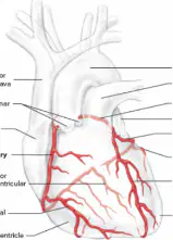
Which color represents the coronary arteries in the diagram?
Red
Blue
Green
Yellow
Name the two main coronary arteries that supply blood to the heart.
Left Coronary Artery and Right Coronary Artery
Pulmonary Artery and Aorta
Carotid Artery and Femoral Artery
Subclavian Artery and Brachial Artery
What is the name of the structure that connects cardiac muscle cells end-to-end and laterally to adjacent cells?
Intercalated disks
Desmosomes
Gap junctions
Sarcomeres
Which layer of the heart wall is responsible for the contraction of the heart chambers?
Endocardium
Myocardium
Epicardium
The outer surface of the heart is called the ________.
Epicardium (Visceral Pericardium)
Endocardium
Myocardium
Pericardial cavity
Which of the following is NOT a vein found in the heart?
Great Cardiac Vein
Small Cardiac Vein
Coronary Sinus
Pulmonary Artery
The surfaces of the interior walls of the ventricles are modified by ridges and columns of cardiac muscle called ________.
trabeculae carneae
papillary muscles
chordae tendineae
intercalated discs
Cardiac muscle contains ________ and ________ myofilaments organized to form sarcomeres.
actin; myosin
collagen; elastin
tubulin; keratin
troponin; tropomyosin
Cardiac muscle relies on Ca2+ and ATP for contraction.
True
False
What is the resting membrane potential described as?
Negative inside the cell (negative sa loob)
Positive inside the cell (positive sa loob)
Neutral inside the cell (neutral sa loob)
No potential difference (walang potential difference)
What is the term for the banded appearance of cardiac muscle due to the organization of actin and myosin?
Striated
Smooth
Fibrous
Non-striated
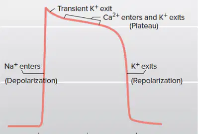
Which ion is primarily responsible for the depolarization phase?
Na+ (Sodium)
K+ (Potassium)
Ca2+ (Calcium)
Cl- (Chloride)
Which node is known as the pacemaker of the heart?
Sinoatrial Node
Atrioventricular Node
Bundle of His
Purkinje Fibers
Match the following components of the cardiac conduction system with their correct descriptions:
Pacemaker, unang naggegenerate ng heart
Pacemaker, unang naggegenerate ng heart
Between artery and ventricular
Between artery and ventricular
Splits in two
(not described)
Contraction begins at the lower, because it contracts the apex first
Splits in two
Rhythmic (arrhythmia = absence ng rhythm)
Contraction begins at the lower, because it contracts the apex first
What is the importance of the repolarization phase in cardiac muscle action potential?
Generating longer tension time; the length of the plateau phase determines the refractory period.
Initiating muscle contraction; it triggers the release of calcium ions.
Increasing heart rate; it accelerates the pacemaker potential.
Preventing depolarization; it blocks sodium channels.
Which ion channel is unique to the nodal cell and is responsible for the pacemaker potential?
F (funny) type calcium channel
L-type calcium channel
T-type calcium channel
Voltage-gated sodium channel
The electrocardiogram (ECG) is a direct measurement of mechanical events in the heart.
True
False
Which of the following is NOT a function of the electrocardiogram (ECG)?
Revealing abnormal heart rates
Measuring mechanical contraction directly
Revealing problems in conduction pathways
Locating damaged cardiac muscle
What happens if the heart stops beating because it is repeatedly stimulated?
The heart stops beating because it is repeatedly stimulated (Di tumitigil ang beating ng heart kasi paulit ulit nag sstimulate ito).
The heart beats faster and stronger each time it is stimulated.
The heart starts to pump more blood than usual.
The heart becomes immune to stimulation and never stops.
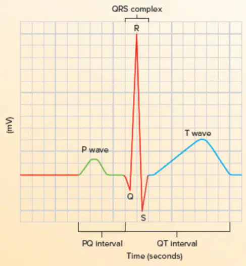
What does the P Wave represent in an ECG?
A) Repolarization of ventricle
B) Depolarization and contraction of atrial myocardium
C) Depolarization and contraction of ventricle
D) Repolarization of atrial myocardium
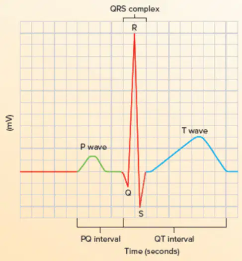
What does the QRS Complex represent in an ECG?
Depolarization and contraction of ventricle
Repolarization only of ventricle
Depolarization and contraction of atrial myocardium
Repolarization of atrial myocardium
What does the T Wave represent in an ECG?
Depolarization and contraction of ventricle
Repolarization only of ventricle
Depolarization and contraction of atrial myocardium
Depolarization only of ventricle
Fill in the blank: Tachycardia is when the heart rate is in excess of ____ bpm.
100
80
120
60
Fill in the blank: Bradycardia is when the heart rate is less than ____ bpm.
60
80
100
50
Which of the following is NOT a characteristic of Sinus Arrhythmia?
Variation in heart rhythm from sinus node
+HR during inhalation
Heart rate is less than 60 bpm
Occurs during inhalation
Paroxysmal Atrial Tachycardia is characterized by:
Slow heart rhythm
Sudden increase in HR to 150-250 bpm
Decreased sympathetic stimulation
No P waves
Atrial Flutter is characterized by as many as ____ P waves/min and ____ QRS complex/min.
300 P waves/min and 125 QRS complex/min
200 P waves/min and 100 QRS complex/min
150 P waves/min and 75 QRS complex/min
400 P waves/min and 200 QRS complex/min
Which arrhythmia is described as rapid and irregular beating of the atria with no P waves, but normal QRS complex and T waves?
Atrial Fibrillation
Ventricular Tachycardia
Atrial Flutter
Supraventricular Tachycardia
Ventricular Tachycardia is often associated with:
Damage to AV node or ventricular muscle
Increased SV in athletes
Ectopic beats in atria
Excessive sympathetic stimulation
Match the following ECG components with their descriptions:
Depolarization and contraction of atrial myocardium
C
Depolarization and contraction of ventricle
A
Repolarization only of ventricle
B
The atrium is called the ______ pump, and the ventricle is called the ______ pump.
Primer; Power
Power; Primer
Primary; Secondary
Secondary; Primary
The mechanical events of the cardiac cycle and their trigger are:
Contraction and relaxation of heart chambers triggered by electrical impulses
Continuous contraction of heart chambers triggered by blood flow
Relaxation of heart chambers triggered by hormone release
Random contraction and relaxation triggered by external temperature changes
List the phases of the cardiac cycle.
Atrial systole, ventricular systole, diastole
Atrial diastole, ventricular diastole, systole
Atrial contraction, ventricular relaxation, repolarization
Depolarization, repolarization, hyperpolarization
Which valves are open and which are closed during isovolumetric ventricular contraction?
AV valves are closed
Aortic valve is closed
AV valves are open
Aortic valve is open
Pulmonary valve is closed
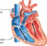
Which valves are open during ventricular filling?
AV valves
Aortic valves
Pulmonary valves
Semilunar valves
A Wigger's diagram is used to represent which of the following?
The cardiac cycle, including pressure, volume, and heart sounds
The structure of the heart chambers
The electrical conduction system of the heart
The blood flow through the coronary arteries
What instrument is used to listen to the sounds of the lungs and heart?
Stethoscope
Thermometer
Sphygmomanometer
Otoscope
Which heart sound occurs at the beginning of ventricular systole and results from the closing of the atrioventricular valve?
lubb
dupp
Which heart sound occurs at the beginning of ventricular diastole from the closing of the semilunar valve?
lubb
dupp
The first heart sound (lubb) has a pitch than the second heart sound (dupp).
lower
higher
same
variable
Identify the sites on the thorax where heart sounds can be best heard with a stethoscope as shown in the figure.
Pulmonary valve area, Aortic valve area, Tricuspid valve area, Mitral valve area
Pulmonary valve area, Carotid artery area, Tricuspid valve area, Mitral valve area
Pulmonary valve area, Aortic valve area, Jugular vein area, Mitral valve area
Pulmonary valve area, Aortic valve area, Tricuspid valve area, Femoral artery area
What is the main function of a heart valve? Choose the correct answer.
To pump blood
To prevent back flow
To increase heart rate
To regulate blood pressure
Fill in the blank: In some individuals, a heart valve does not close completely and is called an ________ valve.
incompetent
efficient
resistant
elastic
Which type of flow is associated with heart murmurs? Choose the correct answer.
Laminar flow
Turbulent flow
One direction flow
No flow
What are the causes of heart murmur?
Stenosis, leaky valve, septal defect
High blood sugar, dehydration, obesity
Asthma, bronchitis, pneumonia
Fracture, sprain, dislocation
Cardiac output (CO) is defined as the volume of blood pumped by the ventricle each minute.
True
False
Fill in the blank: Stroke volume (SV) is the volume of blood pumped per ventricle each time the heart ________.
contracts
relaxes
beats irregularly
stops
Heart rate (HR) is the number of times a heart contracts each ________.
minute
hour
second
day

