
 Worksheets
Exam Review Anatomy
Total questions: 223
Worksheet time: 3hrs 2mins
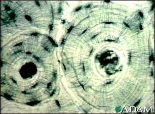
smooth muscle
bone
nerve
blood


The tissue shown is called ______.
Skeletal muscle
Cardiac muscle
Smooth muscle
Dense connective

Which type of muscle cells are voluntary?
Skeletal
Cardiac
Medial
Smooth

Name this tissue:
Stratified squamous
Smooth muscle
Skeletal muscle
Hyaline cartilage


The pituitary gland puts growth hormone into the blood. What type of gland is it?
Endocrine Gland
Exocrine Gland
Thyroid Gland
Capillary Gland
Which tissue type functions and secretion, absorption, protection, and excretion
Epithelial
connective
Arelolar
muscle
Nervous
There are four primary tissue types that interweave to form the "fabric" of the body, they are;
epithelial,connctive,muscle,nervous
skeletal,cardiac,smooth,adipose
epithelial,skeletal,nervous,squamous
cartilage,muscle,epithelial,areolar
Which term means, more than one layer of cells?
Stratified
Squamous
Simple
Cuboidal
Columnar
Fibers that form a thick protein matting in a dense irregular connective tissue include
collagen fibers
recticular fibers
elastic fibers
cartilage fibers
Which membrane has goblet cells?
nervous
muscular
mucous
serous
Which membrane is your skin?
serous
synovial
cutaneous
mucous
What type of gland is the sebaceous glad of the skin?
Apocrine
Merocrine
Holocrine
The function of the integumentary system includes all of these except what?
Homeostasis
Cooling system
Synthesis of Vitamin A
Excretion of wastes
What layer of the skin contains sweat glands?
Dermis
Epidermis
Hypodermis
Subcutaneous
What part of a hair can be seen?
Follicle
Matrix
Root
Shaft
What part of the hair provides cells for new growth?
Bulb
Matrix
Papilla
Root
The area where there is active nail growth is the ______.

List the layers of the Epidermis in order from top to bottom.
5,2,3,4,1
1,2,3,4,5
4,5,3,1,2
1,4,3,2,5

What is number 10?
Pore
Hair Shaft
Epidermis
Nerve
What is number 11?
Dermis
Epidermis
Hair Shaft
Artery
What is number 5?
Dermis
Epidermis
Sebaceous Gland
Sudoriferous Gland
What is number 4?
Hair Follicle
Hair Shaft
Sebaceous gland
Sudoriferous gland
What is number 3?
Dermis
Epidermis
Hypodermis/subcutaneous
Artery
What is number 2?
Dermis
Epidermis
Hypodermis/subcutaneous
Artery
What is number 1?
Dermis
Epidermis
Hypodermis/subcutaneous
Artery
How do first-degree burns differ from second degree burns?
First-degree burns only affect the epidermis; second-degree burns can extend into the dermis
First degree-burns are painless; second-degree burns hurt a lot
First degree-burns can extend into the dermis; second-degree burns only affect the epidermis
First-degree burns hurt a lot; second-degree burns are painless
Why can you often not feel third-degree burns?
Because they damage the nerve endings in your skin
Because they aren' t very damaging
Because they cause scabs to form
Because they only affect the inner part of your skin

Number 4 on the diagram
arrector pili muscle
hair shaft
sebaceous gland
nerve
A second degree burn is one that only affects the epidermis and dermis and is also called a ________ burn.
full- thickness
partial thickness
superficial
simple

Identify structure number 9.
Pacinian corpuscle
freen nerve endings
Meissner's corpuscle
sebaceous gland
During vasoconstriction, where blood cells assemble together to close the wound, what is formed?
Carbon Dioxide
Oxygen
Nitrogen
Blood clot
The nurse recognizes that which of the following are classic signs of inflammation? (select all that apply)
redness
heat
fever
swelling
pain
What do sebaceous glands secrete?
Oil
Milk
Sweat
Earwax
Dermis
Deep layer of the skin
Epidermis
Superficial layer of the skin
Hypodermis
Also known as subcutaneous tissue
1st degree burn
superficial; involves the epidermis
2nd-degree burn
partial thickness; involves the epidermis and dermis
3rd degree burn
full-thickness; involves the epidermis, dermis, and subcutaneous


Articular cartilage is found in what part of the long bone?
inside of the medullary cavity
on the outer surface of the diaphysis
the outer surface of the epiphyses
in the spaces of the spongy bone
The function of Osteoclasts is
To breakdown calcified matrix of bone
Secrete acids that dissolve the inorganic component of calcified matrix
Phagocytize components of the bony matrix
All the above

The shaft of long bones in the skeletal system is called:
periosteum
epiphysis
diaphysis
cartilage

Which bones are part of the appendicular skeleton?
scapula, radius, pelvis
scapula, skull, phalanges
hyoid bone, radius
brain case, thoracic cage, lower limbs
Fibrous, cartilaginous, and synovial are types of tissues found in:
joints
interior of bones
ends of fingers
all responses are incorrect
Process of creating bone is is called_____.
Periosteum
Bone resorption
Ossification
Hematopoeisis
A female patient is brought to the emergency room with an injury to her lower leg from an ATV accident. You notice a white tip of a bone protruding through the skin as you cut her pant leg off. Her injury is described as what kind of fracture.
Comminuted
Pathologic
Compound
Stress
Which of the following is not a function of bones?
Protection
Blood cell formation
Storage
Absorption
Which part of the osteon contains the osteocytes?
Lacunae
Lamellae
Canaliculi
Central Canal


Compact bone is made of
trabeculae
osteons
canaliculi
central canals

This type of bone is porous, allowing blood vessels to move through it and containing bone marrow.
Compact Bone
Spongy bone

The hardest, most dense part of the bone tissue is the
periosteum
spongy bone
osteoblast
compact bone

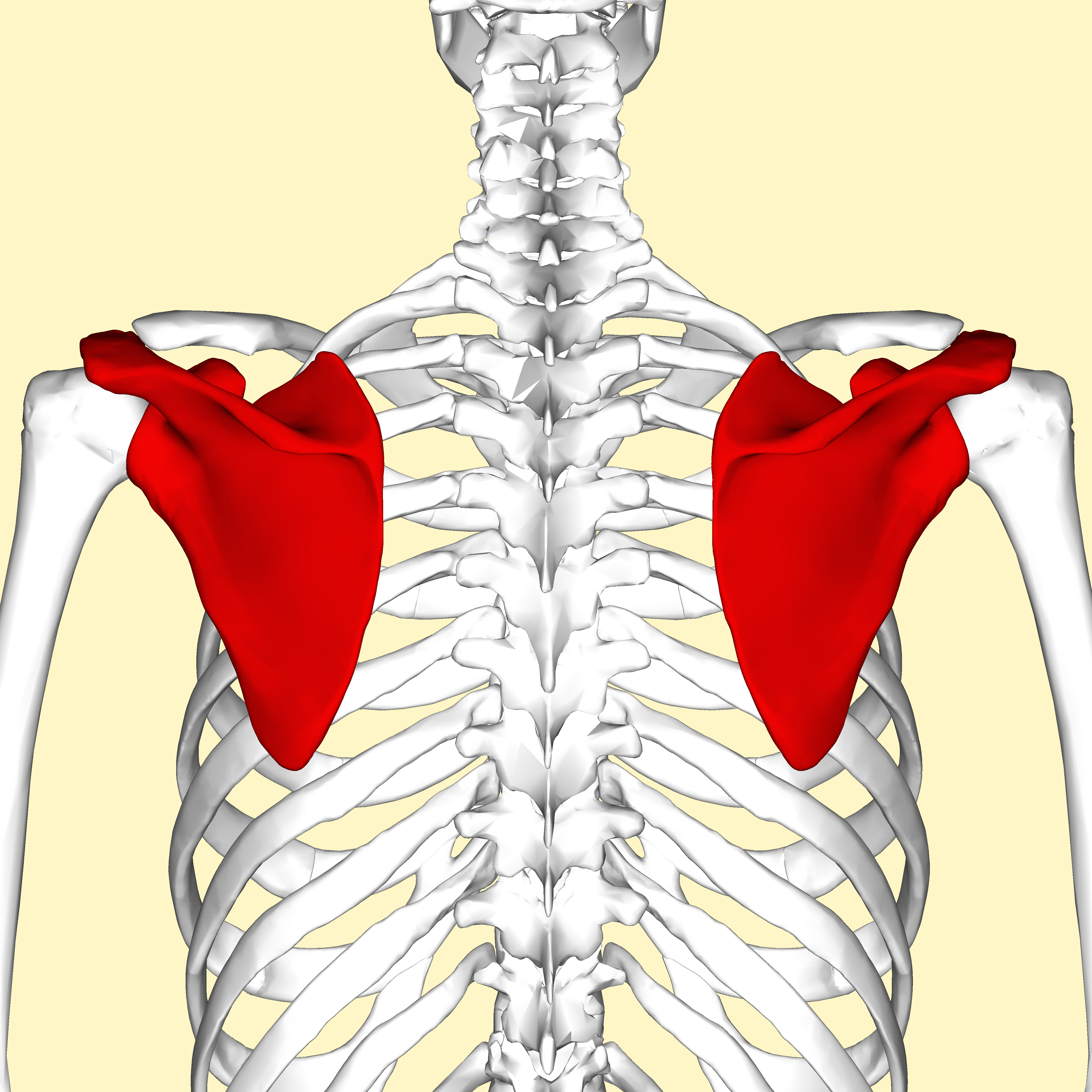
What bone is the arrow pointing to?
rib
spine
scapula
clavicle

What bone is the arrow pointing to (the bone closest to the thumb)?
ulna
humerus
radius
vertebrae


The section of this picture highlighted in green is called the _______________?
Tibia
Patella
Ilium
Fibula

The name of the blue bone under the eye
parietal bone
zygomatic bone
sphenoid bone
mandible

The orange bone on the front of the skull
parietal bone
frontal bone
nasal bone
lacrimal bone

The pink bone on the top of the skull
parietal bone
occipital bone
frontal bone
lacrimal bone

The purple bone on the side of the head
lacrimal bone
sphenoid bone
ethmoid bone
temporal bone

8.?
mandible
maxilla
zygotmatic
coronal suture

18?
lambdoid suture
coronal suture
squamous suture
There are (a) groups of sinuses (number)
At what part of the bone does growth occur in children and adolescents?
Marrow cavity
Epiphyseal plate
Periosteum
Endosteum
Which type of joint allows for the greatest range of motion?
Pivot joint
Ball and socket joint
Gliding joint
Hinge joint
How many bones are in an adult human body?
365
206
270
302
Fluid-filled sacs that help cushion the friction in a joint is called what?
Ligaments
Cartilage
Bursa
Tendons
The bones that make up the ankle are known as the:
carpals
tarsals
clavicles
phalanges
Some marrow of long bones is termed "red" marrow. The function of this is to:
manufacture blood cells
manufacture tissue cells for the skin
store adipose tissue
store bone forming cells

The functions are the skeletal system are
To support the body, protect the organs, store calcium, and produce blood cells
to regulate the composition of body fluids by removing metabolic wastes and retain the balance of water, salt, and other nutrients
to regulate and maintain bodily functions by producing hormones and releasing them into the blood stream
to enable the diffusion of oxygen into the bloodstream and the removal of carbon dioxide from the bloodstream

The bone (in green) is known as the:
C1 Vertebra
Mandible
Hyoid
Trachea
What is the primary function of the muscular system?
To produce hormones
To facilitate movement
To protect internal organs
To transport nutrients
Which type of muscle is under voluntary control?
Cardiac muscle
Smooth muscle
Skeletal muscle
Involuntary muscle
Explain how muscles work in pairs to move bones.
Muscles work independently to move bones.
One muscle contracts while the other relaxes to create movement.
Both muscles contract simultaneously to move bones.
Muscles do not work in pairs.
Describe the role of tendons in the muscular system.
Tendons connect muscles to bones.
Tendons connect bones to bones.
Tendons produce red blood cells.
Tendons store calcium.
What is the basic contractile unit of a muscle called?
Neuron
Sarcomere
Osteon
Alveolus
Which muscle type is responsible for pumping blood throughout the body?
Skeletal muscle
Smooth muscle
Cardiac muscle
Voluntary muscle
Which is NOT a function of the muscular system?
Regulating body temperature
Movement
Protecting delicate organs
Preventing disease
Muscle pairs that oppose each other's movement
antagonistic muscles
opposable muscles
opposite muscles
rivals
What does the root word 'myo' mean?
water
life
earth
muscle

Skeletal muscles consist of numerous _____________ that contain a bundle of _______________ which are composed of repeating contractile units called _____________?
muscle fibers, sarcomeres, myofibrils
sarcomeres, muscle fibers, myofibrils
sarcomeres, myofibrils, muscle fibers
muscle fibers, myofibrils, sarcomeres

What is the correct order of structure of Skeletal Muscle?
Muscle Fiber, Endomysium, Fascicle, Epimysium, Perimysium, Tendon
Muscle Fiber, Endomysium, Epimysium, Fascicle, Perimysium, Tendon
Muscle Fiber, Fascicle, Endomysium, Perimysium, Epimysium, Tendon
Muscle Fiber Fascicle, Endomysium, Epimysium, Perimysium, Tendon
The epimysium covers
the entire muscle
the fascicle
the muscle fiber
the myofibril
Which connective tissue surrounds the fascicle?
epimysium
perimysium
sarcolemma
endomysium
A bundle of fascicles is a
myofibril
muscle fiber
sarcomere
muscle
A bundle of muscle fibers is a
muscle
fascicle
myofibril
sarcomere

What is another name #2? (Hint: It is a muscle layer)
Skeletal muscle
Epimysium
Perimysium
Endomysium
What does calcium do to the actin?
Covers myosin binding sites
Uncovers myosin binding sites
Covers actin binding sites
Uncovers actin binding sites
What is the name of the protein that covers the binding sites on actin?
Myosin
Titin
Troponin
Tropomyosin
How does the sarcomere shorten (contract) ?
The myosin filaments contract
The actin filaments contract
The myosin is pulled by the actin
The actin is pulled by the myosin
___ and ___ are regulatory proteins bound to actin.
Myosin and actin
Glycogen and hemoglobin
Calcium and oxygen
Troponin and tropomyosin

Name the muscle
pectoralis major
pectoralis minor
Deltoid
Trapezius

Name the muscle
pectoralis major
subscapularis
deltoid
triceps

Name the muscle
biceps brachii
triceps brachii
rectus femoris
deltoid

name the muscle in green
masseter
frontalis
temporalis
orbicularis occuli

extension
flexion
circumduction
adduction
abduction

supination
pronation
circumduction
adduction
rotation

supination
pronation
abduction
adduction
opposition

inversion
plantar flexion
extension
dorsiflexion
circumduction

inversion
supination
extension
pronation
eversion


Name the highlighted muscle.
Trapezius
Latissimus dorsi
Gluteus maximus
Triceps brachii


Name the muscle shown in the diagram.
Hamstring
Quadricep
Tibialis Anterior
Gastrocnemius
What two chemicals are necessary for muscle contraction to occur?
Niacin and Potassium
Calcium and ATP
Sodium and Potassium
Acetylcholine and Potassium
___are thin filaments.
troponin
tropomyosin
actin
myosin
___are thick filaments.
actin
myosin
troponin
tropomyosin
The gap between the nerve cell and sarcolemma is the ____ ___.
Synaptic cleft
Neurotransmitter
Motor unit
Neuromuscular junction
What is the specific neurotransmitter that stimulates the movement of skeletal muscles?
GABA
Acetylcholine
Serotonin
Dopamine
Which of the following is a component of the central nervous system (CNS)?
Spinal cord
Brain
Nerve
Which of the following is NOT a part of the peripheral nervous system?
cranial nerve
ganglion
spinal cord
spinal nerve
The sensory division of the peripheral nervous system (PNS) is known as the _________ division.
efferent
sympathetic
parasympathetic
afferent
Which of the following effectors is NOT innervated by the autonomic nervous system?
cardiac muscle
glands
skeletal muscle
smooth muscle
Which one of the following types of neuroglial cells is found in the peripheral nervous system?
microglia
Schwann cells
oligodendrocytes
astrocytes
Bipolar neurons:
are specialized to carry voluntary motor impulses.
conduct action potentials from one neuron to another in the central nervous system.
have a single process extending from the cell body.
are located in special sensory organs such as the retina and have one axon and one dendrite.
Microglia are:
specialized motor neurons in the peripheral nervous system.
star-shaped cells that help form the blood-brain barrier.
specialized macrophages of the CNS that mobilize in response to inflammation.
found in choroid plexuses, and produce cerebrospinal fluid.
Nodes of Ranvier are:
interruptions in the myelin sheath along the course of a myelinated axon
collections of immune cells in the CNS
satellite cells that support neuron cell bodies in ganglia
gaps between choroids plexuses where cerebral spinal fluid emerges
White matter consists of:
myelinated axons and nerve tracks
unmyelinated axons
neuron cell bodies
unmyelinated axons and neuron cell bodies
Gray matter consists of: (pick 2)
neuron cell bodies
unmyelinated axons
nerve tracts
Collections of neuron cell bodies outside the CNS are called:
ganglia.
nerves.
nerve tracts.
nuclei.
This type of neuron is located in the Brain and Spinal Cord.
Sensory Neuron
Interneuron
Motor Neuron
This type of neuron processes information from Sensory Neurons and passes the information onto Motor Neurons.
Sensory Neurons
Motor Neurons
Interneurons
What is it called when your body reacts to something without you having to think about it.
Stimulus
Reflex
Nerves
This thick band of nerves connects the right and left hemispheres of the cerebrum.
corpus callosum
pituitary gland
thalamus
hypothalamus
What part of the nervous system is responsible for slowing down breathing and pulse?
Spinal Nerves
Sympathetic
Parasympathetic
Spinal Nerves
Hallucinations, seizures, and temporary loss of consciousness is a symptom of:
hydorcephalus.
meningitis.
neuritis.
epilepsy.
What is dopamine's role in the brain?
It helps us sleep better.
It motivates us and encourages certain behaviors.
It makes us feel hungry.
It helps us remember names.

Which part of the brain is designated with a star.
Brainstem
Frontal lobe
Medulla oblongata
Temporal lobe
Cerebellum

Which part of the brain is designated with a star.
Cerebrum
Parietal lobe
Medulla oblongata
Frontal lobe
Cerebellum

Which part of the brain is designated with a star.
Frontal lobe
Parietal lobe
Pons
Temporal lobe
Occipital lobe

Which part of the brain is designated with a star.
Frontal lobe
Parietal lobe
Medulla oblongata
Temporal lobe
Cerebellum

Which part of the brain is designated with a star.
Frontal lobe
Brainstem
Medulla oblongata
Temporal lobe
Cerebellum

Look at the image of the neuron. Identify the part numbered 1
Dendrites
Cell Body
Axon
Axon Terminal
Look at the image of the neuron. Identify the part numbered 2
Dendrites
Cell Body
Axon
Axon Terminal
Look at the image of the neuron. Identify the part numbered 4
Dendrites
Cell Body
Axon
Axon Terminal
In which sleep disorder does a person temporarily stop breathing at night?
insomnia
sleep apnea
narcolepsy
night terrors
Sleep Deprivation
Which disorder is characterized by uncontrollable sleep attacks, in which a sufferer may slip directly into REM sleep?
Insomnia
Narcolepsy
Sleep Apnea
Night Terrors


Produce cerebrospinal fluid in the brain & spinal cord
ependymal cells
oligodendrocytes
microglia
astrocytes
Is a concussion considered a brain injury?
Yes
No
Which of the following is NOT a possible symptom of brain injury?
moodiness
slurred speech
hallucination
difficult doing math
All are possible
What is Alzheimer's Disease?
poor blood flow in the brain that causes cell death
short term loss of brain function following a head injury
a brain disease that causes cognitive dysfunction in elderly
a movement disorder that impairs voluntary movements


Vertical plane that divides the body into RIGHT and LEFT parts
Sagittal
Frontal
Transverse
Oblique
[The eyes are _______ to the brain].


