

The Heart

Presentation
•
Biology
•
9th Grade - University
•
Hard

Nikki Chenevert
FREE Resource
15 Slides • 37 Questions
1
by Nikki Chenevert
2
The heart pumps 7,000 liters of blood through the body each day, contracting some 2.5 billion times in an average lifetime. This muscular pump forces blood through arteries, which connect to smaller-diameter vessels called arterioles. Arterioles branch into the tiniest tubes, the capillaries, which are sites of nutrient, electrolyte, gas, and waste exchange. Capillaries converge into venules, which in turn converge into veins that return blood to the heart, completing the closed system of blood circulation. These structures—the pump and its vessels—form the cardiovascular system
3
Multiple Choice
4
Multiple Choice
5
Multiple Choice
6
Multiple Choice
Which of the following is the smallest blood vessel?
Venules
Arterioles
Veins
Capillaries
7
Multiple Choice
8
Multiple Choice
9
The pulmonary (pul′mo-ner′′e) circuit sends oxygen depleted (deoxygenated) blood to the lungs to pick up oxygen and unload carbon dioxide.
The systemic (sistem′ik) circuit sends oxygen-rich (oxygenated) blood and nutrients to all body cells and removes wastes.
Without circulation, tissues would lack a supply of oxygen and nutrients, and wastes would accumulate. Such deprived cells soon begin irreversible change, which quickly leads to their death.
Figure 13.1 shows the general pattern of blood transport in the cardiovascular system.
10
Multiple Choice
11
Multiple Choice
12
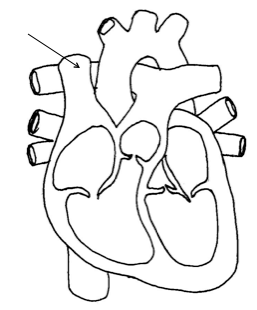
The heart is a hollow, cone-shaped, muscular pump. It lies within the thoracic cavity and rests on the diaphragm (fig. 13.2).

13
The heart is
(a) within the mediastinum and
(b) enclosed by a layered pericardium

14
Size and Location of the Heart Heart size varies with body size. An average adult’s heart is about 14 centimeters long and 9 centimeters wide (fig. 13.3).
- The average size of a human heart is approximately the size of a closed fist.
- An increased size of the heart does not always represent trauma.
Many athletes have an enlarged heart because just as their skeletal muscles are conditioned and developed so is their cardiac muscle. Contrastingly, if a sedentary person has an enlarged heart this might be a symptom of trauma, blockage, etc. to the heart or blood vessels.
15
Multiple Select
if a sedentary person has an enlarged heart this might be a symptom of _______ to the heart or blood vessels. Select all that apply.
trauma
blockage
conditioned cardiac muscles
conditioned skeletal muscle
16
Multiple Choice
17
The heart is within the mediastinum, bordered laterally by the lungs, posteriorly by the vertebral column, and anteriorly by the sternum. The base of the heart, which attaches to several large blood vessels, lies beneath the second rib. The heart’s distal end extends downward and to the left, terminating as a bluntly pointed apex at the level of the fifth intercostal space.
18
- A Term given to refer to this area is Middle Mediastinum.
Middle Mediastinum has 4 landmarks to define its area:
Anterior landmark - Sternum
Posterior landmark – Vertebral Column
Lateral landmarks – 2 lungs
Therefore, the mediastinum is area behind (posterior) the sternum but in front (anterior) of the vertebral column and between the 2 lungs. It doesn’t include the lungs themselves.
- 2/3 of the heart’s mass lies to the left of the body's midline.
19
Multiple Choice
20
Multiple Choice
21
* The heart is enclosed in a membranous sac with 2 layers: parietal and visceral.
1. Parietal pericardium – the outer serous membrane
* Fibrous pericardium – a layer of tough fibrous CT located on the outside of parietal pericardium.
It is attached to the diaphragm, posterior portion of the sternum, the vertebra and the large vessels at the base of the heart.
22
The pericardium (per′′˘-kar ı ′de-um) encloses the heart and the proximal ends of the large blood vessels to which it attaches. The pericardium consists of an outer bag, the fibrous pericardium. The fibrous pericardium is dense connective tissue.
It is attached to the central portion of the diaphragm, the posterior of the sternum, the vertebral column, and the large blood vessels emerging from the heart.
23
the inner serous membrane made of simple epithelium.
* This layer adheres to the myocardium itself.
* Also, blood vessels that nourish the heart are located in the epicardium.
24
The fibrous pericardium surrounds a more delicate, double-layered sac.
The innermost layer of this sac, the visceral pericardium (epicardium), covers the heart. At the base of the heart, the visceral pericardium turns back on itself to become the parietal pericardium, which forms the inner lining of the fibrous pericardium (figs. 13.2 and 13.4; see reference plate 3, p. 25). Between the parietal and visceral layers of the pericardium is a space, the pericardial cavity, that contains a small volume of serous fluid (fig. 13.4). This fluid reduces friction between the pericardial membranes as the heart moves within them.
25
* Between the layers of the parietal sac is pericardial fluid which acts as a lubricant.
Pericardial fluid is filtered plasma and it prevents friction between the membranes of the heart as it moves.
* An over accumulation of fluid in the parietal sac caused by excessive over production of the pericardial fluid or failure to reabsorb the fluid can result in abnormal heart movements called Cardiac Tamponade.
Note: Cardiac Tamponade can also be associated with any trauma that results in bleeding into the pericardial sac.
26
In pericarditis, inflammation of the pericardium due to viral or bacterial infection produces adhesions that attach the layers of the pericardium to each other. This condition is very painful and interferes with heart movements.
27
1. Where is the heart located?
2. Distinguish between the visceral pericardium and the parietal pericardium.
28
Multiple Choice

29
Multiple Choice

30
Multiple Choice
31
Multiple Choice

32
Multiple Choice

33
Multiple Choice

34
Multiple Choice
35
Multiple Choice

36
Multiple Choice

37
Multiple Choice

38
Multiple Choice

39
Multiple Choice

40
Multiple Choice
41
Multiple Choice

42
Multiple Choice

43
Multiple Choice

44
Multiple Choice

45
Multiple Choice

46
Multiple Choice
47
Multiple Choice
48
Multiple Choice
49
Multiple Choice
50
Multiple Choice
51
Multiple Choice
52
Multiple Choice

by Nikki Chenevert
Show answer
Auto Play
Slide 1 / 52
SLIDE
Similar Resources on Wayground

47 questions
Elkhorn History Images
Presentation
•
KG
46 questions
Homeostasis & Feedback
Presentation
•
9th Grade
48 questions
Mendel Experimental Design
Presentation
•
9th - 12th Grade
46 questions
CH 10/23 Writing Communication
Presentation
•
KG - University
46 questions
Trigonometric Ratios
Presentation
•
9th - 12th Grade
49 questions
SOL Review: Scientific Investigation
Presentation
•
9th - 12th Grade
48 questions
Cell Cycle and Mitosis Lesson
Presentation
•
9th - 12th Grade
50 questions
Photosynthesis vs Cellular respiration (regular biology)
Presentation
•
9th - 12th Grade
Popular Resources on Wayground
10 questions
Safety Day 2026 – Student Review

Quiz
•
9th Grade
21 questions
Continents and Oceans
Quiz
•
6th Grade
20 questions
Parts of Speech
Quiz
•
5th Grade
16 questions
Subject & Predicate
Quiz
•
5th Grade
25 questions
Multiplication Facts
Quiz
•
5th Grade
12 questions
Map Skills
Quiz
•
3rd Grade
22 questions
Continents and Oceans
Quiz
•
5th Grade
15 questions
Rounding Decimals
Quiz
•
5th Grade
Discover more resources for Biology
20 questions
Cell organelles and functions
Quiz
•
10th Grade
20 questions
Cell Transport
Quiz
•
9th - 12th Grade
25 questions
photosynthesis and cellular respiration
Quiz
•
9th Grade
20 questions
Cell Cycle
Quiz
•
9th Grade
8 questions
DNA vs. RNA
Presentation
•
9th - 12th Grade
14 questions
Ecological Succession: Primary and Secondary
Quiz
•
9th Grade
20 questions
Macromolecules
Quiz
•
10th Grade
20 questions
Characteristics of Life
Quiz
•
9th - 10th Grade