
Anatomy of the eye
Presentation
•
Biology
•
6th - 8th Grade
•
Medium
Standards-aligned

David Wolf
Used 78+ times
FREE Resource
13 Slides • 25 Questions
1

Eye Structure
and How we see
2

The eye works like a camera: Light enters, is focused on a surface, and a picture is made.
Light enters your eye through a clear portion of
the sclera (the tough, white, outer covering of the eye), called the cornea.
3
Multiple Choice
Pupil
Sclera
Dyslexia
Retina
4

The iris is a muscle that controls the size of the
pupil. The iris is the colored part of the eye.
5
Multiple Choice
The pupil is located at the center of the
sclera
iris
retina
nothing
6
Multiple Choice
The opening of the eye where light enters
Retina
Sclera
Pupil
cornea
7
Multiple Choice

iris
lens
cornea
retina
8
Multiple Choice

cornea
lens
iris
cilicary body
9
Multiple Choice
Explain why the pupil contracts (becomes smaller).
To restrict the amount of light entering the eye
To allow more light into the eye
To focus light onto the retina
To stop waste entering the eye
10
Multiple Choice
What is the purpose of the pupil?
It regulates the amount of light that enters the eye
it serves as the center of vision
it gives the center of the eye its color
to protect the eye
11
Multiple Choice
What part of the eye makes the pupil larger and smaller?
Retina
Iris
Lens
Cornea
12
Multiple Choice
The thick, gelatinous fluid that fills most of the eyeball is called...
Aqueous humor
tapectum lucidum
vitreous humor
conjunctiva
13
Multiple Choice
Why do pupils look black?
because they are just a hole in the eyeball
light passes through it and doesn't reflect at all
14
Multiple Choice
What gives the eye its color?
cornea
optic nerve
pupil
iris
15
Multiple Choice

After light passes through the cornea it then passes through the
Cornea
Lens
Vitreous humor
Pupil
16

The cornea is curved, so it slightly bends the light
as it goes through.
Light then passes through the aqueous humor (a
clear fluid for eye nourishment) and through the pupil.
The pupil is simply a hole in the iris.
17
Multiple Choice
When does the pupil dilate (expand)?
When there is bright light such as on a sunny day
When there is dim light such as in a dark room
18
Multiple Choice
The purpose of this part of the eye is to keep the eye from drying out and free of irritants.
rods
conjunctiva
iris
sclera
19

The iris is a muscle that controls the size of the
pupil. The iris is the colored part of the eye.
• In bright light, iris expands and pupil gets smaller
• In low light, iris contracts and pupil gets bigger
20
Multiple Choice
What gives the eye its color?
cornea
optic nerve
pupil
iris
21

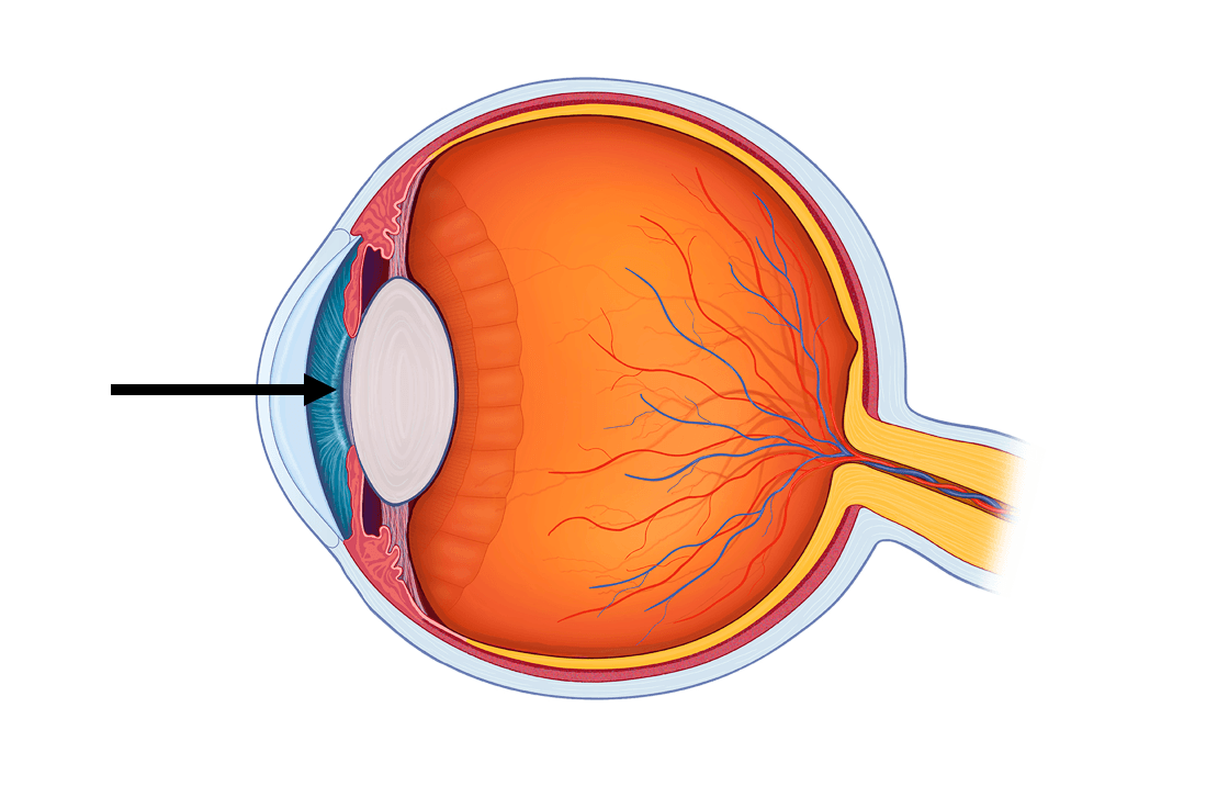
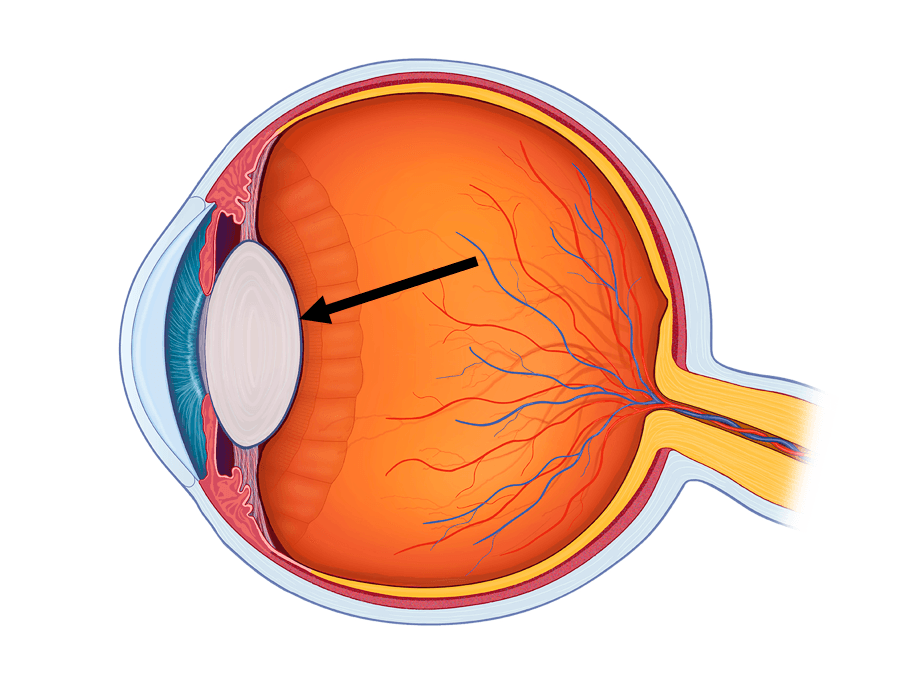
Directly behind the iris is the lens. This structure
changes shape to focus the light so that we can
see clearly. Its shape is convex, meaning it curves
outward on both sides.
The ciliary muscles above and below the lens
control the shape of the lens.
22
Multiple Choice
The optic nerve is located in what part of the eye?
in the front
right behind the lens
at the back
underneath the iris
23
Multiple Choice

choroid
sclera
cornea
retina
24

Behind the lens is a clear gel called the vitreous
humor. After moving through the vitreous humor,
the light strikes the retina. The retina is the lining
on the inside of the back of the eye that contains
two types of light-sensitive cells: rods and cones.
25
Multiple Choice
What provides nutrients and oxygen to the eye and remove waste?
Cornea
Aqueous humour
Pupil
Lens
26
Multiple Choice
This part of the eye focuses light onto the retina.
Retina
Optic Nerve
Lens
ciliary body
27
Multiple Choice
What part of the eye makes the pupil larger and smaller?
Retina
Iris
Lens
Cornea
28

Rods sense black and white and work in low light.
• L-cones sense long wavelengths in the red range
• M-cones sense mid-range wavelengths in green range
• S-cones sense short wavelengths in the blue range
Cones sense
color and must
have more light
than rods to
work. Three
kinds of cones:
29

The rods and cones send messages to the brain
through the optic nerve. The brain makes sense of
all the information it is receives.
In your brain, the sight
center is in the back,
between your ears. This
location explains why a
blow to the back of your
head might cause
blindness, even though
your eyes are fine.
30
Multiple Choice
Cones are light sensitive cells in the retina that
give you color vision in bright light
respond in dim light
give you color vision in dim light
respond in bright light
31

Like the pinhole viewer, images that appear on
your retina are upside down and backward. Your
brain interprets them in the right way.
I
32

The rods and cones send messages to the brain
through the optic nerve. The brain makes sense of
all the information it is receives.
33
Multiple Choice
The bundle of neural fibers that travel from the retina to the brain is called
conjunctiva
rods
cones
optic nerve
34
Multiple Choice
What is the purpose of the pupil?
It regulates the amount of light that enters the eye
it serves as the center of vision
it gives the center of the eye its color
to protect the eye
35
Multiple Choice
The eye has a blind spot. Why?
Because each eye is is damaged in some way
We all watch too much Netflix and it damages a part of our eye
The optic nerve connects to the retina and there are no photoreceptors there
The lens doesn't reflect light onto certain points of the retina
36

Eye Anatomy Review
• cornea
• pupil
• iris
• anterior chamber
• aqueous humor
• lens
• vitreous humor
• retina
• fovea
• choroid
• sclera
• optic nerve
37
Multiple Choice
The lens is most like what?
a kidney bean
a green pea
a marble
a small pebble
38

Wikipedia http://en.wikipedia.org/wiki/File:Schematic_diagram_of_the_human_eye_en.svg
2004 Microsoft Corporation, One Microsoft Way,
Redmond, WA 98052-6399 USA.
National Cancer Institute at the National Institutes of Health
http://www.cancer.gov/cancertopics/pdq/treatment/retinoblastoma/patient/page1/AllPages/Print
Image Sources
National Eye Institute, National Institutes of Health
http://www.nei.nih.gov/healthyeyes/eyeexam.asp
MedLine Plus, National Institutes of Health
http://www.nlm.nih.gov/medlineplus/ency/presentations/100206_1.htm
MedLine Plus, National Institutes of Health
http://www.nlm.nih.gov/medlineplus/ency/imagepages/9482.htm
Federal Aviation Administration
http://www.hf.faa.gov/webtraining/VisualDisplays/HumanVisSys2.htm
Federal Aviation Administration
http://www.hf.faa.gov/webtraining/visualdisplays/HumanVisSys6.htm
Glaucoma, MedLine Plus, National Institutes of Health
http://www.nlm.nih.gov/medlineplus/ency/imagepages/9349.htm
Eye Structure
and How we see
Show answer
Auto Play
Slide 1 / 38
SLIDE
Similar Resources on Wayground

33 questions
Part of the Cell
Presentation
•
6th - 8th Grade
36 questions
Safety & Lab Equipment
Presentation
•
6th - 8th Grade
34 questions
Evidence for Evolution
Presentation
•
6th - 8th Grade
35 questions
4 Types of Tissue
Presentation
•
6th - 8th Grade
34 questions
Box and Whisker Plots
Presentation
•
6th - 8th Grade
34 questions
Online Safety
Presentation
•
6th - 8th Grade
34 questions
Basics of Cybersecurity
Presentation
•
6th - 8th Grade
33 questions
The Skeletal System
Presentation
•
6th - 8th Grade
Popular Resources on Wayground
11 questions
Hallway & Bathroom Expectations
Quiz
•
6th - 8th Grade
10 questions
HCS SCI 03 Summer School Assessment 2
Quiz
•
3rd Grade
11 questions
Home Scope
Quiz
•
7th - 8th Grade
12 questions
2026 TAP Technology in the Classroom
Presentation
•
Professional Development
15 questions
HCS SCI 05 Summer School Assessment 2 Review
Quiz
•
5th Grade
15 questions
HCS SCI 04 Summer School Review 2
Quiz
•
4th Grade
59 questions
Geometry Unit 3 Review
Quiz
•
9th - 12th Grade
14 questions
FAST ELA READING SMAPLE TEST MATERIALS
Passage
•
3rd Grade