
Skeletal System Functions and Structures Review
Presentation
•
Physical Ed
•
12th Grade
•
Easy
Arthur EnloeHS
Used 687+ times
FREE Resource
20 Slides • 58 Questions
1
Skeletal System Functions Review
2
3
OSTEOCLASTS - a large multinucleated bone cells that absorb bone tissue during growth and healing.
OSTEOBLASTS - cells that form new bones and grow and heal existing bones.
OSTEOCYTES - these cells are derived from osteoblasts, they exist within the bone matrix and help to nourish bone tissue and regulate other bone cells
Osteoid is an unmineralized organic tissue that eventually undergoes calcification and is deposited as lamellae or layers in the bone matrix.
4
The skull is made up of many bones. There are 8 bones in the skull itself and 14 bones in the face area. They join together to form a solid, bony cavity that protects and supports the brain. The areas where the bones join together are called the sutures.
The bones are not joined together firmly at birth. This allows the head to change shape to help it pass through the birth canal. The sutures gradually gain minerals and harden, firmly joining the skull bones together. This process is called ossification.
In an infant, the space where 2 sutures join forms a membrane-covered "soft spot" called a fontanelle (fontanel). The fontanelles allow the brain and skull to grow during an infant's first year.
medlineplus.org
5
Osteoblasts group into clusters and form an ossification center. Osteoblasts begin secreting osteoid, an unmineralized collagen-proteoglycan matrix that can bind calcium. The binding of calcium to osteoid results in the hardening of the matrix and entrapment of osteoblasts. This entrapment results in the transformation of osteoblasts to osteocytes. As osteoid continues to be secreted by osteoblasts, it surrounds blood vessels, forming trabecular/cancellous/spongy bone. These vessels will eventually form the red bone marrow. Mesenchymal cells on the surface of the bone form a membrane called the periosteum. Cells on the inner surface of the periosteum differentiate into osteoblasts and secrete osteoid parallel to that of the existing matrix, thus forming layers. These layers are collectively called the compact/cortical bone.
nih.gov
The fontanel closes via ossification. During ossification the following happens
6
Multiple Choice
Special bone-forming cells that begin the mineralization process are referred to as what?
osteoporosis
osteoclast
ossification
osteoblast
7
Multiple Choice
8
Multiple Choice
Hemopoesis refers to what?
the production of blood
The storage of calcium and phosphorus
Red marrow
Yellow marrow
9
Multiple Choice
The skeletal system contains (or stores) what minerals ?
the production of blood
calcium and phosphorus
Red marrow
Yellow marrow
10
11
Multiple Choice
12
Multiple Choice
13
Multiple Choice
14
Multiple Choice
Which part of the bone is indicated by the number 9?
Spongy bone
Yellow bone marrow
Compact bone
Epiphysis
15
Multiple Choice
Which part of the bone is indicated by the number 2?
spongy bone
epiphysis
diaphysis
compact bone
16
Multiple Choice
What is the soft spot on an infant's head, where the bones have not yet grown together, called?
furuncle
foramina
fontanel
foramen
17
Multiple Choice
Age, diet, exercise, and hormones affect the production of calcium in bones.
true
false
18
Multiple Choice
During ossification osteoblasts become trapped in the bone matrix and become what kind of cells?
Osteoclasts
Osteocytes
Osteobons
Osteophages
19
20
There are flat bones in the skull (occipital, parietal, frontal, nasal, lacrimal, and vomer), the thoracic cage (sternum and ribs), and the pelvis (ilium, ischium, and pubis). The function of flat bones is to protect internal organs such as the brain, heart, and pelvic organs.
21
You will not need to know all of the irregular bones. You just need to know that they have unusual shapes that are associated with their anatomical functions.
The complex anatomy of the vertebrae is directly related to its' complex function
22
Long bones are the most common bones in the body. they are longer than they are wide.
Short bones include the carpal bones of the hands that allow movement of the wrist, and the tarsal bones of the feet that allow movement of the foot. Short bones are shaped roughly as a cube and contain mostly spongy bone.
23
A sesamoid bone is a small bone commonly found embedded within a muscle or tendon near joint surfaces, existing as focal areas of ossification and functioning as a pulley to alleviate stress on that particular muscle or tendon.
24
Multiple Choice
What kind of bone is this?
Long Bone
Short Bone
Sesamoid Bone
Irregular Bone
25
Multiple Choice
What kind of bone is this?
Long Bone
Short Bone
Sesamoid Bone
Irregular Bone
26
Multiple Choice
What kind of bone is this?
Long Bone
Short Bone
Sesamoid Bone
Irregular Bone
27
Multiple Choice
What kind of bone is this?
Long Bone
Short Bone
Sesamoid Bone
Irregular Bone
28
Multiple Choice
What kind of bone is this?
Long Bone
Short Bone
Sesamoid Bone
Irregular Bone
29
Multiple Choice
What kind of bone is this?
Long Bone
Short Bone
Sesamoid Bone
Irregular Bone
30
Multiple Choice
What kind of bone is this?
Long Bone
Short Bone
Sesamoid Bone
Irregular Bone
31
Epiphysis - end of a long bone.
Diaphysis - shaft of a long bone.
Medullary (Marrow) Cavity - space in the middle of the diaphysis.
Periosteum - Lining of the outside of the bone.
Endosteum - Lining of the medullary cavity. Contains yellow marrow.
Spongy Bone - porous bone at the epiphysis. Contains red marrow.
Compact Bone - dense and strong bone that makes up the diaphysis.
32
Multiple Choice
Red bone marrow is found in what type of bone?
33
Multiple Choice
34
Multiple Choice
35
Multiple Choice
36
Multiple Choice
37
38
39
40
41
Drag and Drop
42
Multiple Choice
The bones of the fingers and toes are called ___.
carpals
phalanges
tarsals
metacarpals
43
Multiple Choice
Which bone is a part of the appendicular skeleton?
Cranium
Spinal Cord
Ethmoid
Patella
44
Multiple Choice
45
Multiple Choice
Which is not part of the vertebral column
Sternum
Cervical
Thoracic
Lumbar
Sacrum
46
Multiple Choice
There are 12 of these
Thoracic Vertebra
Ribs
Cervical Vertebra
Transverse Foramen
47
Multiple Choice
48
Multiple Choice
49
Multiple Choice
How many pairs of ribs are found in the typical human?
50
Multiple Choice
51
Multiple Choice
52
Multiple Choice
How many vertebra are found in the typical cervical spine?
5
7
10
12
53
Multiple Choice
54
Multiple Choice
Cervical
Thoracic
Lumbar
55
Multiple Choice
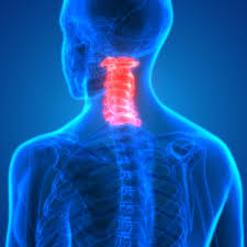
Which section of the vertebrae is highlighted
thoracic
cervical
lumbar
sacral
56
Multiple Choice
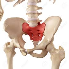
Which section of the vertebral column is highlighted
lumbar
cervical
coccyx
sacrum
57
Synarthrosis are immovable joints such as the sutures in the skull. Diarthrosis are freely movable (ball and socket, pivot, hinge joints fit here). Amphiarthroses are slightly movable joins. Examples of amphiarthroses include the vertebral articulations and the symphisis pubis in the pelvis.
58
Ball and socket joint. Permitting movement in all directions, the ball and socket joint features the rounded head of one bone sitting in the cup of another bone. Examples include your shoulder joint and your hip joint.
Hinge joint. The hinge joint is like a door, opening and closing in one direction, along one plane. Examples include your elbow joint and your knee joint.
Condyloid joint. The condyloid joint allows movement, but no rotation. Examples include your finger joints and your jaw.
Pivot joint. The pivot joint, also called the rotary joint or trochoid joint, is characterized by one bone that can swivel in a ring formed from a second bone. Examples are the joints between your ulna and radius bones that rotate your forearm, and the joint between the first and second vertebrae in your neck.
Gliding joint. The gliding joint is also called the plane join. Although it only permits limited movement, it’s characterized by smooth surfaces that can slip over one another. An example is the joint in your wrist.
Saddle joint. Although the saddle joint does not allow rotation, it does enable movement back and forth and side to side. An example is the joint at the base of your thumb.
59
Bone, muscle, cartilage, tendons, ligaments, membranes, and synovial fluid all come together to make our joints stable and functional.
60
61
Multiple Choice
62
Multiple Choice
63
Multiple Choice
64
Multiple Choice
Carpals and tarsals are examples of this type of joint
65
Multiple Choice
66
Multiple Choice
67
Multiple Choice
68
Multiple Choice
69
Multiple Choice
70
Multiple Choice
71
Multiple Choice
72
Multiple Choice
73
Multiple Choice
74
Multiple Select
Which of the following is part of a Synovial joint
Synovial membrane
Joint cavity
Ligament
Cartilage
75
Multiple Choice
When kicking a football the knee
Flexes
Extends
Abducts
Adducts
76
Multiple Choice
77
Multiple Choice
78
Multiple Choice
Skeletal System Functions Review
Show answer
Auto Play
Slide 1 / 78
SLIDE
Similar Resources on Wayground
74 questions
Heart/ Diseases
Presentation
•
KG - University
72 questions
Bài thuyết trình tính chuyên nghiệp
Presentation
•
KG - University
70 questions
Unit 4 level 3
Presentation
•
University
73 questions
Oligopolies
Presentation
•
12th Grade
73 questions
week 1
Presentation
•
KG - University
70 questions
Berry - Gilded Age STAAR review
Presentation
•
11th Grade
76 questions
Price Floor
Presentation
•
12th Grade
72 questions
Q1W3 EALS Types of Rocks & Weathering
Presentation
•
11th Grade
Popular Resources on Wayground
20 questions
STAAR Review Quiz #3
Quiz
•
8th Grade
20 questions
Equivalent Fractions
Quiz
•
3rd Grade
6 questions
Marshmallow Farm Quiz
Quiz
•
2nd - 5th Grade
20 questions
Main Idea and Details
Quiz
•
5th Grade
20 questions
Context Clues
Quiz
•
6th Grade
20 questions
Inferences
Quiz
•
4th Grade
19 questions
Classifying Quadrilaterals
Quiz
•
3rd Grade
12 questions
What makes Nebraska's government unique?
Quiz
•
4th - 5th Grade