Radiology: Lung Patterns and Conditions
Interactive Video
•
Science, Biology
•
10th Grade - University
•
Practice Problem
•
Hard

Ethan Morris
FREE Resource
Read more
10 questions
Show all answers
1.
MULTIPLE CHOICE QUESTION
30 sec • 1 pt
What is the most common cause of reduced lung volumes on an X-ray?
Asthma
Pneumothorax
Pulmonary embolism
Poor inspiratory effort
2.
MULTIPLE CHOICE QUESTION
30 sec • 1 pt
Which condition is most commonly associated with hyperinflation on an X-ray?
Pulmonary fibrosis
COPD
Pneumonia
Pleural effusion
3.
MULTIPLE CHOICE QUESTION
30 sec • 1 pt
What type of fluid accumulation is associated with alveolar opacities?
Air
Edema, pus, or blood
Fat
Bone
4.
MULTIPLE CHOICE QUESTION
30 sec • 1 pt
Which of the following is a feature of non-cardiogenic pulmonary edema?
Pleural effusions
Air bronchograms
Homogeneous opacities
Enlarged cardiac size
5.
MULTIPLE CHOICE QUESTION
30 sec • 1 pt
What is the significance of Kerley B lines on an X-ray?
Indicate lung hyperinflation
Show pleural effusion
Suggest interstitial edema
Indicate pneumothorax
6.
MULTIPLE CHOICE QUESTION
30 sec • 1 pt
Which pattern is associated with cardiogenic pulmonary edema?
Nodular pattern
Miliary pattern
Batwing pattern
Patchy distribution
7.
MULTIPLE CHOICE QUESTION
30 sec • 1 pt
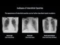
What is a characteristic feature of reticular opacities?
Too many lines
Too much air
Too much fluid
Too many dots
Access all questions and much more by creating a free account
Create resources
Host any resource
Get auto-graded reports
Continue with Google

Continue with Email
Continue with Classlink
Continue with Clever
or continue with
Microsoft
%20(1).png)
Apple
Others
Already have an account?
