Knee Anatomy and X-ray Interpretation
Interactive Video
•
Science, Biology, Other
•
9th - 12th Grade
•
Practice Problem
•
Hard

Patricia Brown
FREE Resource
Read more
10 questions
Show all answers
1.
MULTIPLE CHOICE QUESTION
30 sec • 1 pt
What are the three standard views used in knee x-ray anatomy?
AP, lateral, and oblique views
AP, lateral, and sunrise views
AP, oblique, and axial views
Lateral, sunrise, and axial views
2.
MULTIPLE CHOICE QUESTION
30 sec • 1 pt
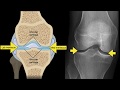
In the AP view of the knee, which bone is identified as looking like a 'T'?
Femur
Fibula
Patella
Tibia
3.
MULTIPLE CHOICE QUESTION
30 sec • 1 pt
What is the function of the joint space in the AP view of the knee?
To accommodate articular cartilage and menisci
To connect the femur and fibula
To provide structural support
To allow for muscle attachment
4.
MULTIPLE CHOICE QUESTION
30 sec • 1 pt
Which bone is described as a sesamoid bone in the lateral view of the knee?
Patella
Fabella
Tibia
Fibula
5.
MULTIPLE CHOICE QUESTION
30 sec • 1 pt
In the lateral view, where is the fabella typically located?
In the medial tendon of the gastrocnemius
In the lateral tendon of the gastrocnemius
In the anterior tendon of the quadriceps
In the posterior tendon of the hamstrings
6.
MULTIPLE CHOICE QUESTION
30 sec • 1 pt
What is the primary focus of the sunrise view of the knee?
The femoral condyles
The patellar tendon
The femoral patellar joint
The tibial plateau
7.
MULTIPLE CHOICE QUESTION
30 sec • 1 pt
What does the trochlear groove resemble in the sunrise view?
A ball
A hinge
A socket
A pulley
Access all questions and much more by creating a free account
Create resources
Host any resource
Get auto-graded reports
Continue with Google

Continue with Email
Continue with Classlink
Continue with Clever
or continue with
Microsoft
%20(1).png)
Apple
Others
Already have an account?
