
 Worksheets
Unit 2 Brain Biology
Total questions: 90
Worksheet time: 2hrs 49mins
Uses powerful magnets, radio waves, and a computer to make detailed pictures of brain; doesn't use radiation
EEG
PET
CT
MRI
The “all or nothing” response of a nerve causing it to “fire” or activate sending a signal to the next neuron.
Activation threshold
Action potential
Refractory period
Polarization
This is a high detailed x-ray scan of the brain and skull. The pictures come in “slices” to form a composite view.
a. MRI (Magnetic Resonance Image)
a. CAT (Computed Tomography) Scan
a. EEG (Electroencephalogram)
a. PET (Positron Emission Tomography)
This scan measures the level of brain activity using magnetic imaging techniques. It connects cerebral blood flow to neural activity.
a. fMRI (functional Magnetic Resonance Imaging)
a. MRI (Magnetic Resonance Image)
a. CAT (Computed Tomography) Scan
a. PET (Positron Emission Tomography)
This test allows doctors to monitor brain wave functions by putting electrodes on the skin around the skull and face.
a. MRI (Magnetic Resonance Image)
a. CAT (Computed Tomography) Scan
a. EEG (Electroencephalogram)
a. PET (Positron Emission Tomography)
This scan measures the level of brain activity using magnetic imaging techniques. It connects cerebral blood flow to neural activity.
a. fMRI (functional Magnetic Resonance Imaging)
a. MRI (Magnetic Resonance Image)
a. CAT (Computed Tomography) Scan
a. PET (Positron Emission Tomography)

Controls arousal (sleepiness and awakefulness)
Reticular Formation
Thalamus
Amygdala
Medulla
What characterizes a neuron in the resting state? Select any that apply
negatively charged
positively charged
polarized
depolarized
What distinguishes the neural pathway of reflexive movement?
motor neurons originate in the spinal cord to carry out response
the sensory neurons travel to the brain first for processing
it never enters a state of repolarization
neurotransmitters often act as inhibitors to movement
This part of the brain is the bridge between two other parts; it has optic nerves running through it, involving it in eye movement and vision
midbrain
hindbrain
olfactory bulb
forebrain
Structuralism, as defined by Edward Titchner, featured
introspection
neurotransmitters
evolutionary theory
consciousness
When the neuron is temporarily incapable of firing again
refractory phase
depolarization
all or nothing principle
ionization
The ventromedial hypothalamus, when lesioned in mice, results in
weight gain
weight loss
starvation
satiation
What looks at how traits adapt to the environment over time?
behavioral genetics
epigenetics
neurogenisis
imprinting
A neurotransmitter, in order to excite or inhibit action potential, must first
cross the synaptic gap/cleft
be in a resting state of polarization
interact with interneurons
initiate from the myelin sheath
Increasing the intensity of excitatory signal above the necessary threshold is not necessary because of the
depolarization status
all or nothing response principle
terminal vesicles
electrical-chemical process
Voluntary movement in the cerebral cortex is represented by the
somatosensory cortex
visual cortex
association areas
primary motor cortex
The parietal lobes specialize in ___, which is a/an
mathematical and spatial reasoning; association area
higher level thinking; prefrontal lobe
creativity and artistic ability; visual association area
physical touch and sensation; hindbrain function
Konrad Lorenz is associated with
critical periods of development and imprinting
modeling and self efficacy
positive and negative conditioning
taste aversion classical conditioning
The process of neural communication is best described as
an electrochemical process
resting potential
transmission of neurotransmitters
dependent on the myelin sheath
The gyri and sulci, which expands the "territory" of the brain, is located in
frontal lobe exclusively
cerebral cortex
corpus callosum
prefrontal cortex
One positive outcome of phrenology was
neural communication understanding
conditioning of taste aversions
neuroplasticity
hemispheric specialization
A source of rich data for studying heritability includes
CRISPR technology
twin studies
epigenetics
correlational studies
Neuroplasticity is facilitated by the growth of new
dendrite receptors
schwann cells
nuclei of the the soma
terminal vesicles
Feeling no pain in your left arm could indicate
right frontal lobe damage
right parietal lobe damage
left parietal lobe damage
right frontal lobe damage
Part of the brain responsible for brain activity during sleep
Medulla
Pons
Thalamus
Cerebellum
Our "little brain"; responsible for perception of time, equilibrium, non-verbal learning
Cerebellum
Pons
Medulla
Thalamus
Fear and Aggression
Hypothalamus
Hippocampus
Amygdala
Central to learning and memory
Hippocampus
Hypothalamus
Amygdala
Responsible for planning, judging, personality aspects
Frontal Lobe
Parietal Lobe
Temporal Lobe
Occipital Lobe
Responsible for sense of touch & perceiving spatial relationships
Frontal Lobe
Parietal Lobe
Temporal Lobe
Occipital Lobe
Responsible for comprehension, sound, speech
Frontal Lobe
Parietal Lobe
Temporal Lobe
Occipital Lobe
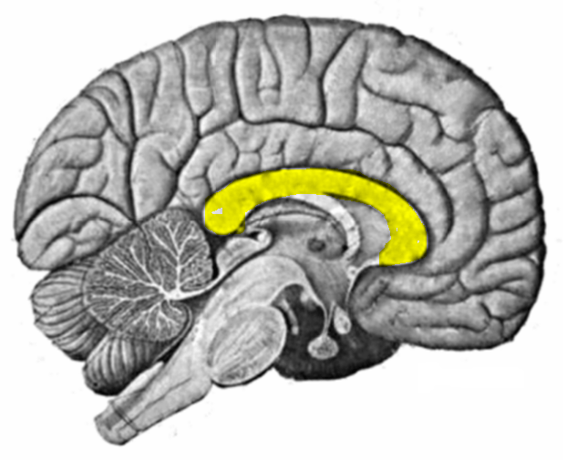
Responsible for connecting the two cerebral hemispheres
Corpus Callosum
Cerebral Cortex
Cerebellum
Cerebrum
What is the part of the neuron that receives information/impulses from other neurons
Dendrites
Axon Buttons
Myelin Sheath
Cell body

What is the Light Pink highlighted region called
(use your Cognitive Dictionaries)
Cerebellum
Occipital Lobe
Temporal Lobe
Frontal Lobe
Parietal lobe

What is the Hot Pink highlighted region called
(use your Cognitive Dictionaries)
Cerebellum
Occipital Lobe
Temporal Lobe
Frontal Lobe
Parietal lobe

What is the Orange highlighted region called
(use your Cognitive Dictionaries)
Cerebellum
Occipital Lobe
Temporal Lobe
Frontal Lobe
Parietal lobe

What is light green highlighted region called
(use your Cognitive Dictionaries)
Cerebellum
Occipital Lobe
Temporal Lobe
Frontal Lobe
Parietal lobe

What is the yellow highlighted region called
(use your Cognitive Dictionaries)
Cerebellum
Occipital Lobe
Temporal Lobe
Frontal Lobe
Parietal lobe

This structure is involved in fear/panic response
Cerebellum
Hippocampus
Amygdala
Parietal Lobe


Which lobe is this?
Occipital Lobe
Parietal Lobe
Temporal Lobe
Frontal Lobe

