
 Worksheets
Nervous System
Total questions: 46
Worksheet time: 32mins
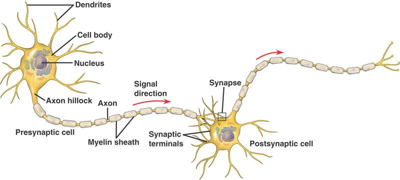
What is the name for a cell in the nervous system?
Corpus Callosum
Neuron
Node of Ranvier
Red Blood Cell
Which part of a neuron receives stimuli?
Dendrites
Axon
Myelin Sheath
Nucleus
Which part of the neuron is a long fiber that SENDS information to other cells?
dendrite
terminal
cell body
axon
the point at which an impulse from one nerve cell is communicated to another cell is the____...
Synapse
collateral branch
receptor
outflux

What is represented by D?
Neurotransmitter
Receptor
Plasma membrane
Vesicle
Synapse
What is represented by C?
Neurotransmitter
Receptor
Plasma membrane
Vesicle
Synapse
What is represented by B?
Neurotransmitter
Receptor
Plasma membrane
Vesicle
Synapse
Neuroglial cells that form the fatty myelin sheath on neurons in the CNS are ___________ in the PNS are ________________?
ganglia and oligodendroglia
astrocytes and schwann cells
schwann cells and oligodendroglia
oligodendroglia and schwann cells
Small cells that engulf and destroy microbes and cellular debris in the CNS
microglia
horns
neuroglia
astrocytes
When a neuron is at REST,
the inside of the neuron is negatively charged compared to the outside.
the inside of the neuron is positively charged compared to the outside.
there is no charge difference established
none of these is true
At resting potential, the ion distribution inside and outside of a neuron is such that __________ ions are most abundant on the outside of the cell, while __________ ions are most abundant on the inside of the cell.
Potassium; sodium
Sodium; potassium
Calcium; potassium
Calcium; sodium
A bundle of neurons is called a...
Super Neuron
Mega Neuron
Nerve
Inter Neuron
Vagus Nerve
swallowing
saliva production
digestive and heart
tongue movements
Which one has two hemispheres?
thalamus
medulla
reticular formation
cerebrum
The right hemisphere of your brain controls _______________.
the left side of your body
the right side of your body
your entire body
the entire brain
The connecting piece for the right hemisphere and the left hemisphere is called the ______________________________.
amygdala
corpus connector
corpus collossum
cortex


A clear fluid that helps the cornea keep its rounded shape
aqueous humor
blind spot
cones
cornea
The place where all nerves from the retina join to form the optic nerve. Each eye has a _____ _____ where there are no light-sensitive cells
aqueous humor
blind spot
cones
cornea
A tough, clear covering over the iris and the pupil that helps protect the eye. Light bends as it passes through the _______ before making its way to the lens.
aqueous humor
blind spot
cones
cornea
A muscle that controls how much light enters the eye. It is suspended between the cornea and the lens. Human ________ come in many colors, including brown, blue, green, and gray.
iris
lens
myelin
optic nerve
A clear flexible structure that makes an image on the eye’s retina. The _____ is flexible so that it can change shape, focusing on objects that are close up and objects that are far away.
iris
lens
myelin
optic nerve
The bundle of nerve fibers that carry information from the retina to the brain.
iris
lens
myelin
optic nerve
The _________ is the dark circle in the center of your iris. It’s a hole that lets light into the inner eye.
pupil
retina
rods
sclera
The layer of light-sensitive cells at the back of the eye. The _________ detects images focused by the cornea and the lens and is connected to the brain by the optic nerve.
pupil
retina
rods
sclera
The thick, tough, white outer covering of the eyeball.
pupil
retina
rods
sclera
The colorful, shiny material located behind the retina. Found in animals with good night vision, the ________ reflects light back through the retina.
tapetum
vitreous humor
The thick clear jelly that helps give the eyeball its shape
aqueous humor
vitreous humor
__________ carry messages between sensory neurons and motor neurons.
Active Neurons
Interneurons
Refraction Neurons
Flower Neurons

