
 Worksheets
FINAL A&P EXAM Radiography
Total questions: 60
Worksheet time: 43mins
Refers to dense, hard bone tissue found primarily in the shafts of long bones and on the outer surfaces of other bones.
Spongy bone
Cancellous bone
Compact bone
Cartilage
Enlarged ends of the long bone. Articulates with a second bone at a joint. covered by cartilage. Spongy bone covered by compact bone.
Diaphysis
Medullary cavity
Endosteum
Epiphysis
The ribs directly below the true ribs that attach indirectly to the sternum by cartilage or do not attach at all
Floating ribs
True ribs
False ribs
Cartilar
Band of hyaline cartilage located near the proximal and distal ends of a growing long bone. also known as the growth plate. Longitudinal bone growth occurs
Ephyphyseal line
Articular Cartilage
Epiphyseal disc
Suture
One of the contractile proteins in muscle; forms the thin filaments
Myosin
Sarcomere
Z-Band
Actin
Consists of a single motor neuron and the muscle fibers that are supplied by the motor neuron.
Motor neuron
Sarcomere
Motor unit
Sarcoplasm
Myosin heads make contact with actin heads. Myosin heads rotate. Actin is pulled to the center of the sarcomere. Sarcomere shortens (muscle contracts).
Action Potential
Threshold
Sliding filament theory
Depolarization
Muscle attaches to the stationary bone
Insertion
Prime mover
Prime mover
Origin
Found in walls of visceral muscle, tubes and passageways, bronchioles, and blood vessels. Involuntary muscle. Not striated. Stretchiness allows expansion of organs. Better capacity for regeneration
Skeletal Muscles
Cardiac Muscles
Striated Muscles
Smooth Muscles
Largest muscle in the body. Forms the buttock and is the muscle on which you sit. Origins on the ilium, and sacrum and inserts on the femur. Rotates thigh laterally and extends the thigh at the hip
Femoral
Gluteus maximus
Romboid
Tibialis anterior
Muscles that oppose the action of another muscle
Prime mover
Agonist muscles
Synergist muscles
Antagonist muscles
Flat muscle that covers the frontal bone. Extends from the cranial aponeurosis and insert in the skin above the eyebrows
Buccinator
Zigomatic
Frontalis
Orbicularis
Overused muscles that increase in size
Atrophy
Muscle fatigue
Hypertrophy
Contracture
Sternocleidomastoid, 3 Scalene muscles, and Trapezius
Hamstrings muscle group
Abdominal muscles
Quadriceps group
Neck muscles
Group of four muscles: subscapularis, supraspinatus, infraspinatus, and teres minor. They attach (insertion) the humerus to the scapula (origin)
Neck muscles
Muscles of the chest
Rotator cuff muscles
Wrist muscles
Dome-shaped muscle that separates the thoracic cavity from the abdominal cavity. Chief muscle of inhalation, or the breathing-in phase of respiration
Intercostal
Sartorius
Diaphragm
Santorini
Long extension that transmits signals away from the cell body. Contains the myelin sheath, the neurilemma, and the node of Ranvier
Dendrites
Axon terminals
Cell bodies
Axon
Helps to interpret sensory information. Ex: visual, auditory, somatosensory
Hypoccampus
Medulla
Association areas
Carpus Callosum
Neuron impulse. A series of events cause the electrical charge inside the cell to move from its negative resting stat (-) to its positive depolarize state (+) and back to its negative resting state
Sliding Filament Theory
Threshold
Action Potential
Interaction of electrolytes
Connects the spinal cord with higher brain structures. Composed of the midbrain, pons, and medulla oblongata
Cerebellum
Ventricles
Brain stem
Spinal cord
Contains the nucleus and is essential for the life of the cell. Receives signals from the dendrites and chooses which ones to send to the axon
Axon
Node of Ranvier
Neuron
Cell body
Concerned with coordination of voluntary muscle activity. Major reflex center. Evaluates sensory input
Medulla Oblongata
Broca's Area
Cerebellum
Astrocytes
Integrating system for the autonomic nervous system; regulation of temperature, water balance, sex, thirst, appetite, and some emotions.
Brain
Hypothalamus
Cerebrum
Meninges
Located in the back of the head, contains the visual cortex
Frontal lobe
Temporal lobe
Corpus Callosum
Occipital lobe
Is the most important in the transmission of electrical signals. Enables their nervous system to act as a vast communication. 3 parts: denrites, cell body, and axon
Neuroglia
Schawn cell
Neuron
Brain
Divided into 2 structures: the central nervous system (CNS) and the peripheral nervous system (PNS). Performs 3 general functions: a sensory function, an integrative function, and a motor function
Autonomic Nervous system
Central Nervous System
Nervous System
Peripheral Nervous System

What muscle is this?
Trapezius
Deltoid
Pectoralis Major
Latissimus Dorsi
Which muscle is in the upper chest and connects the sternum to the proximal humerus?
pectoralis major
external oblique
rectus abdominus
internal oblique
Basic unit of the nervous system; another name for nerve cell.
brain stem
axon
dendrite
neuron

What muscle is located on the anterior arm?
biceps brachii
triceps brachii
brachialis
brachioradialis
Besides being able to hear, ears also help with balance
True
False
Photoreceptors respond to what?
chemicals in a solution
changes in light
changes in pressure and movement
changes in temperature
What connects bones together?
ligaments
brains
trachea
heart

Bone forming cells:
osteocytes
osteoblasts
osteoclasts
osteomasts
Process of bone formation before birth:
ossification
hematopoiesis
appositional growth
atrophy
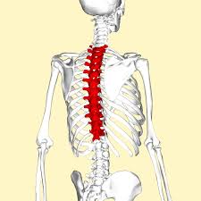

Which of the following is not a function of bones?
Protection
Blood cell formation
Storage
Absorption
Articular cartilage is found:
at the ends of bones
between sutures
surrounding long bones
at the growth plate



