
 Worksheets
CET Midterm Review
Total questions: 100
Worksheet time: 3hrs 20mins
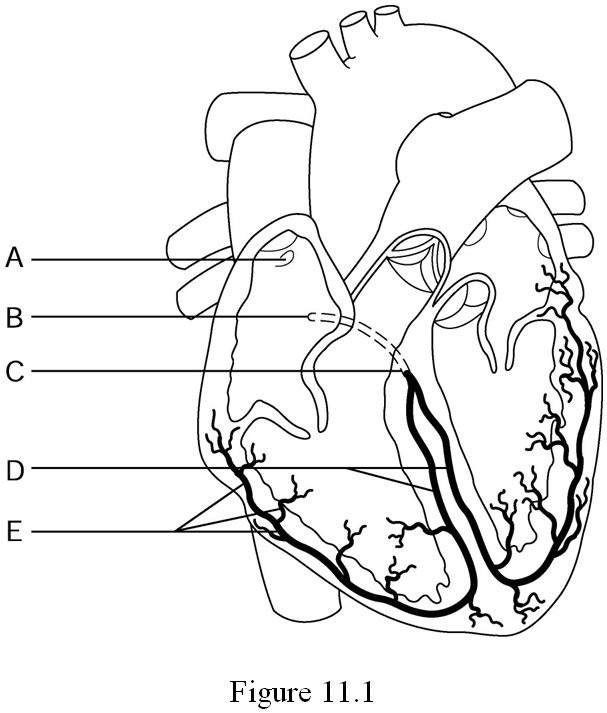
SA node, AV node, Bundle of His, Purkinje fibers
What are random parts of the heart
What are the four major parts of the hearts electrical system
What are the four major parts of the brain
What are the four random parts of the digestive system
What is the intrinsic rate of the Sinoatrial node (SA node)
20-40 BPM
40-60 BPM
60-100 BPM
What is the intrinsic rate of the Atrioventricular node (AV node)
20-40 BPM
40-60 BPM
60-100 BPM
What is the intrinsic rate of the Bundle of His
20-40 BPM
30-50 BPM
60-100 BPM
What is the intrinsic rate of the Purkinje Fibers
20-40 BPM
40-60 BPM
60-100 BPM
What mechanical action is seen during depolarization?
Contraction / Pumping
Contraction / Filling
Relaxation / Filling
Relaxation / Pumping
What mechanical action is seen during repolarization?
Contraction / Pumping
Contraction / Filling
Relaxation / Filling
Relaxation / Pumping
What time period is represented by a small block on EKG graph paper?
0.02 seconds
0.04 seconds
0.20 seconds
0.40 seconds
What time period is represented by a large block on EKG graph paper?
0.02 seconds
0.04 seconds
0.20 seconds
0.40 seconds
The number of beats per minute
Heart Rhythm
Heart Rate
Heart Pump
Heart Interval
Six second method, Small block method, Large block method
What is three methods of counting squares
What is three methods of wasting time
What is three methods used to determine heart rate
What is three methods used to determine heart rhythm
Count the number of complete QRS complexes in a six second strip and multiply by 10
What is the six second method
What is the Small block method
What is the Large block method
Count the number of large blocks between two consecutive R waves and use chart to determine rate
What is the six second method
What is the Small block method
What is the Large block method
Count the number of small blocks between two consecutive R waves and divide 1500 by the number of small blocks to determine heart rate
What is the six second method
What is the Small block method
What is the Large block method
Using the large block method - 1 block = a heart rate of ________
50
60
100
150
300
Using the large block method - 3 blocks = a heart rate of ________
50
60
100
150
300
Using the large block method - 5 blocks = a heart rate of ________
50
60
100
150
300
Using the large block method - 4 blocks = a heart rate of ________
50
60
75
150
300
Using the large block method - 2 blocks = a heart rate of ________
50
60
75
150
300
Using the large block method - 6 blocks = a heart rate of ________
50
60
75
150
300
Find the beginning of the P wave and the first deflection of the QRS, count the number of small blocks between and multiply by 0.04 seconds
What is how to determine the QRS interval
What is how to determine the R-R interval
What is how to determine the PR interval
What is how to determine the ST segment
A measurement of time taken from the start of the Q wave, through the R wave, and to the end of the S wave
What is how to determine the QRS interval
What is how to determine the R-R interval
What is how to determine the PR interval
What is how to determine the ST segment
The cardiac conduction systems ability for each part to conduct electrical impulses at its own particular rate
Regularity
Intrinsic
Interval
Deflection
What does the P wave represent?
Atrial depolarization
Ventricular depolarization
Atrial repolarization
Ventricular repolarization
What does the QRS complex represent?
Atrial depolarization
Ventricular depolarization
Atrial repolarization
Ventricular repolarization
What is the first step in analyzing a heart rhythm?
Measure the length of the PR interval
Measure the length of the QRS interval
Determine the heart rate in BPM
Examine the rhythm for regularity
Note the presence or absence of identical P waves before each QRS interval
What is the second step in analyzing a heart rhythm?
Measure the length of the PR interval
Measure the length of the QRS interval
Determine the heart rate in BPM
Examine the rhythm for regularity
Note the presence or absence of identical P waves before each QRS interval
What is the third step in analyzing a heart rhythm?
Measure the length of the PR interval
Measure the length of the QRS interval
Determine the heart rate in BPM
Examine the rhythm for regularity
Note the presence or absence of identical P waves before each QRS interval
What is the fourth step in analyzing a heart rhythm?
Measure the length of the PR interval
Measure the length of the QRS interval
Determine the heart rate in BPM
Examine the rhythm for regularity
Identify the Rhythm
What is the fifth step in analyzing a heart rhythm?
Measure the length of the PR interval
Measure the length of the QRS interval
Determine the heart rate in BPM
Examine the rhythm for regularity
Identify the Rhythm
What is the sixth step in analyzing a heart rhythm?
Measure the length of the PR interval
Measure the length of the QRS interval
Determine the heart rate in BPM
Examine the rhythm for regularity
Identify the Rhythm
The term used to describe the shape and direction of waves, complexes, and segments on an EKG tracing
Regularity
Intervals
Relaxation
Morphology
The isoelectric line is also known as the
Top line
Bottom line
Normal Sinus Rhythm
Baseline
What is the "normal" measurement of time for a PR interval
0.08 - 0.20 seconds
0.12 - 0.20 seconds
Less than 0.12 seconds
More than 0.20 seconds
What is the "normal" measurement of time for a QRS interval
0.08 - 0.20 seconds
0.12 - 0.20 seconds
Less than 0.12 seconds
More than 0.20 seconds
When evaluating the ____ wave we are looking for an upward deflection, and uniformity
P
Q
R
S
T
The QRS complex can look very different from one tracing or rhythm to another, so mastering measurement requires a lot of practice
True
False
When assessing regularity -- you want to measure from the point of one ____ wave to the point of the next ____ wave and then compare that measurement to each on the strip
P to P
Q to Q
R to R
S to S
T to T
A surgical procedure in which an instrument with a tiny balloon is inserted into a blocked artery to clear a blockage.
heart attack
hypertension
stroke
angioplasty
V1
LEFT side of the sternum, directly across from V1 at the FOURTH intercostal space
LEFT side of the chest, FIFTH intercostal space, midclavicular line
RIGHT side of the sternum at the FOURTH intercostal space
LEFT side of the chest, FIFTH intercostal space, midaxillary line
V2
LEFT side of the chest, FIFTH intercostal space, midclavicular line
LEFT side of the sternum, directly across from V1 at the FOURTH intercostal space
LEFT side of the chest, FIFTH intercostal space, midclavicular line
LEFT side of the chest, FIFTH intercostal space, anterior axillary line
V3
LEFT side of the chest, FIFTH intercostal space, midaxillary line
RIGHT side of the sternum at the FOURTH intercostal space
LEFT side of the chest, FIFTH intercostal space, anterior axillary line
LEFT side of the chest, midway between V2 and V4

Einthoven's triangle is formed by using which of the following placement sites
right arm, left arm, left leg
right arm, left leg, right leg
right arm, middle of chest, left leg
none of the above
Activity that causes ECG interference
artifact
impulse
interval
voltage
Any irregularity in heart rhythm
PVC
V-Fib
A-Fib
Arrhythmia
This controls the height of the ECG waveform
speed
gain
hertz
lead
The nurse is reviewing a patient’s electrocardiogram. Which wave should the nurse recognize as being the first positive wave in a normal cardiac cycle?
P wave
Q wave
R wave
S wave
After reviewing an electrocardiogram the nurse determines that an electrical impulse originated in a patient’s the sinoatrial node. What did the nurse see on this tracing?
An upright T wave
An inverted T wave
A positive P wave before a QRS complex
A negative P wave before a QRS complex
The nurse is determining the functioning of a patient’s sinoatrial node. How many times per minute should the nurse expect the node to normally fire?
20 to 40
25 to 55
40 to 85
60 to 100
Rate: less than 60 bpm
Rhythm: regular
Sinus Rhythm
Sinus Arrhythmia
Sinus Bradycardia
Sinus Tachycardia
What is the time frame occupied by one lead on a regular 12-lead ECG?
0.3 seconds
3 seconds
13 seconds
30 seconds
A wave that is 10 small boxes high and three small boxes wide is described as being:
1.0mm by 0.3 seconds
10mm by 0.12 seconds
12mm by 0.3 seconds
12mm by 0.10 seconds

EKG interpretation:
Normal Sinus Rhythm
Sinus Bradycardia
Sinus Tachycardia
Sinus Arrhythmia

EKG interpretation:
Normal Sinus Rhythm
Sinus Bradycardia
Sinus Tachycardia
Sinus Arrhythmia

EKG interpretation:
Normal Sinus Rhythm
Sinus Bradycardia
Sinus Tachycardia
Sinus Arrhythmia

EKG interpretation:
Normal Sinus Rhythm
Sinus Bradycardia
Sinus Tachycardia
Sinus Arrhythmia

EKG interpretation:
Normal Sinus Rhythm
Sinus Bradycardia
Sinus Tachycardia
Sinus Arrhythmia

EKG interpretation:
Normal Sinus Rhythm
Sinus Bradycardia
Sinus Tachycardia
Sinus Arrhythmia
Lead II is the standard lead in viewing the EKG.
True
False
The paper on an ECG normally moves at:
50mm/sec
75mm/sec
25cm/sec
25mm/sec
The entire ECG is:
3 seconds long
6 seconds long
9 seconds long
12 seconds long
The small boxes on ECG paper measure:
1 cm by 0.20 seconds
1mm by 0.20 seconds
1cm by 0.04 seconds
1mm by 0.04 seconds
The big boxes on the ECG paper measure:
5mm by 0.20 seconds
1mm by 0.20 seconds
5mm by 0.04 seconds
1mm by 0.04 seconds
A distance of 2 big boxes and 2 little boxes wide is described as being:
22mm wide
0.22 seconds wide
0.48 seconds wide
4.8 seconds wide
Also known as the baseline, it is best determined by drawing a line between the end of the T and beginning of the P wave.
P wave
isoelectric line
SA node
Av node
The interval from the peak of one QRS complex to the next. It is used as an indicator of regularity when evaluating the heart's rhythm on an EKG.
long QT interval
R-R interval
PR interval
P-P interval
The electrical sensors placed on the chest to record the bioelectrical activity of the heart.
electrode
node
wave
interval
Corresponds to rapid ventricular repolarization.
A wave
P wave
T wave
Q wave
V6
LEFT side of the chest, FIFTH intercostal space, midclavicular line
LEFT side of the chest, FIFTH intercostal space, anterior axillary line
LEFT side of the chest, FIFTH intercostal space, midaxillary line
LEFT side of the sternum, directly across from V1 at the FOURTH intercostal space
P, Q, R, S, T, U are known as:
Waves
Intervals
Segments
Complex
Usually the central and most visually obvious part of the tracing.
P Wave
QRS Complex
R-R Interval
T Wave
Lines between two waves
Complex
Amplitude
Duration
Segment
"Snow over Grass; Smoke over Fire" is one way we remember the placement for:
V1
V2
aVR
Limb Leads

What is A?
QRS Complex
P wave
ST segment
T wave
QT segment

What is C?
T wave
R wave
Q wave
U wave
P wave

What is D?
PR interval
QRS Complex
ST segment
T wave

What is F?
P wave
Q wave
R wave
S wave
T wave

What is G?
QT segment
P wave
QRS Complex
ST segment
PR interval

What is H?
ST segment
T wave
P wave
QRS complex
QT interval

What is I?
P wave
PR Interval
QRS Complex
ST segment
QT Interval
ECG stand for
Electrocardiograph
Electrocardiogram
Electrocardiopraphy
Electronicardiogram
ECG is a reading that shows
the activity of heart
the electrical activity of cardiac muscle
the systole and diastole of cardiac muscle
the initiation of heartbeat
The correct sequence of the three events in ECG
P wave , T wave , QRS complex
QRS complex , P wave , T wave
T wave , QRS complex , P wave
P wave, QRS complex, T wave
The ECG is the most commonly used clinical tool for diagnosing what?
Cardiac dysfunction
Cardiac Failure
Congestive Heart Failure
COPD
When disrobing the patient will be undressed from ?
Waist down with gown open to the front
Waist up and no lower leg access , gown open to the front
Waist up, lower leg access and gown open to the front
Gown open to the front, Waist up and no lower leg access
The normal heart rhythm is called?
Sinus brady
ventricle fibrillation
Normal sinus rhythm
Junctional Rhytms
The measurement of force exerted on the wall of the arteries when the heart contracts is
systolic pressure
diastolic pressure
pulse pressure
At which arterial site do we normally assess a patients blood pressure?
femoral
brachial
coronal
radial

What is "blood pressure"
how stressed your heart is
how hard your muscles are pushing on the heart
the force of the blood pumping through your veins

