
 Worksheets
Unit 4 Quizizz Skeletal System
Total questions: 53
Worksheet time: 12hrs 51mins

Area #5 is made up of -
spongy bone
compact bone
diaphysis bone
yellow matter

Area #1 nodules are called the
mastoid process
occipital condyles
auditory meatus
none of these

The Forearm bone located on the pinky side is called the
Radius
Ulna
Clavicle
Humerus

Synchondrosis means "held by hyaline cartilage" which allows for some movement, (like the rib cage). This type of joint would be -
synarthosis
amphiarthrosis
diarthrosis
redduarthrosis

An autoimmune disorder where the body immune system attacks its own joints.
anorexia nervosa
fracture
sprain
rheumatoid arthritis

Area "B" is called a -
Central Haversian canal
Osteon
Osteocyte
Perforating Volkman's canal

Area #12 is known as the
Styloid process
Mastoid process
External auditory meatus
Condyloid process

Area #3 of the Pelvic girdle is called the -
Illium
Ishium
Pubic bone
Sacrum

This type of joint found in the phalanges or metacarples of the hands has an oval convex shape while the other is convex, but can move in simple directions.
saddle joint
condyloid joint
ball and socket joint
hinge joint

A complete breakage and splintered into smaller pieces.
greenstick fracture
stress fracture
comminuted fracture
spiral fracture

The covering that surrounds the end of the bones like #8 is called the -
spongy bone
articular cartilage
yellow marrow
epiphyseal plate.

suture #2 is called the
coronal suture
lamboidal suture
squamous suture
sagittal suture

Area #2 of the foot is called the -
tarsals
carples
metatarsals
metacarples

Articular tissues are tissues that cushion the joint and reduced wear and tear. Another one of them is called a ligament which connects -
bone to bone
bone to tissue
bone to muscle
bone to cartilage

A type of fracture when a ligament or tendon pulls away from the bone, taking part of the bone with it.
avulsion
apophysis
bursitis
arthritis

Area #2 - the "shaft" of the bone is known as the -
epiphysis
medullary cavity
articulate cartilage
diaphysis

Area #5 is known as the
Frontal bone
Sphenoid bone
Zygomatic bone
Mandible

The Pectoral (shoulder) girdle is made up of all of the following EXCEPT
Clavicle
Scapula
Ribs

This type of joint found in the carpals and tarsals (wrist and ankle) are nearly flat and can only glide over each other.
ball and socket
hinge joint
gliding joint
condyloid joint

A condition of having no period or menstrual flow. Normally found in the female athlete triad.
anorexia nervosa
amenorrhea
bulemia nervosa
osteopenia

Area #7 Is the site of bone growth and is known as the
yellow marrow
periosteum
epiphyseal plate
compact bone

Area #1 is called the
Temporal bone
Parietal bone
Sphenoid bone
Occipital bone

Of the two bones of the lower leg, the one on the big toe side is called the -
humerus
femur
fibula
tibia

a suture of the skull is an example of a -
synarthrosis
amphiarthrosis
diarthrosis
reddusarthrosis

Where quads in the front thigh attach to the upper tibia and when overused during growth. Believed to be caused by overuse before area has finished growing
Osteoporosis
Osteochrondrosis
Osteopenia
Osteonervosa

Area "D" is called the
Osteocyte
Lacunae
Central Haversian canal
Perforating Volksman canal

Area #6 is known as the
Sphenoid bone
Zygomatic bone
Maxilla
Mandible

Area #2 of the pelvic girdle is called -
ishium
illiac crest
acetabulum
pubic symphysis

This type of joint can be found around the radius and ulna. It allows you to twist your lower arm while keeping your upper arm straight. It is called a -
hinge joint
gliding joint
saddle joint
pivot joint

An incomplete fracture caused by a bone bending or twisting but doesn’t break all the way through.
greenstick fracture
stress fracture
comminuted fractures
spiral fracture

Area #6, the outer lining of the bone is called the.
periosteum
spongy bone
epiphysis
compact bone

Ribs #8 - #10 are commonly called -
false ribs
true ribs
floating ribs
fake ribs
The Appendicular Skeleton is made up of all of the following EXCEPT!!!
The arms & legs
The Pelvic girdle
The hands and feet
The vertebrae

These structures are found surrounding tendons because they are so close to bones when moving. They secrete synovial fluid to promote free movement.
bursae
ligaments
tendon sheaths
fibrocartilage

A reduction in bone mass without the presence of a fracture. Normally due to osteoporosis in older people.
osteoarthritis
rheumatoid arthritis
osteopenia
osteoporosis

Area #9 is called -
compact bone
yellow marrow
spongy bone
periosteum

Area "D" has 5 fused vertebrae called the
cervical vertebrae
lumbar vertebrae
sacrum
coccyx

Area #4 of the pelvic girdle is called the -
Illium
Pubic bone
Ishium
Sacrum

Symphyses – are thin plates of hyaline cartilage separate a disc of fibrocartilage in the vertebra, and also in the pubic symphysis shown above. this type of joint is known as a -
synarthrosis
amphiarthrosis
diarthrosis
reddudarthrosis

A breakage of bone where tiny cracks result from overuse.
greenstick fracture
avluslsion
stress fracture
spiral fracture

The location of the ribs demonstrates which function of the skeletal system?
Mineral storage
Lever for muscle contraction
Protection of organs
Regulation of calcium levels

The periosteum covers the long bones and contain blood vessels, lymph nodes, and nerves. Damage to the periosteum could result in the loss of —
feeling in fingers and toes
protection for organs
leverage for movement
nutrients to the bone

A teacher highlighted a specific bone feature from a skeletal model.
Femur
Ilium
Pubis
Ischium

Computer models allow you to view individual structures separate in isolation.
Identify the bone in the image.
Frontal
Mandible
Temporal
Maxilla

What do scientists call the hollow space filed with marrow that is found inside the shaft of a long bone?
Diaphysis
Epiphyseal plate
Medullary cavity
Periosteum

If the body is in the anatomical position, what is the medial bone of the forearm?
Humerus
Radius
Scapula
Ulna

What is the main function of the appendicular skeleton?
Cover the brain
Protect the spinal cord
Support skeletal muscles of the extremeties
Provide the framework for the trunk

Area #4 is called the _______ bone
Parietal
Temporal
Frontal
Sphenoid
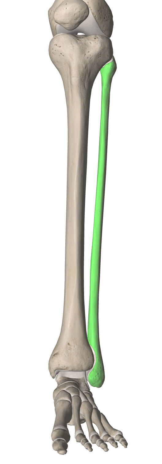

The bone colored red is called the ....

Identify the bones highlighted blue.
carpals
tarsals
metacarpals
metatarsals
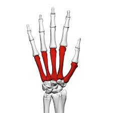
Identify the bones highlighted red.
metacarpals
metatarsals
carpals
tarsals

The bone colored red is called the ....

