
Font size
 Worksheets
MICROSCOPE
Total questions: 87
Worksheet time: 41mins
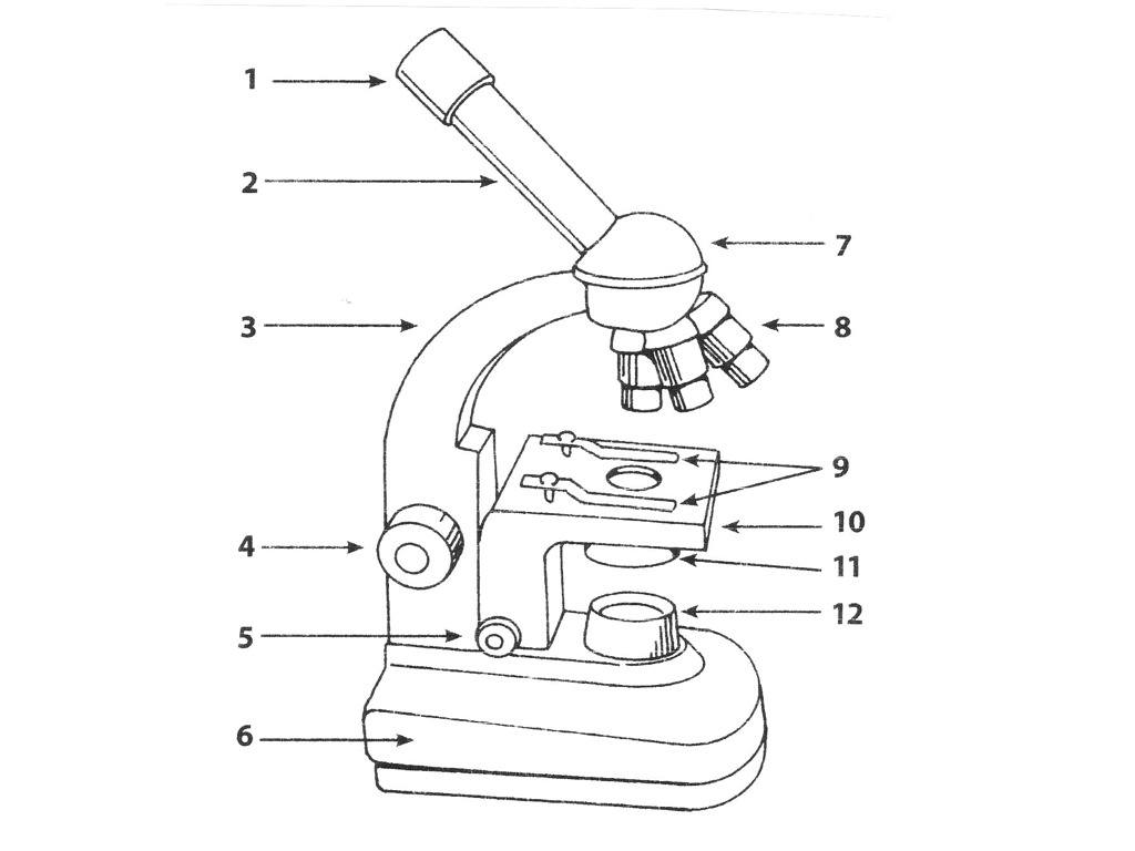


You should always carry the microscope with just one hand and let it swing freely.
True
False
Which part of the microscope should you use to sharpen the image?
Diaphragm
Coarse adjustment
Stage
Fine adjustment

Which part connects to the base and supports the microscope head?
back
spine
arm
handle

Which part supports the microscope?
base
bottom
stand
foot

The primary optical lenses on a microscope are called ___
condensors
objective lenses
stage clips
ocular lenses

Which part holds the eyepiece in place above the objective lens?
condensor
objective lense
barrel
body tube

The specimen slide is placed on the ___
platform
table
stage
base

Typically located in the base of the microscope, it provides light:
condensor
illuminator
diaphragm
adjustment knob

This part houses the objectives and allows for easy selection:
revolving nosepiece
condensor
diaphragm
adjustment knob

This part is used to collect and focus the light from the illuminator on to the specimen:
revolving nosepiece
coarse focus knob
condensor
fine focus knob

The hole in the stage, which allows light to reach the specimen, is called the ___
aperture
iris diaphragm
condensor
objective

The part you look through at the top of the microscope is called the eyepiece or ___
objective
ocular
aperture
diaphragm

These are used to hold the slide in place:
rotating nosepiece
objective lenses
stage clips
adjustment knobs


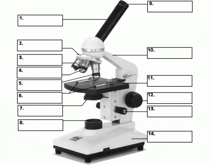

Identify the microscope part:
revolving nosepiece
coarse focus knob
diaphragm
fine focus knob

The lens assembly with the longest body is the ___
low power objective
high power objective
low power ocular
high power ocular

Typical objective lenses come in magnifications of ___
6x, 15x, 60x
60x, 150x, 600x
4x, 10x, 40x
40x, 100x, 400x

Using a 10x eyepiece and a 40x objective would give a total magnification of ___
40x
50x
400x
500x

When transporting the microscope it should be held at the ___
arm and body tube
base and stage
base and arm
arm and stage

A thin flat piece of glass used to hold objects for examination is called a ___
sheet
slide
cover slip
objective
Lenses used to change the magnification of the specimen
Body Tube
Eyepiece
Objective lenses
Diaphragm
Holds objective lenses, used to change magnification
Objective lenses
body tube
light source
revolving nosepiece
Connects eyepiece to other lenses
Body tube
Arm
Base
Diaphragm
Changes the amount of light shinning through a specimen
Light source
Diaphragm
Arm
Fine adjustment
Holds the slide in place
Stage Clips
Stage
Eyepiece
Coarse adjustment
Top lens you look through to see the specimen
Body tube
Stage
Light source
Eyepiece
Top lens you look through to see the specimen
Body tube
Stage
Light source
Eyepiece
Holds the slide in place
Stage Clips
Stage
Eyepiece
Coarse adjustment
Moves stage up and down to focus specimen
Stage
Stage clips
Diaphragm
Coarse adjustment
Used to focus specimen, smaller adjustment than coarse adjustment
Fine adjustment
Stage
Arm
Base
Supports the microscope and connects pieces
Base
Body Tube
Arm
Light source
Bottom of microscope, used for support
Arm
Stage
Light source
Base
Shines light through the specimen, making it visible
Diaphragm
Light Source
Eyepiece
Objective lens
What parts should you use to carry the microscope?
Body tube
Base
Arm
Eyepiece
Can you use the coarse adjustment on high power?
Yes
No
What is the total magnification on high power?
4
100
40
400
How many hands do you use to carry a microscope?
1
2
3
4

Label the part of the microscope. What is part B?
eyepiece lense
turret
eyepiece tube
objective lens

Label the part of the microscope. What is part A?
eyepiece lens
eyepiece tube
turret
objective lens

Label the part of the microscope. What is part D?
body
foot
objective lens
turret

Label the part of the microscope. What is part E?
stage
stage clips
focus knob
eyepiece tube

Label the part of the microscope. What is part F?
stage
stage clips
body
focus knob

Label the part of the microscope. What is part J?
focus knob
turret
eyepiece lens
miror
The __________________ reflects light into the microscope.
mirror
stage clips
field
focus knob
The smallest objective we used in class was _____X.
two
three
four
five
Tissues embedded in celloidin are usually sectioned with a _______ microtome.
Rotary
Sliding
Base Sledge
Cambridge rocking
2 of these microtomes are applicable
Study of diseases caused to tissues
Histopathology
Histology
Both 1st & 2nd
None
TRUE about freezing microtome (more than 1 answer required)
otherwise known as cold knife
uses carbon dioxide for freezing
otherwise known as cold chamber
uses liquid nitrogen for freezing
TRUE about rotary microtome (more than 1 answer required)
most common
invented by Adams
incorporated in cryostat
cuts 4-6 um sections
TRUE about standard sliding microtome (more than one answer required)
most dangerous
used for celloidin blocks
used for paraffin blocks
block remains stationary
Instrument that involves fixation, dehydration, clearing and infiltration phases:
microtome
tissue embedding system
floatation bath
automated tissue processor

Which part is A?
body tube
eyepiece
objective lens
stage clip

Which part is B?
body tube
eyepiece
objective lens
diaphragm

Which part is C?
Stage
Fine Adjustment knob
Coarse Adjustment Knob
diaphragm

What part is E?
Diaphragm
Eyepiece
base
Light source

What part is AB?
Stage
Diaphragm
Light Source
base

What part is # 9?
Body tube
base
arm
stage
This part contains the lens you look through.
nosepiece
eyepiece
low power objective lens
high power objective lens
If you want to control the amount of light entering the field of view, you would move this part to make the hole smaller or larger.
eyepiece
nosepiece
mirror/light
diaphragm
The two parts we hold to carry the microscope safely.
arm and base
arm and body tube
eyepiece and base
stage and arm
Which scenario is correct?
Focusing a slide under high power with the coarse adjustment knob.
Focusing a slide under low power with the coarse adjustment knob
Carrying the microscope by the base and body tube
Focusing a slide by moving the diaphragm
What is the type of microscope we use in the lab?
transmission electron microscope
phase contrast microscope
compound light microscope
fluorescence microscope
This type of microscope shows high magnification of a surface of a specimen using electron beams.
Dissecting microscope
Compound light microscope
Transmission Electron Microscope (TEM)
Scanning Electron Microscope (SEM)
Which provides stronger magnification....light or electron beams?
light
electron beams

Light or mirror that projects light through the diaphram
stage clips
light source
tube
arm

Small platform where the specimen is mounted for examination
base
arm
tube
stage

Holds the slide in place on the stage
nosepiece
adjustment knob
tube
stage clips

