
 Worksheets
Conduction of the Heart
Total questions: 86
Worksheet time: 58mins
The muscular middle layer of the heart is called the
epicardium
myocardium
pericardium
endocardium
What area of the heart's electrical conduction is known as the "pacemaker" of the heart?
Purknife fibers
SA node
Bundle of his
Bachmann's bundle
What area of the heart is responsible for the delay of conduction between the atrium and ventricles?
Right bundle branch
AV node
Bundle of his
Bachmann bundle
True or False: Depolarization of the heart muscle is when the muscle contracts and repolarization is when the heart muscle rests.
True
False
The SA node fires at a rate of
40-60 bpm
80-90 bpm
60-80 bpm
60-100 bpm
What part of the heart's electrical system is known as the "gatekeeper?"
AV node
Gap junction
Tetany
SA node
True and False: The SA node is located in the left atrium.
True
False
What is the correct sequence of electrical conduction of the heart?
AV node, internodal pathways, SA node, Bundle of his, right and left bundle branch, Purkinje fibers.
SA node, internodal pathways, AV node, Purkinje fibers, right and left bundle branches, Bundle of his.
SA node, internodal pathways, AV node, Bundle of his, right and left bundle branches, Purkinje fibers.
None of the options are correct.
The sounds made by the heart are due the contractions of the atria and ventricles.
True
False
What is the delivery of blood to and from the heart itself called?
coronary circulation
pulmonary circulation
systemic circulation
cardiac circulation
What is the volume of blood ejected by the ventricle in one minute called?
stroke volume
cardiac cycle
coronary volume
cardiac output

What part of the hearts conduction system is shown in the diagram?
SA node
AV Node
AV bundle
Purkinje Fibers

What part of the hearts conduction system is shown in the diagram?
SA node
AV Node
AV bundle
Purkinje Fibers

What part of the hearts conduction system is shown in the diagram?
SA node
AV Node
AV bundle
Purkinje Fibers

What part of the hearts conduction system is shown in the diagram?
SA node
AV Node
AV bundle
Purkinje Fibers
What part of the hearts electrical conduction system "receives" the impulse from the nervous system telling it to contract?
SA Node
AV Node
AV Bundle
Purkinje Fibers
What part of the hearts electrical conduction system "delays" the impulse from the atria to the ventricles?
SA Node
AV Node
AV Bundle
Purkinje Fibers
What part of the hearts electrical conduction system is found in the hearts septum and conveys impulses toward the ventricles?
SA Node
AV Node
AV Bundle
Purkinje Fibers
What part of the hearts electrical conduction system is found in the ventricle muscle walls and is what causes them to contract?
SA Node
AV Node
AV Bundle
Purkinje Fibers


True or False: The mitral valve is located on the left side of the heart.
True
False

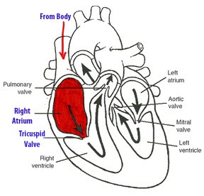


The blood enters on the left side of the heart through the ______________ and enters the left atrium. It then passes through the _____________to enter the left ventricle.*
pulmonary artery, mitral valve
pulmonary vein, tricuspid valve
pulmonary artery, aortic valve
. pulmonary vein, bicuspid valve
On the right side of the heart, blood flows from the right ventricle to the?
Tricuspid valve to the pulmonary vein
Pulmonic valve to the pulmonary artery
Tricuspid valve to pulmonary artery
Inferior vena cava to the aorta
Blood goes to the systemic circulation
through the aorta
through the pulmonary artery
through the pulmonary vein
through the right ventricle
The tricuspid valve is between
the right ventricle and the pulmonary artery
the right atrium and right ventricle
the pulmonary artery and left ventricle
the left atrium and left ventricle
The left side of the heart
separates oxygenated and deoxygenated blood before bringing it to the lungs
pumps only deoxygenated blood
pumps oxygenated blood only
brings deoxygenated blood from the lungs to the body where it is oxygenated
Which ventricle is larger?
left
right
upper
lower
The left ventricle leads to the
pulmonary veins and then the systemic circulation
pulmonary artery and then the lungs
aorta and then the lungs
aorta and then the systemic circulation
Which of the below is correct? Note you have a full minute for this question.
Pulmonary artery - left atrium - left ventricle - aorta; Vena cava - right atrium - right ventricle - pulmonary vein
Vena cava - left atrium - left ventricle - pulmonary vein; Aorta - right atrium - right ventricle - pulmonary artery
Pulmonary vein - left ventricle - left atrium; Aorta - right atrium - right ventricle
Pulmonary vein - left atrium - left ventricle - aorta; Vena cava - right atrium - right ventricle - pulmonary artery

Which side of the heart carries poorly oxygenated (deoxygenated) blood?
Right
Left
Vein that empties blood from the upper body into the upper right heart chamber
Inferior Vena Cava
Superior Vena Cava
Pulmonary Veins
Aortic veins
The term "auscultate" means
to touch
to eat
to listen
to test
Protective sac around the heart
pleural cavity
pericardium
bubble wrap
water sac
Natural pace maker in the heart
pace maker
systemic maker
pulmonary node
sinoatrial node
Sound of heart is caused by these close
nodes
vessels
valves
chambers
Any sound the heart makes besides lub-dub is called a
heart attack
echo
murmur
twitch
Largest artery is the
aorta
venae cavae
capillary
What puts O2 into the blood?
heart
lungs
nose
The nurse is assessing the client’s electrocardiography (ECG). What does the P wave on the ECG tracing represent?
Contraction of the atria
Contraction of the ventricles
Depolarization of the atria
Depolarization of the ventricles
A patient has a normal heartbeat of 72 beats per minute. What area of the conduction system should the nurse consider as functioning normally in this patient?
Bundle of His
Sinoatrial node
Purkinje’s fibers
Atrioventricular node
The nurse is determining the functioning of a patient’s sinoatrial node. How many times per minute should the nurse expect the node to normally fire?
20 to 40
25 to 55
40 to 85
60 to 100

The rate of the Sinoatrial node is
40-60 bpm
60-80 bpm
60-100 bpm
20-40 bpm
The nickname for the Atrioventricular node is
the pacemaker
the terminator
the gatekeeper
the baby maker

What is commonly referred to as the hearts 'pacemaker'?
Bundle of His
Atrioventricular Node
Sinoatrial Node
Purkinje fibres

What is the job of the atrioventricular node?
Acts as a buffer/gate to slow the signal from the SAN
Acts as the hearts pacemaker
Makes the impulse 5x stronger

The atrioventricular node (AVN) acts as a buffer and delays the impulse for approximately ....... to allow the atria to finish contracting
0.2 seconds
0.3 secons
0.5 seconds
0.1 seconds

From the AV node the message then runs down the Bundle of His which is located in the....
Atrial walls
Aorta
Septum & atrial walls
Purkinje fibres

The Purkinje fibres distribute the impulse causing the ...................... to contract.
Atrial walls
Ventricle
Atrium
Septum
What prevents blood from flowing back into the atria when the ventricles contract?
semilunar valves
sinoatrial node
atrioventricular node
tricuspid and bicuspid valves
One of the body's largest blood vessels that transports blood from the body region below the diaphragm to the heart
superior vena cava
inferior vena cava
pulmonary trunk
pulmonary veins

