
NEW
Font size
 Worksheets
Electrical Conduction of Heart and EKG
Total questions: 45
Worksheet time: 39mins
Which part starts the first step in the electrical conduction system?
sinoatrial node
AV node
AV Bundle branches
Purkinge fibers
Which part would be second in the electrical conduction system?
sinoatrial node
AV node
AV Bundle branches
Purkinge fibers
Unlike skeletal mucle tissue which is also striated, cardiac muscle is branched. What does this allow?
contraction of the entire heart in a coordinated way
contraction of the heart in one direction
contraction of the heart all at once
contraction of the heart from side to side
Which part would be third in the electrical conduction system?
sinoatrial node
AV node
AV Bundle (bundle if His)
Purkinge fibers
Contraction of the ventricles is associated with what part of the electrical conduction system?
sinoatrial node
AV node
AV Bundle branches
Purkinge fibers
Diastole is associated with
contraction of the heart
relaxation of the heart
myocardial infarction
angina
What is a cardiac cycle?
the number of beats per minute
the volume of blood ejected with each left ventricular stroke
everything that goes on in the heart from one heart beat to the next
the ability of the heart to turn around
Which of the following is represents a cardiac cycle?
PQRST
QT interval
QRS Complex
PR Interval
Relaxation of the heart corresponds to
T wave and baseline
P wave
QRS Complex
PR Interval
On an ECG, the P wave represents __________________________.
Contraction of the Atria
Contration of the Ventricle
The pause at the AV node
The impulse traveling through the bundle branches
What wave on a rhythm strip indicates the repolarization of the ventricles?
P
Q
R
S
T

Which of the following best describes this strip?
Tachycardia
Normal Sinus Rhythm
Bradycardia
Asystole
The PCT is monitoring the client’s electrocardiography (ECG). What does the P wave on the ECG tracing represent?
Contraction of the atria
Contraction of the ventricles
Depolarization of the atria
Depolarization of the ventricles
QRS
Repolarization or recovery of the ventricles
Impulses hit av bundle branch off into two bundles down front of heart into purkinje fibers
SA Nodes contracts
Both atria contracts
Impulses reach the av node 0.10 delay allows atria
The pacemaker of the heart
AV node
SA node
Bundle of His
Bundle branches
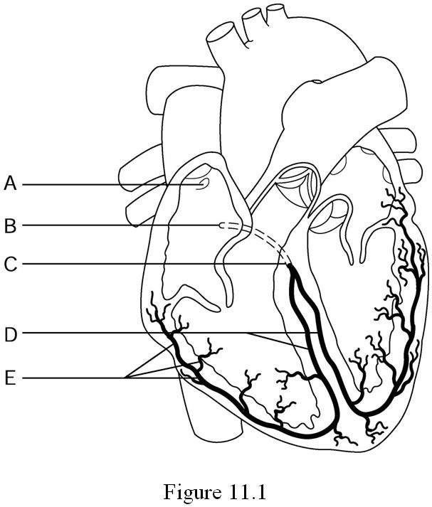
1.atria contract
2. AV node
3. Bundle of His
4.Purkinje fibers
5. SA node
6. ventricles contract
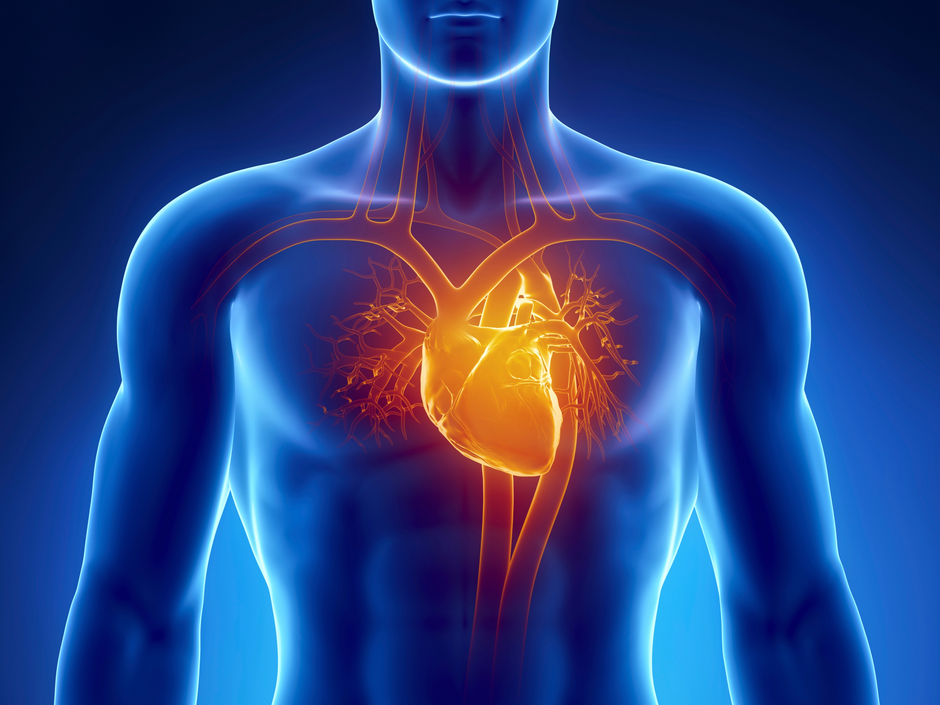
Recording of electrical changes that occur during the cardiac cycle is referred to as
blood pressure reading
ECG
QRS wave
T Wave
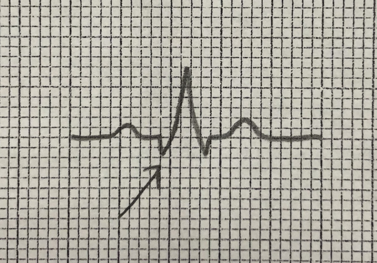
Name this part of the ekg tracing.
P
R
Q
T
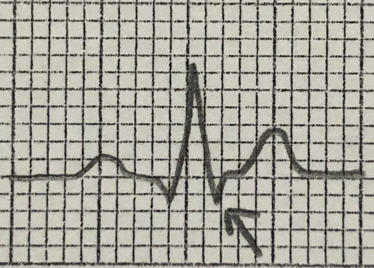
Name this part of the EKG tracing.
P
Q
R
S
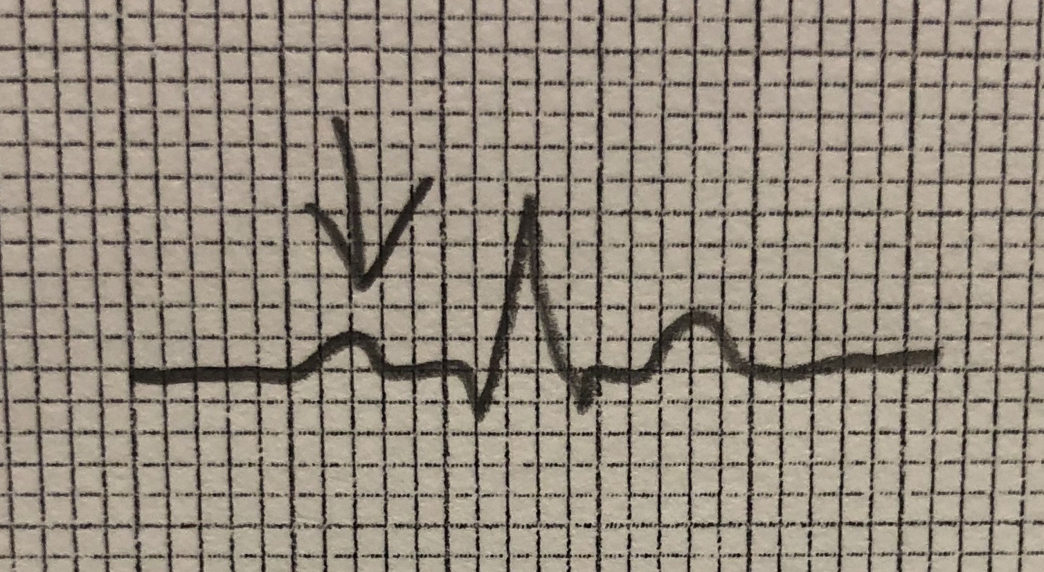
Name this part of the ekg tracing.
P
Q
R
S
If the P wave on an EKG looks abnormal, which structures in the heart are likely damaged?
SA/AV node
Aortic valve
Bundle of His
Purkinje fibers
What does depolarization result in?
relaxation
contraction
repulsion
attraction

Intrinsic electrical impulses start at which of the following locations?
AV Node
Bundle of His
Purkinje fibers
SA Node
Which of the following is most likely to occur if there is a blockage to the AV bundle nerve impulse?
the AV Node will not produce electrical impulses
the SA Node will not produce electrical impulses
no effect
the Purkinje fibers will not produce electrical impulses
A cardiac cycle is:
ventricular relaxation
ventricular contraction
equivalent to 1 complete heart beat
a heart attack
A heart rate 100 bpm or above is called _______.
bradycardia
tachycardia
arrhythmia
fibrillation
What is the effect of narrowed arteries on a person's blood flow and pressure?
increased blood flow and increased pressure
increased blood flow and decreased pressure
decreased blood flow and increased pressure
decreased blood flow and decreased pressure
A person has a blood pressure of 145/90 mmHg. How would you categorize their blood pressure?
normal range
prehypertension
hypertension
hypotension
Normal blood pressure is
80/140mmHg
140/90mmHg
120/80mmHg
80/120mmHg

What's wrong with this EKG?
Atria aren't contracting
Ventricles aren't contracting
Ventricles aren't relaxing
Nothing is wrong. This is a normal EKG.
True or False: Your heart needs a signal from your brain in order for it to keep its normal rhythm.
True
False
What makes the "lub" sound of your heart?
The aorta sending blood to the rest of your body
The Superior and Inferior Vena Cava sending blood into the atria
The SL valves closing
The AV valves closing
The sounds made by the heart are due the contractions of the atria and ventricles.
True
False
The heart makes a LUB / DUB sound. What causes the DUB sound?
Atria Contracting
AV Valves Closing
Ventricles Contracting
SL Valves Closing
What is the volume of blood ejected by the ventricle in one minute called?
stroke volume
cardiac cycle
coronary volume
cardiac output



