
 Worksheets
PART A: Sectional Anatomy Quiz
Total questions: 30
Worksheet time: 1hrs 7mins

This is an axial plane of CT brain (bone window). 1 is parietal bone. 2 is a suture that devide right and left patietal bone. What is 2?
coronal suture
lambdoid suture
sagittal suture
metopic suture

Which number is mastoid air cells?
2
3
4
7

Number 6 is one of the paranasal sinus. Its appear dark (lucence) because its contain air. What is 6?
Frontal sinus
Maxillary sinus
Ethmoid sinus
Sphenoid sinus

Number 3 is a group of __________ sinuses.
frontal
ethmoid
sphenoid
maxillary

As we look on axial plane image from feet to head, structure 5 is _______ maxillary sinus.
right
left

This is a _______ plane of CT paranasal sinuses.
axial
coronal
sagittal

This axial CT image demonstrate a pathology known as sialolithiasis. Suggest the anatomy structure involve?
Submandibular gland
Left Parotid gland
Sublingual gland
Thyroid gland

This is sagittal neck CT showing a fracture (yellow arrows). This involve vertebral body of _________
C4
C5
C6
C7

This is an axial CT neck passing through the cervical vertebra. What is X?
Spinous process
Transverse process
Vertebral body
Intervertebral foramina
The following are the MRI images of brain region. Which one is coronal plane?


Which number shows "interhemispheric fissure"?
1
2
3
4

Which number shows "lateral ventricle"?
1
2
3
4

This a coronal plane of MRI brain. Which number represent "cerebellum"
1
2
3
4

The structure contour by red line is ____
cerebrum
cerebellum
corpus callosum
pons

The structure contour by red line is _____
ventricle
pons
corpus callosum
sphenoid sinus

The structure contour by red line is _____
medulla oblongata
pineal gland
frontal sinus
pituitary gland

X represent ________
medial rectus muscle
lateral rectus muscle
optic nerve
lacrimal gland

These are radiotherapy planning to treat a cancer located posterior to nasal cavity or known as __________
larynx
nasopharynx
oropharynx
hypopharynx

These images shows a pathology involving the enlargement of ___________
corpus callosum
ventricles
medulla oblongata
cerebellum

X is one of the ventricle system in the brain that contain cerebrospinal fluid (CSF). X is _________
Left lateral ventricles
Right lateral ventricles
3rd ventricle
4th ventricle
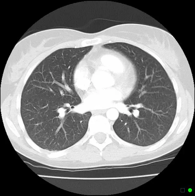
This is _______ window of axial CT chest.
mediastinal
lung
bone
soft tissue
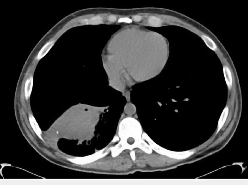
This is _______ window of axial CT chest.
mediastinal
lung
bone
soft tissue
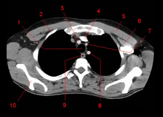
Which number represent an esophagus?
1
3
4
9
Which number represent trachea?
1
3
4
9
Number 10 is __________ scapula.
right
left
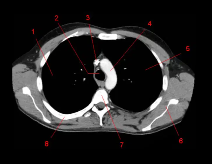
Number 4 is ___________
sternum
vertebra body
superior vena cava
arch of aorta

X is _____
Pulmonary trunk
Ascending aorta
Descending aorta
Superior vena cava
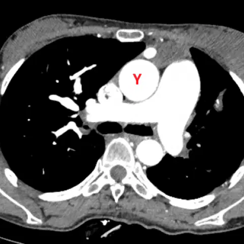
Y is _____
Pulmonary trunk
Ascending aorta
Descending aorta
Superior vena cava
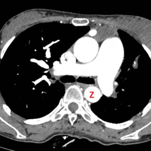
Z is _____
Pulmonary trunk
Ascending aorta
Descending aorta
Superior vena cava
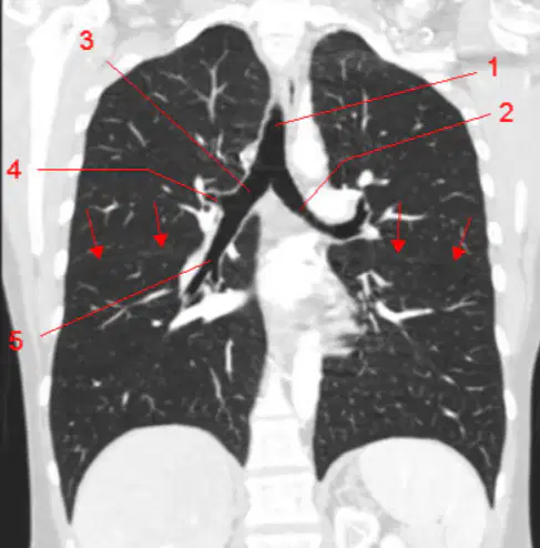
Number 1 is ________
esophagus
ascending aorta
esophagus
trachea

