
 Worksheets
NCM 112: MIDTERMS- CARDIOVASCULAR DISEASES
Total questions: 100
Worksheet time: 2hrs 40mins

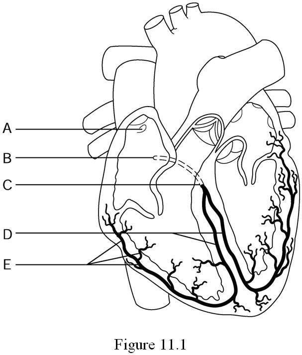
Which one is represent P wave
A
B
C
E
Which one is represent T wave
A
B
C
E
Which one is represent QRS complex
A
B
C
E
By relating events in ECG with phases in cardiac cycle, at which event the AV valve will be closed?
A
B
C
E
By relating events in ECG with phases in cardiac cycle, at which event both atria and ventricles will be relaxed?
A
B
C
E
By relating events in ECG with phases in cardiac cycle, at which event atria contract and ventricles relax?
A
B
C
E

How many cardiac cycle shown in the diagram
2
4
5
6
The ECG is the most commonly used clinical tool for diagnosing what?
Cardiac dysfunction
Cardiac Failure
Congestive Heart Failure
COPD
Where is the placement of V1 lead?
LEFT side of the sternum, directly across from V1 at the FOURTH intercostal space
LEFT side of the chest, FIFTH intercostal space, midclavicular line
RIGHT side of the sternum at the FOURTH intercostal space
LEFT side of the chest, FIFTH intercostal space, midaxillary line
Where is the placement of V2 lead?
LEFT side of the chest, FIFTH intercostal space, midclavicular line
LEFT side of the sternum, directly across from V1 at the FOURTH intercostal space
LEFT side of the chest, FIFTH intercostal space, midclavicular line
LEFT side of the chest, FIFTH intercostal space, anterior axillary line
Where is the placement of V3 lead?
LEFT side of the chest, FIFTH intercostal space, midaxillary line
RIGHT side of the sternum at the FOURTH intercostal space
LEFT side of the chest, FIFTH intercostal space, anterior axillary line
LEFT side of the chest, midway between V2 and V4
Where is the placement of V4 lead?
LEFT side of the chest, FIFTH intercostal space, midclavicular line
LEFT side of the chest, midway between V2 and V4
LEFT side of the chest, FIFTH intercostal space, midaxillary line
LEFT side of the sternum, directly across from V1 at the FOURTH intercostal space
Where is the placement of V5 lead?
LEFT side of the chest, FIFTH intercostal space, anterior axillary line
LEFT side of the chest, FIFTH intercostal space, midaxillary line
LEFT side of the chest, FIFTH intercostal space, midclavicular line
LEFT side of the sternum, directly across from V1 at the FOURTH intercostal space
V6
LEFT side of the chest, FIFTH intercostal space, midclavicular line
LEFT side of the chest, FIFTH intercostal space, anterior axillary line
LEFT side of the chest, FIFTH intercostal space, midaxillary line
LEFT side of the sternum, directly across from V1 at the FOURTH intercostal space
The paper on an ECG normally moves at:
50mm/sec
75mm/sec
25cm/sec
25mm/sec
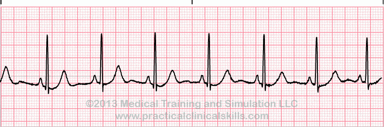
What is the ECG showing
Atrial Fibrillation
Ventricular Fibrillation
normal
Premature Ventricular Contraction
Heart Block

What is the ECG showing
Atrial Fibrillation
Ventricular Fibrillation
normal
Premature Ventricular Contraction
Heart Block

What is the ECG showing (arrow)
Atrial Fibrillation
Ventricular Fibrillation
normal
Premature Ventricular Contraction
Heart Block
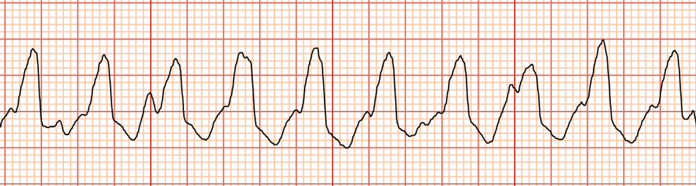
Identify the tracing
Atrial Fibrillation
Sinus Tachycardia
Ventricular tachycardia
Supraventricular tachycardia
These are the characteristics of the normal sinus rhythm except
P wave should be a rounded shape
P-R interval should be 5-6 small square
P wave should be the same shape
The rhythm should be regular
What does ST elevation suggest?
Myocardial infarction
Heart murmur
Heart block – Mobitz type II
Bundle branch block
What is the physiological correspondence of ECG P wave occurrence?
Atrial depolarization
The depolarization wave slowly passes the AV node
Repolarization of atrium
Atrioventricular conduction
The sinus bradycardia
Shows irregular rhythm
Heart rate is more than 65/min
Occurs due to anxiety
Heart rate is less than 60/min
The sinus tachycardia
Shows irregular rhythm
Occurs due to suppression of SA
Saw tooth appearance
Heart rate is more than 100/min
Regarding Atrial fibrillation
Conditions where atria beats rapidly and irregularly
QRS complex more than 120 mili second
There is P wave
Irregularly regular rhythm
The outstanding characteristic of sinus arrest is
fast heart rate
a long pause with no heart beat
a long P-R interval
everything is normal
With sinus dysrhythmia
the rate is abnormal
the rhythm is abnormal
the p wave is abnormal
the t wave is abnormal
If our patient's heart rate is 75bpm, rhythm is regular, P-R interval is normal, QRS is normal, then our diagnosis is
Sinus tachycardia
Sinus dysrhythmia
normal sinus rhythm
Sinus arrest
Medical condition where SA node ceases to generate the electrical impulses that normally stimulate the tissues for heart conduction.
Sinus Arrhythmia
1st Degree AV Block
Sinus Arrest
Sino-Atrial Block
ST Depression
NSTEMI
STEMI
ST Elevation
NSTEMI
STEMI
CARDIAC TAMPONADE
NONE OF THE ABOVE

Which best describes this rhythm?
Asystole
PEA
Ventricular Tachycardia
Ventricular fibrillation
2. Chest pain may cause?
A. permanent damage to the heart muscle
B. Heart muscle get enough oxygen in the blood
C. Heart muscle doesn't get enough oxygen in the blood
D. All above
7. Myocardial Infarction is ?
A. Heart muscle get enough oxygen in the blood
B. Permanent damage to the heart muscle / death of tissue due to lack of blood
C. Heart muscle doesn't get enough oxygen in the blood
D. Discomfort at chest feel like burning
Bleeding from blood vessels within the brain
Myocardial Infarction
Cerebral Hemorrhage
Arrhythmia
Aneurysm
The ballooning out of an artery, accompanied by the thinning arterial wall, caused by weakening of the blood vessel
Aneurysm
Hematoma
Sickle cell anemia
Coronary Artery Disease
Disease that occurs when deposits of fatty substances form along the walls of the arteries.
Atherosclerosis
Heart Failure
Myocardial Infarction
Claudication
Occurs when the ventricles of the heart are unable to contract effectively and blood pools in the heart
Heart Failure
Peripheral Vascular Disease
Arteriosclerosis
Claudication
A circulatory condition in which narrowed blood vessels reduce blood flow to the limbs
Claudication
Coronary Artery Disease
Peripheral artery disease
Arteriosclerosis
Caused by blockage of the arteries, usually in the legs.
Peripheral Vascular Disease
Myocardial Infarction
Tachycardia
Angina Pectoris
When a stroke happened
It happens when there is enough oxygen getting to the brain.
It happens when there is a lack of oxygen getting to the brain.
Both of them
What are some common symptoms of a stroke?
Paralysis of one side of body, slurring speech, facial droop
Headache
Chest pain
Shortness of breath
A patient with a history of coronary artery disease is being treated for a myocardial infarction (MI). During treatment, acute mitral valve regurgitation occurs. What is the most likely cause of the acute mitral valve dysfunction?
Infective endocarditis
Rupture of chordae tendinae
Atherosclerosis
Increased preload
Which of the following conditions occurs most commonly with cardiomyopathies?
Pericardial effusion
Heart failure
Diabetes
Myocardial infarction
A 82 year old client presents to the emergency room with dizziness, weakness, and palpitations. His heart rate is 152. The nurse demonstrates an understanding of EKG changes that occur in the elderly when she makes what correct statement about the client’s condition?
Ventricular fibrillation is a common arrhythmia in older patients.
It is normal for an aging heart to go into complete heart block.
Atrial fibrillation is a common dysrhythmia in older patients.
Sinus bradycardia can occur due to decreased baroreceptor reactivity.
Upon physical assessment of a patient diagnosed with pericarditis, the nurse anticipates what type of heart sound?
Pericardial friction rub
Diastolic murmur
S3 or S4
Pleural friction rub
A nurse is caring for a patient diagnosed with bacterial endocarditis and assessment reveals nontender hemorrhagic macules found on the palms and soles. What are these abnormalities known as?
Osler nodes
Roth spots
Splinter hemorrhages
Janeway lesions
A patient with carotid artery disease is at high risk for what complication?
Bradycardia
Stroke
Hypertension
Neck pain
Which statement from the nurse demonstrates understanding about mitral valve regurgitation?
MV regurgitation causes blood to back up into the right atrium
MV regurgitation causes an increased cardiac output
MV regurgitation causes a narrowing in the valve opening
MV regurgitation causes blood to back up into the left atrium
Which of the following is true about the semi-lunar valves in the heart?
They are between atria and ventricles
They close when pressure in the arteries is higher than pressure in the atria
They prevent backflow of blood into the atria
They open when pressure in the ventricles is higher than pressure in the arteries
During ventricular systole, which of the following occurs?
Atrioventricular valves close and semilunar valves close
Atrioventricular valves close and semilunar valves open
Atrioventricular valves open and semilunar valves are open
Atrioventricular valves open and semilunar valves close
What causes the changes in pressure in the ventricles?
Opening and closing of atrioventricular valves
Opening and closing of semi-lunar valves
Contraction and relaxation of muscles in ventricles
Contraction and relaxation of ventricles
Which of the following is NOT one of the three basic problems that generally cause cardiac conditions?
Interruption of blood flow to the heart muscle
Interruption of nerve pulses from the brain
Problems with the heart’s rhythm
Problems with the heart’s pumping
A 55-year-old patient is complaining of pain in his neck, back, and jaw. He also states that he feels nauseated and light-headed. You should suspect a(n) ________ emergency.
cardiac
hyperventilation
respiratory
ulcer
In the condition known as ________, a patient has difficulty breathing because inefficient beating of the heart causes fluid to build up in the lungs.
chronic obstructive pulmonary disease
asthma
congestive heart failure
pneumonia
A myocardial infarction occurs when one of the ________ arteries is blocked, preventing blood from getting to the heart muscle.
pulmonary
descending
coronary
carotid
You are caring for a 44-year-old man who began experiencing chest pain and shortness of breath while jogging. His pain went away completely about 20 minutes after stopping to rest. The most likely cause of his chest pain is:
myocardial infarction.
heart failure.
angina.
muscle strain.
You have completed your secondary assessment for a 65-year-old woman who has a long history of cardiac problems. She is complaining of chest pain and difficulty breathing. You should:
lay her in a supine position.
administer high-flow oxygen.
obtain a medical history.
obtain her vital signs.
You have responded to a 53-year-old man who suddenly collapsed while moving his lawn. Your assessment reveals that he is pulseless and apneic. He is likely suffering from:
angina pectoris.
acute fatigue.
cardiac arrest.
heart failure.
Which one of the following statements best describes the relationship between a heart attack and sudden cardiac arrest?
A heart attack and sudden cardiac arrest are the same thing.
Sudden cardiac arrest is a leading cause of heart attack.
Heart attack results in tissue damage; sudden cardiac arrest does not.
Heart attack is a leading cause of sudden cardiac arrest.
You are caring for a 72-year-old woman with a chief complaint of shortness of breath. You observe pedal edema and distented neck veins. She states that it is more difficult to breathe while lying down. Her signs and symptoms are most likely caused by:
a myocardial infarction.
angina pectoris.
heart failure.
congestive infarction.
For patients with the signs and symptoms of cardiac compromise, use a ______________________ to monitor peripheral oxygen saturation.
blood pressure cuff
pulse oximeter
myocardial incidator
oxygen monitor
Your patient is a 52-year-old woman who was found unresponsive on her kitchen floor by a neighbor.
She brought to the Emergency Department by ambulance.
As you approach the patient, you observe that she is supine on a stretcher. Her eyes are closed, her
lips are blue, and her skin is pale. What should be done next?
check response
check pulse
perform head tilt and chin lift
scan the chest for breathing
Atrial & Ventricular diastole takes place for about?
0.1 second
0.4 second
0.3 second
0.8 second
Atrial systole & Ventricular diastole phase takes place for about?
0.1 second
0.4 second
0.3 second
0.8 second
What does depolarization result in?
relaxation
contraction
repulsion
attraction
An ECG has been ordered for a newly admitted client. What should the nurse do prior to electrode placement?
Clean the skin with providone–iodine solution.
Ensure that the area for electrode placement is dry.
Apply tincture of benzoin to the electrode sites and wait for it to become “tacky.”
Gently abrade the skin by rubbing the electrode sites with dry gauze or cloth.
Surgeon's Expectations from YOU
preparation
anticipation
active participation -1st assist
all of the above
Heart Attack, Stroke & Atherosclerosis are examples of?
Respiratory Diseases
Cardiovascular Diseases (CVD)
Cholesterol
Hypertension
The nurse would evaluate the patient as understanding teaching for prevention of coronary artery disease if the patient stated that which of the following is a risk factor for coronary artery disease that can be controlled?
“Family history of cardiovascular disease.”
“Hypertension.”
“Ethnicity.”
“Family history of diabetes mellitus.”
The nurse would evaluate the patient as understanding teaching on the purpose of coronary artery bypass graft surgery if the patient made which of the following statements?
“It cures coronary artery disease.”
“It is done to increase blood flow to the myocardium.”
“It prevents spasms of the coronary arteries.”
“It will decrease blood flow to the coronary arteries.”
The nurse is caring for a patient who has peripheral arterial disease. Which of the following statements by the patient indicates understanding of how to manage the pain of peripheral arterial disease?
“I will sit with my legs down.”
“I will use a reclining chair.”
“I will lie down frequently.”
“I will do knee flexion exercises.”
The nurse is analyzing the data from the assessment of a client's heart and neck vessels. The client's first heart sound corresponds with what event in the cardiac cycle?
Isometric contraction
Closure of the semilunar valves
Beginning of diastole
Closure of the atrioventricular valves
A group of students is reviewing the structures of the heart, noting that the thickest layer of the heart is made up of contractile muscle cells. How would the students identify this layer?
Myocardium
Epicardium
Endocardium
Pericardium
A nurse is reviewing the electrical conduction system of the heart in preparation for assessing a client with a conduction problem. The nurse should be aware that the electrical signal originates in which location?
Bundle of His
Purkinje Fibers
Sinoatrial node
AV node
During a health visit, a client says, “I know that arteries and veins are both blood vessels, but what's the difference?” Which statement would the nurse include in the response?
“Arteries have thicker walls than veins.”
“Arteries carry 70% of the body's blood volume.”
“Arteries have a lower pressure than veins.”
“Arteries carry waste from the tissues.”
The jugular venous pressure is an indirect reflection of the:
Heart’s efficiency as a pump.
Cardiac cycle.
Conduction effectiveness.
Synchronization of mechanical activity.
A student nurse will be palpating a patient's point of maximum impulse. At which location should the student nurse palpate?
Second intercostal space, left sternal border
Third intercostal space, left axillary line
Fourth intercostal space, left sternal border
Fifth intercostal space, left midclavicular line
It is the central compartment of the thoracic cavity surrounded by loose connective tissue, as region that contains a group of structures within the thorax.
Mediastinum
Precordium
Pericardium
Sternum
Electrical impulses can be detected on the surface of the skin. This electrical activity can be measured and recorded by
ECG
X-RAY
ULTRASOUND
MRI
1. Streptokinase, Metalyse & Alteplase are thrombolytic treatment use to break down clots in some cases of --------,EXCEPT?
A. Myocardial Infarction
B. Pulmonary Embolism
C. Arterial thromboembolism
D. Deep Vein Thrombosis
Bleeding from blood vessels within the brain
Myocardial Infarction
Cerebral Hemorrhage
Arrhythmia
Aneurysm
The swollen veins that result from a slowing of blood flow back to the heart
Peripheral Vascular Disease
Transient Ischemic Attacks (TIAs)
Varicose Veins
Arrhythmia
-------- that may or may not radiate to other parts of the body is the most typical and significant symptom of myocardial infarction.
Body pain
Leg pain
Chest pain
Head pain
Which of the following is NOT one of the three basic problems that generally cause cardiac conditions?
Interruption of blood flow to the heart muscle
Interruption of nerve pulses from the brain
Problems with the heart’s rhythm
Problems with the heart’s pumping
A 55-year-old patient is complaining of pain in his neck, back, and jaw. He also states that he feels nauseated and light-headed. You should suspect a(n) ________ emergency.
cardiac
hyperventilation
respiratory
ulcer
In the mnemonic OPQRST, used to assist with the evaluation of a patient in discomfort, the “P” stands for:
Pulse
Palpitation
Pain
Provocation
In the condition known as ________, a patient has difficulty breathing because inefficient beating of the heart causes fluid to build up in the lungs.
chronic obstructive pulmonary disease
asthma
congestive heart failure
pneumonia
It is also known as "chest pain", signifying an inadequate supply of oxygen to the heart.
angina pectoris
atherosclerosis
myocardial infarction
arteriosclerosis
Which of the following major signs will lead to a severe complication?
Carditis
St. Vitus’ dance
Subcutaneous nodules
Polyarthritis
Which of the following statements about coronory arteries is wrong?
Right coronory artery supplies the right side of the heart.
Left anterior descending artery supplies the anterior wall of the left ventricle
Circumflex artery supplies the left atrium and lateral wall of the left ventricle
Right coronory artery supplies the left ventricle.

You were assessing your patient's hands and noticed that your patient's hands change in color in response to cold or stress. What do you call this disorder?
Lymphedema
Superficial Varicose Veins
Raynaud’s Syndrome
Deep Vein Thrombophlebitis

A medical condition in which superficial veins become enlarged and twisted. These veins typically develop in the legs, just under the skin.
Superficial Varicose Veins
Venous (stasis) Ulcer
Raynaud’s Syndrome
Lymphedema

