
 Worksheets
NCHSE Anatomy Review
Total questions: 213
Worksheet time: 5hrs 54mins
Basic unit of all forms of life
Cell
Nucleus
Prokaryote
Eukaryote
Thin, flexible barrier that surrounds all cells; regulates what enters and leaves the cell
Cell Wall
Cell Membrane
Nucleus
Golgi apparatus
This organelle acts like a gatekeeper, allowing some materials to pass through it, but not others.
Cell Membrane
Vacuoles
Ribsomes
Cytoplasm
A group of similar cells that perform a specific function.
Cells
Tissues
Organ
Cell Differentiation
A group of organs that work together to perform vital body functions.
Cells
Tissues
Organ
Organ System
An individual living thing, made up of one or many cells, and possibly tissues, organs and organ systems, that is capable of growing and reproducing.
Cells
Tissues
Organ
Organism
A system of organs responsible for circulating blood and oxygen through the body.
Circulatory System
Respiratory System
Digestive System
Muscular System
A system of organs responsible for taking in oxygen and releasing carbon dioxide.
Circulatory System
Respiratory System
Digestive System
Muscular System
A system of organs responsible for breaking down food and absorbing nutrients. Also responsible for disposing of solid waste.
Circulatory System
Respiratory System
Digestive System
Muscular System
System of tissues that allows movement of the body and internal organs.
Muscular System
Circulatory Systems
Respiratory System
Digestive System
Skeletal, cardiac, and smooth
Major organs of the circulatory system
Major organ of the respiratory system
Major organs of the digestive system
3 main kinds of muscles in the muscular system
Stomach, pancreas, small intestine, large intestine.
Major organs of the circulatory system
Major organ of the respiratory system
Major organs of the digestive system
3 main kinds of muscles in the muscular system
Lungs, and diaphragm
Major organs of the circulatory system
Major organ of the respiratory system
Major organs of the digestive system
3 main kinds of muscles in the muscular system
What is the correct order of the levels of cell organization from simplest (smallest) to most complex (biggest)?
Cell, Organ, Tissue, Organ System, Organism
Organism, Tissue, Cell, Organ system, Organ
Cell, Tissue, Organ, Organ System, Organism
Tissue, Cell, Organ, Organ System, Organism
above another part of the body
superior
inferior
distal
proximal
below another part of the body
superior
inferior
distal
proximal
away from point of attachment
superior
inferior
distal
proximal
close to point of attachment
superior
inferior
distal
proximal
front of the body
ventral
dorsal
medial
lateral
back of the body
ventral
dorsal
medial
lateral
toward midline
ventral
dorsal
medial
lateral
toward side of the body
ventral
dorsal
medial
lateral
toward the surface
superficial
deep
medial
lateral
away from the surface of the body
superficial
deep
medial
lateral
The elbow is __________ to the hand.
Superior
Distal
Proximal
Superficial
The chest is __________ to the head.
Inferior
Deep
Distal
Superior
The foot is _____________ to the knee.
Inferior
Distal
Proximal
Lateral
_____________________: closer to the trunk of the body.
Superior
Inferior
Distal
Proximal
______________: Further away from the trunk of the body.
Proximal
Distal
Lateral
Medial
Front Side of the body
Posterior
Anterior
Lateral
Superior
Further from the midline:
Lateral
Medial
Superficial
Posterior
Toward the midline:
Medial
Lateral
Distal
Superior
The skin is __________ to the bone.
Superficial
Deep
Superior
Inferior
The sternum (chest bone) is __________ to the shoulder.
Distal
Lateral
Medial
Superior
The Pelvic Cavity is _____ to the Abdominal Cavity.
Superior
Proximal
Inferior
Lateral
The backside of the body is also referred to as what?
Posterior
Anterior
Dorsal
Both Posterior and Dorsal
Both Anterior and Posterior
Movement of hand/foot so that the thumb/big toe moves away from the midline
inversion
eversion
adduction
abduction
Movement of the arm/leg away from the midline
adduction
eversion
inversion
abduction
Movement of the forearm/hand so the palm faces up
pronation
supination
extension
flexion
Movement of arm/leg toward the midline
flexion
extension
adduction
abduction
Movement of the hand/foot so that the thumb/big toe moves toward the midline
inversion
eversion
adduction
abduction
Movement to bend a joint
extension
circumduction
eversion
flexion
Rotating away from the center of the body
rotation
internal rotation
external rotation
eversion
Body part movement in a circular or conical motion
ovicular
pronation
rotation
circumduction
Movement to straighten a joint
extension
flexion
dorsiflexion
pronation
Rotating toward the center of the body
transverse rotation
circumduction
external rotation
internal rotation
Movement of the forearm/hand so palm faces downward or backwards
supination
pronation
palmar
flexion
The neuron is part of the
nervous tissue
connective tissue
epithelial tissue
muscular tissue
Epithelial tissue is NOT classified by shape.
True
False
Which is NOT a type of connective tissue?
Dense connective tissue
Areolar tissue
Compact bone
Smooth muscle
Which of the following is a type of epithelial tissue?
smooth muscle
transitional
nervous
blood
the functions of muscle tissue include
binding structures together
forms glands
help move substances and structures in the body
conduct impulses
Epithelial cells are classified in three shapes.
True
False
The connective tissues can be loose or dense in form.
True
False
Muscles are located in all the following EXCEPT
in the digestive tract
in the heart
attached to bones
inside bones
Epithelial cells are found in the skin.
True
False
The two ways to classify epithelium cells are
number and size
shape and color
shape and number of layers
color and number
All the following are epithelial cell shapes EXCEPT
cubodial
squamous
columnar
triangular
Tissues work together and make up
cells
organelles
molecules
organs

What is number 1?
Dermis
Epidermis
Hypodermis/subcutaneous
Artery
What is number 2?
Dermis
Epidermis
Hypodermis/subcutaneous
Artery


Identify the structure indicated
Hair shaft
Hair root
Hair folicle
Hair papilla

Identify the structure indicated by the red arrow
Blood vessels
Dermal Papillae
Hair Papilla
Adipose tissue

Identify the structure indicated by the red arrow
Sebaceous Gland
Hair follicle
Arrector pili muscle
Sudoriferous gland

Identify the structure indicated by the red arrow
blood vessels
sebaceous glands
sudoriferous glands
nerves

Identify the structure indicated by the red arrow
blood vessels
sebaceous glands
sudoriferous glands
nerves

What type of fracture is this?
greenstick
comminuted
spiral
impacted

AB
cancellous bone
compact bone
red marrow
yellow marrow

C
haversian canal
volkman canal
periosteum
endosteum

A
epiphysis
diaphysis
epiphyseal line
medullary cavity

B
epiphysis
diaphysis
epiphyseal line
medullary cavity

I
cancellous bone
compact bone
blood
osseous

J
periosteum
endosteum
yellow marrow
red marrow
The point of muscle attachment to an immovable or less movable bone is known as the ________.
innervation
origin
insertion
action
The prime mover of arm abduction is the ________ muscle.
trapezius
deltoid
triceps brachii
biceps brachii
What is the main function of the quadriceps group?
arm flexion
hand supination
thigh abduction
knee extension
foot inversion
Which muscle forms the curved calf of the posterior leg?
fibularis longus
gastrocnemius
rectus femoris
tibialis anterior
soleus
Which of these muscles is the prime mover of elbow extension?
brachialis
latissumus dorsi
deltoid
triceps brachii
biceps brachii
Which of the following is a type isometric exercise in which muscles increase in size and strength?
swimming
biking
jogging
lifting weights
dancing
Which type(s) of muscle are involuntary? Check all that apply.
Cardiac
Skeletal
Smooth
Tendons attach...
bone to bone
muscle to bone
muscle to muscle
muscle to nerves
The function of skeletal muscle is to...
move and stabilize bones
pump the heart
put pressure on organs
provide energy
Myofibrils are made of...
muscle cells
myocytes
sarcomeres
Z lines

When myosin pulls on the actin filaments, this causes the sarcomere too...
dilate
get longer
grow
shorten

In the diagram, #4 is labeling a(n)...
actin filament
myosin filament
Z line
microfibril

The arrow is pointing to the
bicep
deltoid
pectoral
tricep

The arrow is pointing to the...
calf muscle
gluteus maximum
oblique muscle
quadricep muscle
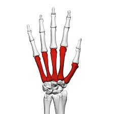
Which bones are highlighted
metacarpals
metatarsals
carpals
tarsals

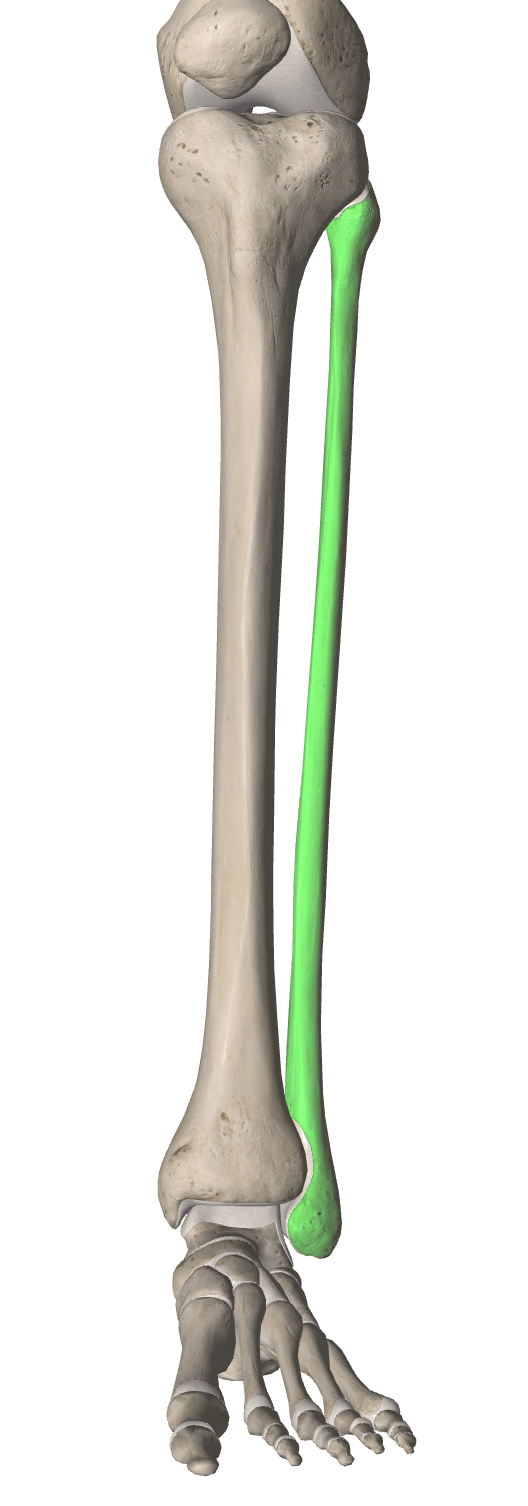
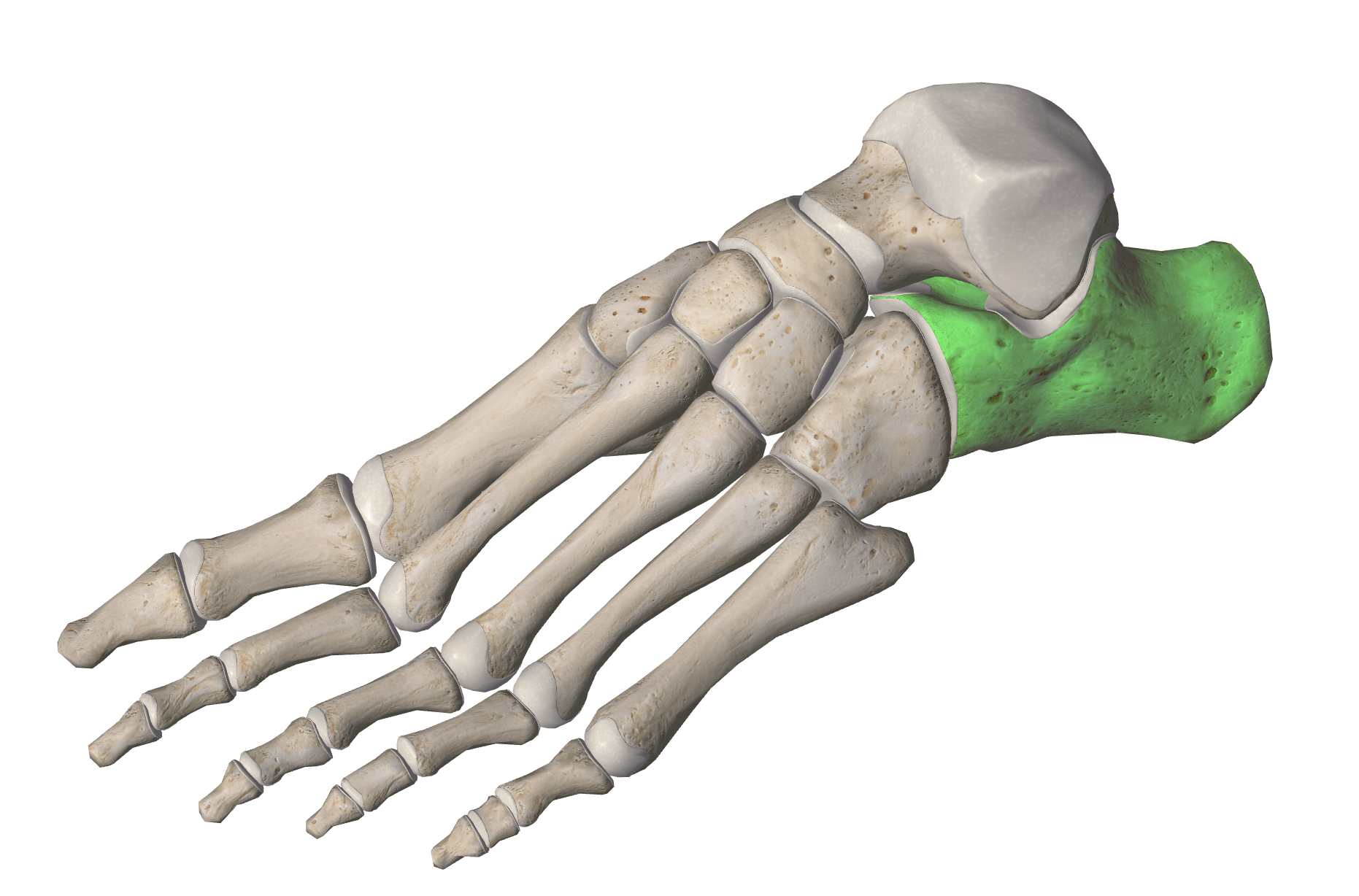
Which bones are highlighted
carpals
tarsals
metacarpals
metatarsals






Contains the medulla oblongata, pons and midbrain.
diencephalon
brain stem
cerebrum
cerebellum
protects the brain
skull
meninges
cerebrum
cerebrospinal fluid


Label the Neuron-What space does the letter "G"
represent?
Axon Terminal
Node of Ranvier
Nucleus
Myelin Sheath


a is pointing to..
nose
nasal cavity
trachea
larynx
b is pointing to..
nose
pharynx
trachea
larynx
c is pointing to..
alveoli
diaphragm
trachea
larynx
d is pointing to..
right lung
left lung
e is pointing to..
alveoli
diaphragm
trachea
larynx
f is pointing to..
nose
nasal cavity
trachea
larynx
g is pointing to..
nose
nasal cavity
trachea
larynx
h is pointing to..
nose
nasal cavity
trachea
larynx
i is pointing to..
bronchi
bronchioles
trachea
larynx
j is pointing to..
bronchi
bronchioles
trachea
larynx
k is pointing to..
right lung
left lung

#1
Renal capsule
Minor calyx
Major calyx
Ureter
#2
Renal capsule
Major calyx
Minor calyx
Ureter
#3
Renal pelvis
Major calyx
Renal medulla
Renal pyramids
#4
Renal pelvis
Major calyx
Renal medulla
Renal pyramids
#6
Renal pelvis
Major calyx
Renal medulla
Renal pyramids
#7
Ureter
Major calyx
Renal medulla
Renal cortex
#8
Ureter
Major calyx
Renal medulla
Renal cortex
#9
Ureter
Major calyx
Renal medulla
Renal cortex


The functional unit of the kidneys which includes the glomerulus, Bowman's capsule and the Loop of Henle.
arteriole
veinule
alveoli
nephron
A nitrogen waste resulting from the breakdown of the amino acids produced during protein digestion.
carbon dioxide
bile
urea
water
A) Urine passes through the ureters
B) Urea enters the kidneys
C) Urine enters the urethra

#2 - salivary gland (under the jaw)

#3 - salivary gland (under the tongue)
#3 - salivary gland (under the tongue)

Structure 16 is the
pancreas
salivary glands
larynx
esophagus
Structure 4 is the
pancreas
liver
stomach
gall bladder
Structure 17 is the
pancreas
liver
stomach
gall bladder
Structure 20 is the
pancreas
stomach
large intestine
small intestine
Structure 8 is the
pancreas
stomach
large intestine
small intestine
Structure 11 is the
gall bladder
appendix
large intestine
small intestine
Structure 22 is the
anus
rectum
large intestine
small intestine
Structure 23 is the
anus
rectum
large intestine
small intestine

What is the first part (A) of small intestine called?
ileum
duodenum
jejunum
pancreas
What is the part B of small intestine called?
ileum
duodenum
jejunum
pancreas
What is the part C of small intestine called?
ileum
duodenum
jejunum
pancreas

What structure is colored in?
ileum
rectum
duodenum
cecum

these 3 section make up the...
mouth
pharynx
esophagus
jejunum

what section is letter D?
jejunum
transverse colon
rectum
ascending colon

what section is letter Z?
descending colon
small intestine
ascending colon
rectum

what section is letter E?
transverse colon
descending colon
small intestine
ascending colon
Which gland secretes estrogen?
Anterior pituitary
Adrenal medulla
Ovaries
Pineal
Which hormone regulates sleep and wake cycles?
Adrenaline
Calcitonin
Estrogen
Melatonin
Which hormone is released when blood/calcium levels rise?
Calcitonin
Follicle stimulating
Parathyroid hormone
Prolactin
Which hormone is antagonistic to insulin?
Calcitonin
Glucagon
Prolactin
Thyroid hormone

Insulin and glucagon are produced by
the liver
the pancreas
the kidneys

Name the structure represented by 1
hypothalamus
pituitary gland
testis
nervous system
Endocrine glands secrete _
Hormones
Neurotransmitters
A wide range of chemical substances

Letter J is pointing to
Ovaries
Pineal Gland
Adrenal
Testes

The gland labeled 4 in the diagram is?
the thymus gland
the thyroid gland
the parathyroid glands
the adrenal glands

What structure(s) shown in the picture above the kidneys?
Thyroids
Adrenal glands
Testes
Parathyroids

What structure is this?
Pancreas
Thyroid
Parathyroid
Adrenal glands
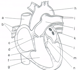
Left atrium receives oxygenated blood through
Coronary artery
Aorta
Pulmonary artery
Pulmonary vein
Tricuspid valve is present between
Left atrium and Left ventricle
Right atrium and Right ventricle
Left atrium and Right atrium
Right ventricle and Left ventricle
Blood is poured into the right atrium through
vena cava
Pulmonary vein
aorta
Pulmonary artery
Bicuspid valve and tricuspid valves are closed during
Ventricular systole
Ventricular diastole
Atrial systole
Late joint diastole
Pulse is the alternate expansion and recoil of the wall of the artery during
Ventricular systole
Ventricular diastole
Auricular diastole
Auricular systole
The second DUBB is produced at the beginning of
Ventricular diastole
Ventricular systole
Auricular systole
Auricular diastole
The first heart sound is produced when
Diastole begins
Semilunar valves close quickly
Interventricular pressure increases
Bicuspid and tricuspid valves close quickly
Left atrium receives oxygenated blood through
Coronary artery
Aorta
Pulmonary artery
Pulmonary vein
Which of the following has the thickest wall?
Right ventricle
Left ventricle
Right auricle
Left auricle
In human beings, the duration of cardiac cycle is
0.08 seconds
0.5 seconds
0.8 seconds
8.0 seconds
Which of the following carries deoxygenated blood?
pulmonary vein
pulmonary artery
aorta
coronary artery

