
 Worksheets
Unit 7 Quizizz Sensory System
Total questions: 43
Worksheet time: 4hrs 35mins
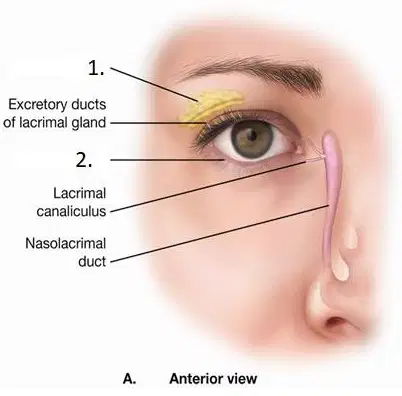
Area #1 - Is the gland that produces tears. This gland is known as the -
Lacrimal gland
Tarsal gland
Ciliary gland
Bonus gland
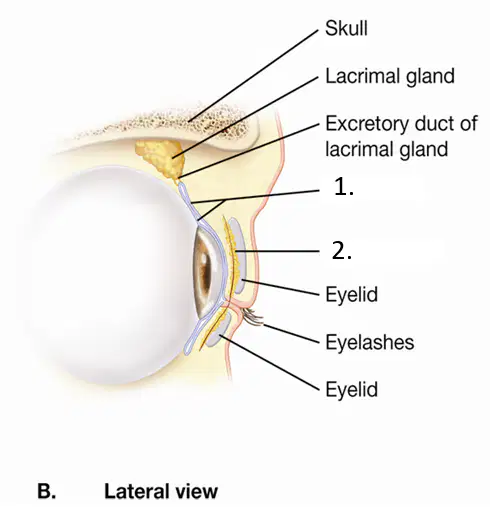
Area #1 - is a delicate external membrane that lines the eyelid and eyeball while it secrets a mucus lubricant.
Conjuctiva
Tarsal glands
Lacrimal gland
Cillary glands
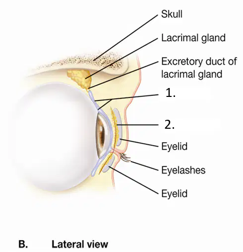
Area #2 - Are secreting glands in the eyelids that produce an oily secretion
Tarsal glands
Conjuctiva
Cliary glands
Lacrimal glands

The Sclera is known as ...
The anterior 1/6 of the eyeball
Muscles to rotate the eyeball
An area containing mostly cone and rod cells
White part of outer eye
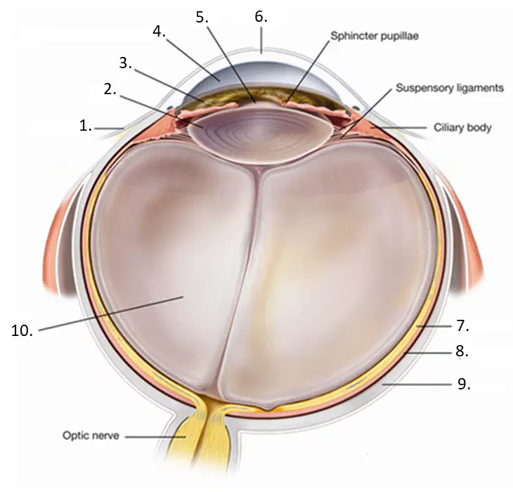
Area #6 - is the transparent part of the Sclera in the antereior part of the eye. It is where Lasik surgery is performed on.
Iris
Lens
Pupil
Cornea

Area #5 - The innermost layer of the eyeball. It contains light sensitive nerve endings that send impulses through the optic nerve to the brain.
Choroid layer
Retina
Sclera
Vitreous humor

Vitreous and Aquous humor are water filled structures in the eye that are which of the followng numbers?
#2 & #10
#4 & #10
#6 & #10
#7 and #10
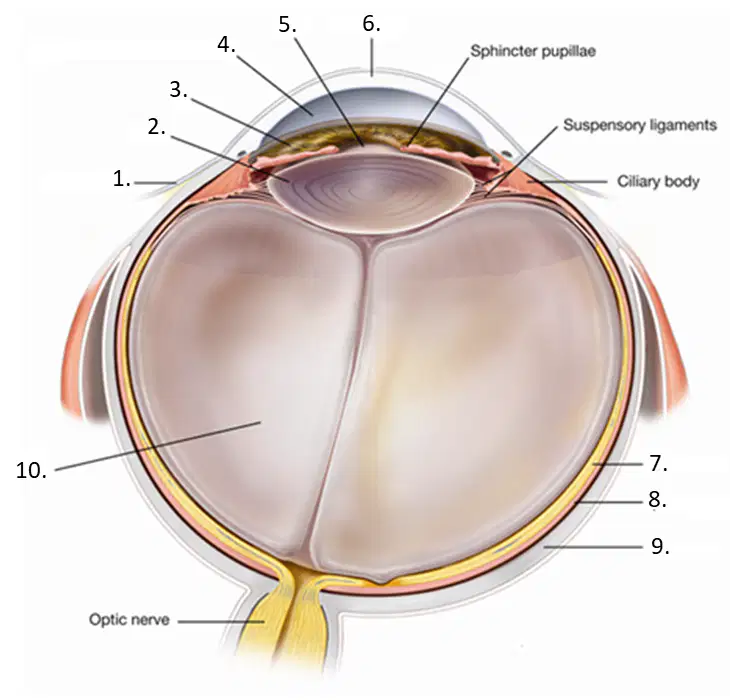
Area #2 - Is a transparent flexible structure that is curved outward on both sides. It is designed to focus light to a certain pont on the retina.
Iris
Cornea
Lens
Fovea

These are used for RGB vision. (Red Green Blue)
rods
cones

The correct path for light to enter the eye is -
Cornea → Pupil → Lens → Retina
Cornea → Lens→ Pupil → Retina
Cornea → Iris → Pupil → Retina
Iris → Pupil → Lens → Retina

LASIK surgery is done to reshape this part of the eye
Lens
Iris
Cornea
Pupil

This results from an elongated eyeball shape which causes the lens to focus objects in front of the retina instead of on it. Commonly called "Nearsightedness".
Hyperopia
Myopia
Presbyopia
Astigmatism
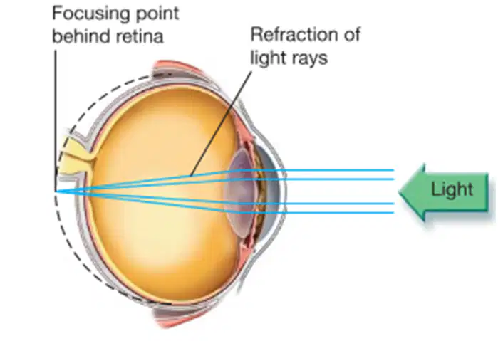
In this disorder, distance from the lens to the retina is shortened because the shape of the eyeball is more flattened. Light rays focus behind the retina instead of directly upon it. commonly caled "Farsightedness:
Myopia
Hyperopia
Presbyopia
Amblyopia

An irregular curvature of the cornea or lens that causes blurred vision.
Hyperopia
Astigmatism
Myopia
Presbyopia
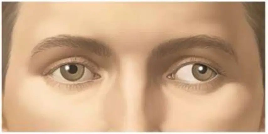
Commonly called "Lazy eye" appears during childhood where one eye is extremely dominant than the other eye. The “lazy eye” develops poor vision so if uncorrected the lazy eye can become blind.
Hyperopia
Amblyopia
Diplopia
Myopia
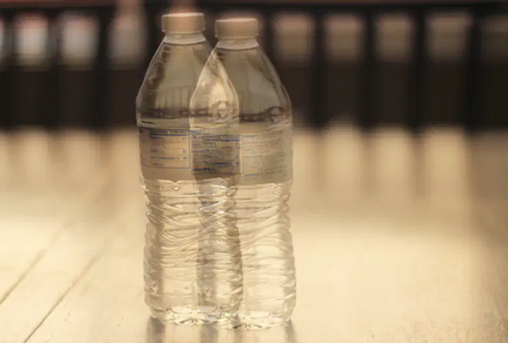
Commonly called "Double vision", this results when one eye is misaligned causing two images to appear at the same time.
Hyperopia
Diplopia
Myopia
Amblyopia
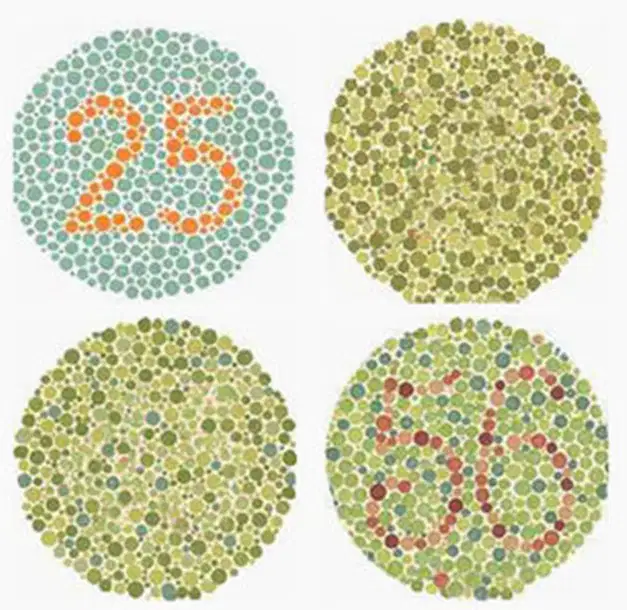
A disorder of the cone cells on the retina. This causes an inability to distinguish colors.
Color blindness
Night blindness
Presbyopia
Glaucoma

Inflammation of the conjunctiva, symptoms include redness, pain, swelling, and mucus discharge.
Glaucoma
Cataracts
Conjunctivitis
Diabetic retinopathy

Clouding of the lens which blocks light. Causes blurred vision, poor night vision, yellowing of colors, and halos around lights.
Glaucoma
Cataracts
Diabetic retinopathy
Amblyopia

Caused either by an overproduction of aqueous humor or blockage of normal aqueous humor drainage.
Cataracts
Glaucoma
Diabetic retinopathy
Vitreous floaters

What is the purpose of the fluid that fills the structure indicated by the arrow?
Stimulate receptors during movement
Detect rotational movement of the head
Produce earwax through glands
Relay auditory information

What condition involves loss of spatial awareness due to equilibrium imbalance?
Accommodation
Glaucoma
Presbyopia
Vertigo

Sound is first funneled into the ear by which of the following structures?
Auricle
External acoustic meatus
Ossicle
Tympanic membrane

A patient is diagnosed with a ruptured eardrum (#3). Which structure is involved in this condition?
Auditory ossicles
Auricle
Cochlea
Tympanic membrane

Which structure does the arrow indicate?
Tympanic membrane
Cochlea
Semicircular canal
Vestibule

Which structure does the arrow in the image indicate?
Auricle
Cochlea
Tympanic membrane
Vestibule

Which of the following functions would be impaired as a result of damage to the structure indicated by the arrow?
Sense of balance
Funneling sound waves
Hearing
Constant ringing

Area #3 is called the -
Malleus (Hammer)
Ossicles
Anvil (Incus)
Stapes (stirrup)

Area #6 is called the
Cochlea
Oval window
Tympanic membrane
Organ of corti

This whole structure is called the
Tectorial membrane
Basilar membrane
Nerve fibers
Organ of Corti

Area #2 The walls of this structure are lined with skin that contains ceruminous glands which secrete cerumen or commonly known as ear wax is called the -
Tympanic membrane
Auricle
Auditory canal
Incus

Area #1 This bone that is attached to the tympanic membrane is called -
Hammer (Malleus)
Anvil (Incus)
Stirrup (Stapes)
Ossicles

This structure equalizes air pressure on either side of the tympanic membrane, and houses the ossicles (#5)
cochlea
vestibule
Eustachian tube
semicircular canal

Area #3 A large nerve coming off of the vestibule is called the
cohlear nerve
vestibular nerve
vestibule
cochlea

Condition that causes sound like ringing in the ear
Labyrinthitis
Deafness
Tinnitus
External Otitis

Area #2 sends the impulse through the Olfactory Nerve into the olfactory cortex of the brain. The area is called
Olfactory nerve
Olfactory bulb
Olfactory receptor cell
Olfactory filaments of the olfactory nerve

An inflammation of the mucous membranes that line the nasal passage
Rhinitis
Septum problems
histamines
antihistamines

These molecules in your body cause a reaction that produces congestion and nasal drainage due to a certain allergic trigger (allergies).
histamines
antihistamines
tastants
Astigmatism

Area #1 A very small opening at the top of the taste bud is called
Gustatory cells
Taste pore
Gustatory hairs

Area #2 Threads that come out of the gustatory cells are called
Taste pore
Gustatory cells
Gustatory hairs

Area #1 is called
bone
olfactory bulb
olfactory nerve
nasal cavity

Area #3 sends impulses into the olfactory cortex of the brain. It is called the
Olfactory nerve
Olfactory bulb
Olfactory hairs
Olfactory of the south

The sensors in your nose are the long yellow cells in the diagram to the right. Area #7 They are called the
Olfactory bulb
Olfactory receptor cell
Olfactory hairs
Olfactory nerve

