
 Worksheets
RAD 117 Upper Extremity Assessment
Total questions: 18
Worksheet time: 7mins
The _____ is found at the distal end of the metacarpals.
Head
Base
Body
Condyle
The "cotton wool" appearance of radiographs is most often caused by:
Osteomyelitis
Paget disease
Marble bone
Osteogenic sarcoma
What is the correct CR for an oblique finger projection?
IP joint
MCP joint
DIP joint
PIP joint

Which joint is letter G?
1st MCP
2nd MCP
4th DIP
3rd PIP
The head of the ulna is located at the ____ end of the ulna.
Proximal
Distal
A Colles fracture involves the __________.
Proximal ulna
Distal radius
5th metacarpal
1st CMC joint
At the elbow, the ulna articulates with the distal humerus at the _____.
Capitulum
Trochlea
Olecranon
Epicondyle
An oblique wrist should be rotated _____ degrees.
15
30
45
90
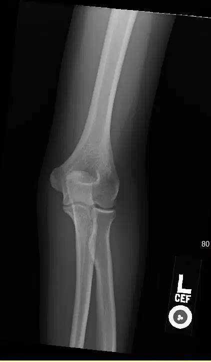
This projection is a(n) _______ elbow.
Medial oblique
Lateral oblique
Lateral
AP
What type of fracture breaks through the skin?
Closed
Simple
Open
Comminuted
The _____ is the hook-shaped process that is part of the hamate bone.
Condyle
Epicondyle
Hamulus
Fossa
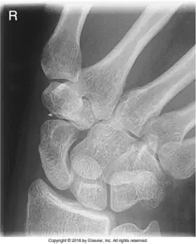
What is the name of this projection?
PA wrist
Oblique wrist
Lateral wrist
PA axial wrist with ulnar deviation
What joint connects a metacarpal to a proximal phalanx?
Proximal interphalangeal joint
Distal interphalangeal joint
Metacarpophalangeal joint
Carpometacarpal joint
The depression found on the posterior side of the distal humerus is the:
Ulnar fossa
Coronoid fossa
Radial fossa
Olecranon fossa
The metacarpophalangeal joint has a(n) ________ movement type classification.
Pivot or trochoidal
Ellipsoidal or condyloid
Hinge or ginglymus
Saddle or sellar

Which carpal is letter F?
Trapezoid
Trapezium
Pisiform
Triquetrum
The lateral elbow projection will place the humeral epicondyles ________ to the IR.
Parallel
Perpendicular
At a 45° angle
Degenerative joint disease can also be called:
Osteopetrosis
Osteomyelitis
Osteoporosis
Osteoarthritis

